Letters to the Editor
Radiol Bras. 2016 Nov/Dez;49(6):406–413
409
http://dx.doi.org/10.1590/0100-3984.2015.0025 (and do not invade) the adjacent structures. Those formations
can compress the kidney cortex, expand the sinus and distort the calyceal system. In some cases, small, predominantly peripheral, hypodense collections can be seen, with attenuation values of 0–15 HU(3). There may be thickening of the renal fasciae and retroperitoneal collections crossing the midline at the level of the renal hilum. After the administration of iodine contrast, there is no enhancement of the collections or of the walls of the cystic formations(6). In MRI, the cysts exhibit a low signal on T1-weighted sequences—although the signal strength can be in-creased if there is bleeding(6)—and a more intense signal on T2-weighted sequences, without enhancement. In addition, RL can be diagnosed on MRI scans by identifying perirenal lymphatic collections with inversion of the corticomedullary signal inten-sity(1,4), as depicted in Figure 1—B,C,D.
To suggest a diagnosis of RL, as well as to devise a treatment strategy and to prevent complications, it is essential to understand the radiological aspects of the disease and to differentiate it from other conditions that mimic cystic kidney disease. Although the combination of RL and renal failure is rare, knowledge of that association is also important to prevent comorbid conditions that can evolve with this complication, such as obesity and high blood pressure.
Andréa Farias de Melo Leite1, Bruna Venturieri1, Rosana Gonçalves de Araújo1, Eduardo Just Costa e Silva1, Jorge Elias Junior2
1. Instituto de Medicina Integral Professor Fernando Figueira de Per-nambuco (IMIP), Recife, PE, Brazil. 2. Faculdade de Medicina de Ri-beirão Preto da Universidade de São Paulo (FMRP-USP), RiRi-beirão Preto, SP, Brazil. Mailing address: Dra. Andréa Farias de Melo Leite. Rua Laura Campelo, 130, Torre. Recife, PE, Brazil, 50710-270. E-mail: [email protected].
REFERENCES
1. Rastoji R, Rastogi V. Computed tomographic scan in the diagnosis of bilateral renal lymphangiectasia. Saudi J Kidney Dis Transpl. 2008;19: 976–9.
2. Ashraf K, Raza SS, Ashraf O, et al. Renal lymphangiectasia. Br J Radiol. 2007;80:e117–8.
3. Vega J, Santamarina M. Linfangiectasia renal unilateral. Caso clínico. Rev Méd Chile. 2012;140:1312–5.
4. Restrepo JM, Amaya JEL, Sepúlveda NA, et al. Renal lymphangiectasia: MDCT and MRI findings. Rev Colomb Radiol. 2011;22:1–8. 5. Ueda S, Yanagida H, Sugimoto K, et al. Chronic renal insufficiency in a
boy with cystic renal lymphangiectasia: morphological findings and long-term follow-up. Clin Nephrol. 2007;68:416–21.
6. Vasconcelos RA, Pereira ES, Bauab Jr T, et al. Renal lymphangiectasia: incidental finding at multislice computed tomography and literature re-view. Radiol Bras. 2012;45:178–80.
Primary undifferentiated sarcoma in the thorax: a rare diagnosis in young patients
Dear Editor,
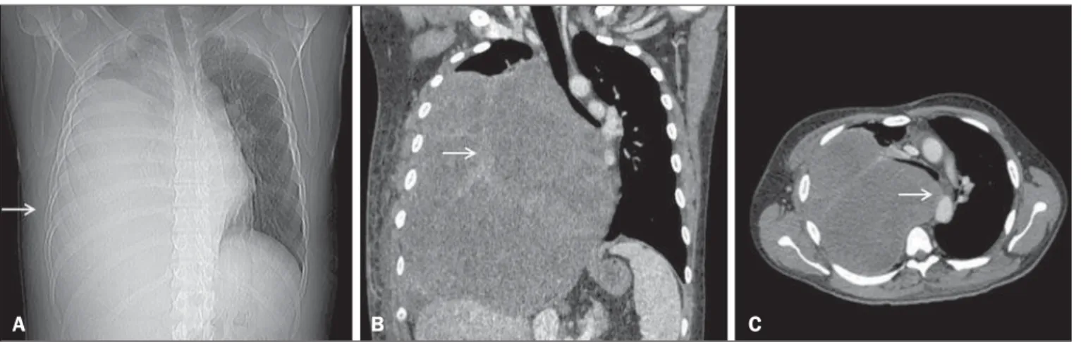
A 30-year-old man was admitted to the thoracic surgery de-partment of a tertiary hospital for investigation of a thoracic mass. Having previously received treatment for pneumonia, he presented with a two-week history of progressively increasing pain in the right hemithorax and right flank, between the anterior axillary line and midaxillary line. On clinical examination, there was an absence of breath sounds in the right hemithorax.
Computed tomography (CT) of the chest showed an exten-sive, heterogeneous, mostly solid mass in right thoracic region (Figure 1), with areas of inner content of low attenuation (21–26 Hounsfield units) and foci of bleeding, without intervening calci-fications and without osteolysis of the rib. Laboratory tests pro-duced results within the limits of normality. The patient under-went percutaneous biopsy, and the pathology examination revealed undifferentiated sarcoma (Figure 2).
Sarcomas represent a heterogeneous group of tumors derived from mesenchymal cells(1–3). They account for 1% of all neo-plasms and occur mainly in the extremities (in 60% of cases), gas-trointestinal tract (in 25%), retroperitoneal space (in 20%), and the head and neck region (in 4.1%). Primary sarcomas of the tho-rax are exceptionally rare, accounting for only 0.2% of lung can-cers and only 5% of all the thoracic neoplasms. Such sarcomas can involve the lungs, mediastinum, pleura, and, mainly, the chest wall. The presence of sarcoma in any other part of the body must be ruled out, because metastasis to the chest is much more com-mon than is primary sarcoma of the thorax(4–7).
The most common histological types of primary sarcomas are angiomyosarcoma, leiomyosarcoma, rhabdomyosarcoma, and sarcomatoid mesothelioma(8). In the chest wall, the most com-mon primary sarcomas are Ewing’s sarcoma, primitive neuroec-todermal tumor, malignant fibrous histiocytoma, chondrosar-coma, osteosarchondrosar-coma, synovial sarchondrosar-coma, and fibrosarcoma(8). Ra-diologically, these tumors typically present as large, heterogeneous masses. However, their appearance can vary from an intrabronchial
Figure 1. CT scan showing a primary sarcoma in the right hemithorax. A: CT scout image showing opacification of the right hemithorax. B: Coronal CT reconstruction with heterogeneous enhancement (arrow). C: Axial CT slice showing contralateral mediastinal deviation.
Letters to the Editor
Radiol Bras. 2016 Nov/Dez;49(6):406–413
410
http://dx.doi.org/10.1590/0100-3984.2015.0165 mass to an intravascular mass or even a solitary pulmonary
nod-ule(8).
In the case reported here, the patient was young, had no comorbidities, and presented with a voluminous mass in the right intrathoracic right region, the initial diagnostic suspicion point-ing to sarcoma.
In accordance with the literature, the analysis of clinical data and CT images obtained can only suggest primary sarcoma of the thorax as one of the differential diagnoses; the differentiation between sarcoma subtypes is only possible through pathological examination of the biopsy sample(8).
Carlos Henrique Simões de Oliveira Waszczynskyi1, Marcos Duarte Guimarães2, Luiz Felipe Sias Franco1, Bruno Hochhegger3, Edson Marchiori4
1. Hospital Heliópolis, São Paulo, SP, Brazil. 2. A.C.Camargo Cancer Cen-ter e Hospital Heliópolis, São Paulo, SP, Brazil. 3. Universidade Federal de Ciências da Saúde de Porto Alegre (UFCSPA), Porto Alegre RS, Brazil. 4. Universidade Federal do Rio de Janeiro (UFRJ), Rio de Janeiro, RJ, Brazil. Mailing address: Dr. Carlos Henrique Simões de Oliveira Waszczynskyi. Hos-pital Heliópolis. Rua Cônego Xavier, 276, Nova Heliópolis. São Paulo, SP, Brazil, 04231-902. E-mail: [email protected].
Therefore, although it is a rare neoplasm, primary sarcoma must be considered among the diagnoses of thoracic tumors, especially when a large heterogeneous mass is identified in a young patient without evidence of malignancy in another part of the body.
REFERENCES
1. Batista MN, Barreto MM, Cavaguti RF, et al. Pulmonary artery sarcoma mimicking chronic pulmonary thromboembolism. Radiol Bras. 2015;48: 333–4.
2. Nascif RL, Antón AGS, Fernandes GL, et al. Leiomyosarcoma of the in-ferior vena cava: a case report. Radiol Bras. 2014;47:384–6. 3. Teixeira VL, Santana Júnior PJ, Teixeira KISS, et al. Gastric Kaposi’s
sarcoma. Radiol Bras. 2015;48:196–7.
4. Alkis N, Muallaoglu S, Koçer M, et al. Primary adult soft tissue sarco-mas: analysis of 294 patients. Med Oncol.2011;28:391–6.
5. Hsu PK, Hsu HS, Lee HC, et al. Management of primary chest wall tu-mors: 14 years’ clinical experience. J Chin Med Assoc.2006;69:377–82. 6. Giaj Levra M, Novello S, Scagliotti GV, et al. Primary pleuropulmonary sarcoma: a rare disease entity. Clin Lung Cancer. 2012;13:399–407. 7. Tzias D, Cassidy HJ, Douraghi-Zadeh D, et al. Imaging characteristics of thoracic sarcomas – an illustration of interesting cases. ECR. 2013 / C-1007.
8. Gladish GW, Sabloff BM, Munden RF, et al. Primary thoracic sarco-mas. Radiographics. 2002;22:621–37.
Figure 2. Undifferentiated sarcoma. Hematoxylin-eosin staining (×100).
Dear Editor,
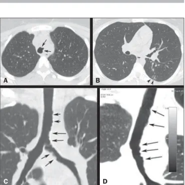
A previously healthy 22-year-old female sought medical at-tention, complaining of productive cough and hoarseness. She re-ported no other respiratory or constitutional symptoms. Physical examination revealed discrete stridor. For diagnostic clarification, computed tomography (CT) of the chest was performed The CT scan showed grouped, branching centrilobular opacities, with the “tree-in-bud” aspect, suggesting distal bronchiolar filling. The trachea and left main bronchus presented irregular internal con-tours, with nodular thickening of the walls (Figure 1), together with a discrete increase in the density of the mediastinal fat adja-cent to those changes. Sputum examination was conducted and was positive for tuberculosis, confirming the clinical and radio-logical suspicion of tracheobronchial tuberculosis. Specific treat-ment was started and resulted in resolution of the findings.
In patients with tuberculosis, tracheal involvement is relatively uncommon, occurring in only 4% of those with the endobronchial form of the disease(1–3). Tracheobronchial tuberculosis mainly af-fects younger, female patients, its incidence peaking in the third decade of life. The disease can affect the greater part of the tra-chea, also affecting the bronchi, or it can affect just a small seg-ment of the trachea or of one bronchus(4,5). The clinical presenta-tion can be insidious, simulating bronchogenic carcinoma, or acute, with a profile similar to that of asthma, foreign body aspiration, or pneumonia. In most cases, patients with tracheobronchial tuber-culosis present a productive cough, hemoptysis, chest pain, gener-alized weakness, fever, dyspnea and bronchorrhea(1,3). In cases that
Tuberculosis: tracheal involvement
Figure 1. A: Axial CT slice showing irregular narrowing of the tracheal lumen (arrows). B: Axial CT slice showing centrilobular opacities, with a tree-in-bud as-pect, in the lower lobe of the left lung, suggesting bronchiolar filling. C,D: Coronal and oblique coronal reconstructions showing irregular internal contours, together with parietal thickening (arrows), in the trachea and the left main bronchus.
A B
C D