Conventional and endovascular treatment for a rare
combination of diseases: superior vena cava syndrome and
aortoiliac aneurysm – control after 12 months
Tratamento convencional e endovascular para rara associação de doenças: síndrome de veia
cava superior e aneurisma aorto-ilíaco – controle após 12 meses
Gustavo Petorossi Solano1, Rodrigo Andrade Vaz de Melo1, Luis Claudio Rosa Arantes1, Daniel Queiroz Neves2,
Márcio Cerbazzi Tavares Cardoso3, Mauro Henrique de Lima3, Sergio Lopes de Azevedo4, Paulo Eduardo Ocke Reis5
Introduction
Superior vena cava and other central veins thrombosis are not rare entities, but benign causes are uncommon, ac-counting for not more than 15 to 22% of such thrombosis1.
Central venous catheter-induced thrombosis has become the leading benign cause of central veins occlusion due to the increasing number of invasive procedures, such as central venous access for dialysis, invasive monitoring and implantation of pacemakers2-4. he association of superior
vena cava syndrome with an aorto-iliac aneurysm makes
the case to be presented rarer still. In this paper, we re-port the conventional and endovascular approaches for the management of a patient with this association, discussing indications and the sequence of procedures, as well as the outcome ater 12 months of follow-up.
Case report
A 56-year-old male patient was admitted at the emergen-cy room complaining of shortness of breath and lushed red face. He had undergone permanent pacemaker implantation
Abstract
he association between superior vena cava syndrome and an aorto-iliac aneurysm is not common. he approach to each of theses diseases can be either by the conventional way with open surgery or by endovascular techniques. We report the two methods of surgical intervention and discuss their indications and results in this particular case.
Keywords: superior vena cava syndrome; iliac aneurysm; blood vessel prosthesis..
Resumo
A associação entre a síndrome de veia cava superior e uma dilatação aneurismática das artérias aorta e ilíacas não é comum. A abordagem de cada uma destas patologias pode ser efetuada através do modo convencional, com cirurgia aberta ou pela técnica endovascular. Neste trabalho, relatamos as duas modalidades de intervenção cirúrgica executadas e discutimos suas indicações e os resultados deste caso em particular.
Palavras-chave: síndrome de veia cava superior; aneurisma ilíaco; prótese vascular.
Study carried out at the Hospital Universitário Antônio Pedro (HUAP) – Universidade Federal Fluminense (UFF) – Niterói (RJ), Brazil.
1 Staf of the Service of Vascular Surgery at HUAP-UFF – Niterói (RJ), Brazil. 2 Resident of the Service of Vascular Surgery at HUAP-UFF – Niterói (RJ), Brazil. 3 Staf Physician (Ph.D) of theService of Vascular Surgery at HUAP-UFF – Niterói (RJ), Brazil. 4 Nurse of the Service of Vascular Surgery at HUAP-UFF – Niterói (RJ), Brazil.
5 Head of the Service of Vascular Surgery at HUAP-UFF – Niterói (RJ), Brazil.
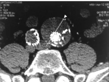
on the right infraclavicular region ater episodes of bradycar-dia and syncope, 11 years earlier. Inframammary migration had led to pulse generator replacement and repositioning of the pacemaker wires 3 years earlier. Six months ater the latter procedure, the patient presented with episodes of intermit-tent feeling of chest oppression, dyspnea, facial edema and plethora. He reported progressive increase in intensity and frequency of such episodes. Physical examination revealed dyspnea at rest, neck, chest and upper-extremity edema, in-tense facial plethora and a pulsatile mass in the hypogastrium extending to the let inguinal region. he results of routine laboratory exams were normal and chest radiography showed mediastinal enlargement caused by previous congestive heart failure. hyroid ultrasonography and coronariography were normal. Upper GI endoscopy showed varices in the upper third of the esophagus. Transesophageal echocardiogram showed images suggestive of thrombus around the pace-maker wire. Computed tomography (CT) scan of the chest did not disclose perihilar adenopathy nor mediastinal or pulmonary mass, conirming the benign origin of the syn-drome. Fibrosis was observed in the juxta-atrial segment of the superior vena cava (Figure 1). Abdominal and pelvic CT scan showed a 3 cm in diameter aneurysm in the distal third of the abdominal aorta and a bilateral common iliac artery aneurysms, measuring 2 cm in the right and 6 cm in the let (Figure 2). Upper extremity phlebography showed slow low in the let brachiocephalic trunk, reversed blood low in the azygous vein and severe stenosis of the superior vena cava at its intra-pericardial portion. hose indings were compatible with type III superior vena cava syndrome (Figure 3).
Figure 1. Fibrosis of the superior vena cava just above the atrium.
Figure 2. Iliac artery aneurysms.
Figure 3. Cavography showing right-atrium stenosis and azygous vein
with reversed blood low.
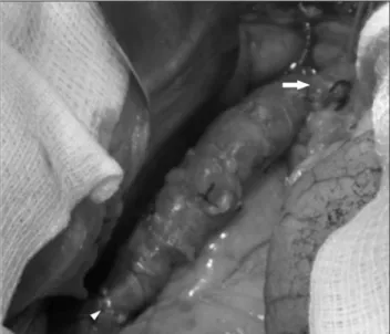
Figure 4. Anastomosis of spiral saphenous vein graft with left
Ater 12 days of thorough investigation, during which the patient was kept in bed rest (with the head elevated at 60o) and received clinical and nutritional support, the
deci-sion was made to repair irst the aorto-iliac aneurysm. With the patient under general anesthesia and orotracheal intu-bation, a Zenith endograt was deployed, the ,let iliac aneu-rysm was embolized and a PTFE femoro-femoral crossover bypass was performed . he procedure went uneventfully. Postoperative period was also uneventful and the lower ex-tremities pulses were bilaterally palpable. he patient was discharged on the 2nd postoperative day, in stable condition.
He was scheduled for readmission in two weeks.
On readmission, a let brachiocephalic vein to right atrial bypass was performed with a spiral saphenous vein grat (Figures 4, 5 and 6). he patient developed sternal wound dehiscence in the 7th postoperative day, and was
re-operated for sternal re-suture. he patient was placed on oral anticoagulation and discharged from the ICU 14 days ater the irst procedure, presenting complete regression of signs and symptoms. Since hospital discharge, he has been followed up at the outpatient clinic. ,He has had no with symptoms or complaints, and has been able to perform his daily activities normally. CT angiography at 12 months of follow-up conirmed patency of the thoracic vein grat (Figure 7) and exclusion of the aneuryms, with reduction of the let common iliac artery aneurysm diameter to 4.8 cm in (Figure 8), with no signs of endoleak (Figure 9).
Discussion
Superior vena cava syndrome of benign origin is a rare condition, although increasingly prevalent due to the ris-ing number of endovascular procedures. As shown in this
Figure 5. Anastomosis of the vein-to-right atrial bypass.
Figure 6. Outcome after low liberation by grafting.
paper, the presence of an associated aorto-iliac aneurysm makes this case even more unusual. Regarding the treat-ment, both conditions may be approached by either con-ventional surgery or endovascular technique. In cases of malignant origin, endovascular recanalization is clearly the treatment of choice5-9, and surgical repair should be
re-served for resectable tumors10-13. In cases of superior vena
casa syndrome of benign cause, the open surgical approach remains the treatment of choice14-17, although some authors
sustain their preference for the endovascular technique, regardless of the costs of additional interventions and the need for longer follow-up18-20.
In this case, we had a young patient with extensive type III chronic venous thrombosis who was not a candidate to endovascular reconstruction due to the juxta-atrial location of the occlusive lesion. Open surgery is also indicated in patients with less extensive lesions (type I or II), who have not beneited from an attempted endovascular treatment1.
On the other hand, we realized that the conventional open treatment of the aorto-iliac aneurysm would dramati-cally increase the surgical risk in this patient. he sequence of interventions in this case is another important issue to be discussed. It is well known that patients with aneurysmal dilatation of the aorta are at an increased risk of rupture during or ater an unrelated surgical procedure. Based on this, we decided to treat the aneurysm irst. he endovas-cular technique was chosen in this case aiming at a lesser surgical trauma and faster recovery of the patient, who would be submitted to another intervention shortly. Both procedures were successful and, 12 months later, the patient remains asymptomatic on follow-up.
Conclusion
Based on what has been presented, we conclude that the surgical techniques for the treatment of superior vena cava syndrome of benign origin associated with aorto-iliac aneurysm should be thoroughly discussed, before patient’s management aiming at better outcomes. he association of these entities requires deciding the best sequence of inter-ventions. Since this a rare case, a longer follow-up is neces-sary. One can conclude, however, that, in some situations, the association of endovascular and conventional tech-niques may clearly beneit the patient.
Acknowledgments
We thank Dr. Helen Christian Pessoni, Lucia Helena Soares, Maria Eduarda Tavares, Celso Luis Chouin, Wellinton Draxler Pereira, Victor Luís Correa and Tiago Mafort for contributing to the conduction of this case.
References
1. Oderich GS, Gloviczki P. Síndrome de veia cava superior. In: Brito CJ. Cirurgia Vascular, Cirurgia Endovascular, Angiologia. 2nd ed. Rio de Janeiro: Revinter; 2008. p. 1618-31.
2. Doty DB, Doty JR, Jones KW. Bypass of superior vena cava. Fifteen years’ experience with spiral vein graft for obstruction of superior vena cava caused by benign disease. J horac Cardiov Surg. 2000; 99(5):889-95.
3. Gloviczki P, Pairolero PC, Toomey BJ, et al. Reconstruction of large veins for nonmalignant venous occlusive disease. J Vasc Surg. 1992; 6(5):750-61.
4. Lin CT, Kuo CT, Lin KH, et al. Superior vena cava syndrome as a complication of transvenous permanent pacemaker implantation. Jpn Heart J. 1999;40(4):477-80.
5. Kastner RJ, Fisher WG, Blacky AR, et al. Pacemaker-induced su-perior vena cava syndrome with successful treatment by ballon venoplaty. Am J Cardiol. 1996;77(9):789-90.
6. Porte H, Metois D, Finzi L et al. Superior vena cava syndrome of malignant origin. Which surgical procedure for which diagnosis? Eur J Cardiothorac Surg. 2000;17(4):384-8.
7. Da Inês D, Chabrot P, Cassagnes L, Merle et al. O tratamento endo-vascular da síndrome do SVC de origem neoplásica: uma revisão de 34 casos. J Radiol. 2008;89(7-8 Pt 1):881-90.
8. Lanciego C, Pangua C, Chacón JI, et al. Endovascular stenting as the irst step in the overall management of malignant superior vena cava syndrome. AJR Am J Roentgenol. 2009;193(2):549-58.
9. Nguyen NP, Borok TL, Welsh J, et al. Safety and efectiveness of vas-cular endoprosthesis for malignant superior vena cava syndrome. horax. 2009; 64(2):174-8.
10. Moore W, Hollier LH. Reconstruction of the superior vena cava and central veins. In Bergan J, Yao JST. (editores). Venous disorders. Philadelphia: W.B. Saunders; 1991. p. 517-27.
Figure 9. Follow-up at 12 months – reduction in the aneurysm
11. Dartevelle PG, Chapelier AR, Pastorino U, et al. Long-term follow-up after prosthetic replacement of the sfollow-uperior vena cava com-bined with resection of mediastinal-pulmonary malignant tumors. J horac Cardiovasc Surg. 1991;102(2):259-65.
12. Magnan PE, homas P, Giudicelli R, et al. Surgical reconstruction of the superior vena cava. Cardiovasc Surg. 1994;2(5):598-604.
13. Gloviczki P, Bower TC, McKusick M, Pailorolero PC. Superiorvena cava syndrome; Endovascular and direct surgical treatment. In: Gloviczki P, Yao JST. (editores). Handbook of venous disorders. London: Chapman & Hall, 1996. p. 580-99.
14. Kalra M, Gloviczki P, Andrews JC, et al. Open surgical and endovas-cular treatment of superior vena cava syndrome caused by non-malignant disease. J Vasc Surg. 2003;38(2):215-23.
15. Alimi YS, Gloviczki P, Vrtiska TJ, et al. Reconstruction of the supe-rior vena cava: beneits of postoperative surveillance and second-ary endovascular interventions. J Vasc Surg. 1998;27(2):287-301.
16. Picquet J, Blin V, Dussaussoy C, et al. Surgical reconstruction of the superior vena cava system: indications and results. Surgery. 2009;145(1):93-9.
17. Erbella J, Hess PJ, Huber TS. Superior vena cava bypass with super-icial femoral vein for benign superior vena cava syndrome. Ann Vasc Surg. 2006;20(6):834-8.
18. Bornak A, Wicky S, Ris HB, et al. Endovascular treatment of stenos-es in the superior vena cava syndrome caused by non-tumoral le-sions. Eur Radiol. 2003;13(5):950-6.
19. Rizvi AZ, Kalra M, Biarnason H, Bower TC, Schleck C, Gloviczki P. Benign superior vena cava syndrome: stenting is now the irst line of treatment. J Vas Surg 2008;47(2):372-80.
20. Cardozo MA, Lichtenfels E, Erling Junior. N, et al. Tratamento endo-vascular da síndrome da veia cava superior: relato de caso e revisão da literatura. J Vasc Bras. 2006;5(4):308-12.
Correspondence
Gustavo Petorossi Solano Rua Doutor Mário Viana 405 – apto 1.006 – Santa Rosa CEP: 24241-000 – Niterói (RJ), Brazil E-mail: [email protected]
Author’s contribution