488
Cordeiro, Samuel Zuinglio de Biasi, et al.
Carcinoid tumor of the skin involving the sternum: Resection and reconstruction
Carcinoid tumor of the skin involving the sternum:
Resection and reconstruction*
SAMUEL ZUÍNGLIO DE BIASI CORDEIRO, PAULO LEAL, MAURO ZAMBONI(TE SBPT)
EMANUEL TORQUATO, PAULO DE BIASI CORDEIRO(TE SBPT)
Key Words: Carcinoma, Merkel cell/surgery. Carcinoid tumor/diagnosis. Carcinoid tumor/surgery. Immunohistochemistry. Sternun/surgery. Neoplasm metastasis.
Carcinoid tumor of the skin, which is a malignant neoplasm originating in the neuroendocrine system and having its pathological substrate in the Merkel cells, is a rare occurrence. It is most frequently seen on the skin around the neck and head and is more common in the elderly. This study relates the case of a 35-year-old woman presenting with a visible and palpable tumor covering the upper third of the sternum. Resection of the tumor was indicated because the patient had experienced two significant episodes of bleeding and osseous invasion had occurred. Penetration of the full thickness of the chest wall at the sternum notch necessitated the implantation of a rigid prosthesis. The definitive histopathological diagnosis was made only through postoperative analysis of the excised section. Reconstruction using a surgical cement plate and interposition of a myocutaneous flap proved its usefulness as an alternative when resection is performed in an area important to the postoperative respiratory dynamic. In this case, the patient required ventilatory support until postoperative day 6. The stability of the chest wall, together with viability of the flap and the recuperation of pulmonary function, allowed the patient to be discharged after 18 days.
*Study conducted in the Thoracic Department of the Hospital do Câncer (Cancer Hospital) in cooperation with the Instituto Nacional de Câncer
Ministério da Saúde (National Cancer Institute Health Ministry) Rio de Janeiro, RJ Brazil
Correspondence to: Estrada Francisco da Cruz Nunes, n.º 11.784, Bloco B 03 - Apto.401, Itaipu Niterói-RJ CEP: 24340-000 - Tel:: 55-21-2608 5537 - E-mail: [email protected]
Submitted: 1 August 2003. Accepted, after review: 20 November 2003.
INTRODUCTION
Primary tumors, as well as metastatic tumors, may involve elements of the chest wall and require that the resection be performed with margins of safety.(1) Replacement of the chest wall has been
used in the treatment of tumors located in this region. The execution of this procedure requires the participation of a multidisciplinary team.
489
Jornal Brasileiro de Pneumologia 30(5) - Set/Out de 2004
Abbreviations used in this paper: T9 Ninth thoracic vertebra
CASE REPORT
A 35-year-old woman reported having noticed the appearance of tumorous tissue in the sternal region one year prior to seeking treatment. Eight years previous, she had undergone a thyroidectomy (due to follicular adenoma).
Upon physical examination, a visible and palpable tumor was observed over the sternal notch. The skin over the surface of the tumor was ulcerous and showed signs of recent bleeding. A biopsy scar was seen over the lesion, and the thyroidectomy scar was noted above the affected area. The tumor was soft and friable, with a Karnofsky Performance Scale score of 100 in the initial clinical evaluation.
A computed tomography scan performed in her home town 8 months prior showed a lobulated, heterogeneous mass, extending from the plane of the thyroid gland to the level of the aortic arch. A second scan showed that the tumor now measured 10 cm along its greatest axis (Figure 1).
The patient presented an episode of bleeding from the tumor and required a blood transfusion while awaiting the completion of the preoperative exams. Bone scintigraphy, respiratory function testing and thyroid hormone level determination results were all normal. A re-evaluation of the original histopathological findings revealed undifferentiated carcinoma. Due to hemodynamic instability, the patient was not subjected to another biopsy.
Figure 1 Computed tomography of the chest showing the tumor at the level of the sternal notch
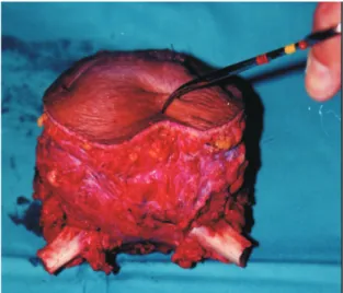
Figure 2 Surgical sample: full thickness of the chest wall at the level of the sternal notch
Figure 3 Aesthetic aspect at 3 months after the surgical procedure: myocutaneous incorporation of the dorsal muscle
methyl-490
Cordeiro, Samuel Zuinglio de Biasi, et al.
Carcinoid tumor of the skin involving the sternum: Resection and reconstruction
methacrylate surgical plate, resulting in a rigid mold to replace the bone. The sandwiching technique consists of inserting the methyl-methacrylate between two layers of Marlex mesh. The plate was fashioned during the operation in order to match the exact dimensions of the parietal defect.
The Marlex mesh was cut into two separate pieces of equal size and shape. A 2-cm, cement-free left margin served as a support for the sutures. The surgical cement (methyl-methacrylate) used to fashion the plate was prepared in water within its own receptacle and, due to the eat released during the procedure, was molded away from the surgical field and subsequently positioned. A myocutaneous pedicle flap of the dorsal muscle was positioned over the plate. Postoperative evolution was monitored in the intensive care unit, where the patient remained for 10 days. Due to thoracic instability, mechanical ventilation was used until postoperative day 6. The patient was discharged on day 18 and monitored as an outpatient until the third postoperative month. The surgical scar healed well (Figure 3). Histopathological diagnosis made through analysis of the surgical sample revealed a carcinoid tumor measuring 10 cm x 6 cm, located within the chest wall and extending from the skin to the bone tissue and deep cartilage. Venous embolism, surgical borders and 4 neoplasm-free lymph nodes were also observed. Postoperative evolution was considered normal, with full recuperation of respiratory function and performance status. Quality of life improved considerably, since the patient no longer experienced the bleeding she had suffered from the ulcerous tumor. However, 6 months after the operation, metastasis was discovered in the spinal column at the T9 level. The patient received radiation therapy, became paraplegic, was admitted to the therapeutic care ward and died 12 months after the surgical intervention.
DISCUSSION
The results of the anatomopathological examination of the surgical sample revealed carcinoid tumor of the skin, invading the bone, cartilage and muscle. Carcinoid tumors originate in the neuroendocrine system, contain neurosecretory granules, and produce peptides and amino acids that, depending on their origin, may act as if they were hormones.(2) In the case under study, the patient
presented no complaints consistent with the carcinoid syndrome. Such a presentation would have motivated the determination of the level of 5-hydroxyindolacetic
acid in urine, which would have elucidated the diagnosis.(3)
Carcinoid tumors are most commonly found in the jejunum (58%), rectum (13%), bronchi (11.5%) and thymus (2%). Other primary sites, such as the ovaries or testicles, are less common. Carcinoid tumors of the skin originate in Merkel cells (related to Kultschitzky cells) and are most often seen on the skin around the head and neck.(4) In the case related herein, the disease
first appeared at the base of the neck, as stated by the patient and evidenced by the biopsy scar.
The 12-month survival observed in this case is in accordance with that reported by Cirillo et al.(2)
in stage-III cases of the disease. Five-year survival of patient in stage I is approximately 60%. Due to the high incidence of local recurrence in lymph nodes and of remote metastasis, adjuvant radiation therapy is indicated after surgical resection.(5-7)
The purpose of the rigid methyl-methacrylate plate is to avoid flaccidity of the thorax and, simultaneously, to protect the intrathoracic structures.(8,9) Currently, such plates are employed
only when the resection involves the upper third of the sternum.(10,11) When using a rigid plate, an
attempt is made to maintain support of the first costal arches and the sternum, promoting synchronization of their movement with that of the chest wall and thereby avoiding hypoventilation. Non-rigid biological materials allow paradoxical movement. Plate-induced complications related to tracheal and vascular compression can be avoided by molding the plate to the exact size of the available space. Other complications, such as infection and local erosion, may require long-term treatment involving irrigation with saline solution and antibiotics. The dorsal muscle myocutaneous flap stretched over the plate(12,13) fills the space created by the removal
of the soft tissues. The most common complications related to this procedure are ischemia and partial necrosis. The entire vascular pedicle maintained its viability.
491
Jornal Brasileiro de Pneumologia 30(5) - Set/Out de 2004
REFERENCES
1. Briccoli A, Manfrini M, Rocca M, Lari S, Giacomini S, Mercuri M. Sternal reconstruction with synthetic mesh and metallic plates for high grade tumors of the chest wall. Eur J Surg. 2002;168:494-9.
2.Cirillo F, Buonomato M, Lima G, Cafaro I, Alquati P. C l i n i c a l e x p e r i e n c e o n e i g h t c a s e s o f M e r k e l c e l l carcinoma. Tumori. 2003;89:146-51.
3. Cimitan M, Buonadonna A, Cannizzaro R, Canzonieri V, Borsatti E, Ruffo R, et al. Somatostatin receptor s c i n t i g r a p h y v e r s u s c h r o m o g r a n i n A a s s a y i n t h e management of patients with neuroendocrine tumors o f d i f f e r e n t t y p e s : c l i n i c a l r o l e . A n n O n c o l . 2003;14:1135-41.
4. Mongardini M, Stagnitti F, Schillaci F, Monacelli G, Di Placido M, Calderisi MD, et al. Cutaneous Merkel cell carcinoma: case report. G Chir. 2002;23:334-6. 5. Muller A, Keus R, Neumann N, Lammering G, Schnabel
T. Management of Merkel cell carcinoma: case series of 36 patients. Oncol Rep. 2003;10:577-85
6. Reichel AO, Mayr D, Issing WJ. Oropharyngeal metastasis o f a M e r k e l c e l l c a r c i n o m a o f t h e s k i n . E u r A r c h Otorhinolaryngol. 2003;260:258-60.
7. D e i c h m a n n M , K u r z e n H , E g n e r U , A l t e v o g t P, Hartschuh W. Adhesion molecules CD 171 (L1 CAM)
and CD 24 are expressed by primary neuroendocrine carcinomas of the skin (Merkel cell carcinomas). J Cutan Pathol. 2003;30:363-8.
8. Monaco M, Barone M, Barresi P, Carditello A, Mondello B, Calabro B, et al. Reconstruction of the thoracic wall. G Chrir. 2000;21:193-5.
9. Wettstein R, Erni D, Berdat P, Rothenfluh D, Banic A. Radical sternectomy and primary musculocutaneous f l a p r e c o n s t r u c t i o n t o c o n t r o l o s t e í t i s . J T h o r a c Cardiosvasc Surg. 2002;123:1185-90.
1 0 . Divisi D, Ferrera R, Montagna P, Hadour G, Tronc F, Boudard C, et al. Chest wall tumors. Report of 17 cases. Rev Mal Respir. 1999;16:369-78.
11. Topalov I, Krustanov P, Iordanov D. Two methods of plastic reconstruction of extensive defects of the chest Wall (experimental study). Khirurgiia (Sofiia). 2002;58:40-4. 1 2 . Koch H, Tomaselli F, Pierer G, Schwarzl F, Haas F,
Smolle-Juttner EM, et al. Thoracic Wall reconstruction using both portions of the latissimus dorsi previourly in the c o u r s e o f p o s t e r o l a t e r a l t h o r a c o t o m y. E u r J Cardiothorac Surg 2002; 21:874-8.