Induced sputum for the diagnosis of lung disease in
HIV-positive patients*
ROSEMERI MAURICI DA SILVA, PAULO JOSÉ ZIMERMANN TEIXEIRA, JOSÉ DA SILVA MOREIRA
Background: Induced sputum is widely used in assessing airway inflammation. However, its utility as a diagnostic tool in the diagnosis of lung disease in immunosuppressed patients merits further investigation.
Objectives: To determinate the diagnostic yield of sputum induction in the diagnosis of lung diseases in HIV-positive patients.
Method: Subjects were selected from among HIV-positive patients older than 14 years who were evaluated at a reference hospital between January 2001 and September 2002. Those with respiratory symptoms for 7 days or longer with normal or abnormal chest X-rays, as well as those without respiratory symptoms but with abnormal chest X-rays, were included. All subjects were submitted to clinical examination, radiologic evaluation, sputum induction and laboratory testing. Subsequently, flexible fiberoptic bronchoscopy, bronchoalveolar lavage and transbronchial lung biopsy were performed. Samples were processed for Gram and Ziehl-Neelsen staining, quantitative culture for pyogenic bacteria, direct staining for fungi, culture for mycobacteria and fungi, silver stain for Pneumocystis jiroveci, as well as for total and differential cellularity determination.
Results: A total of 54 patients were included. Upon testing negative for any etiologic agent, 7 patients were excluded, resulting in a total of 54 patients studied. A total of 60 infectious agents were isolated. Among the etiologic agents isolated, 46.7% were P. jiroveci, 33.5 were pyogenic bacteria and 16.7% were Mycobacterium tuberculosis. Sputum induction presented 57.5% sensitivity, 42.9% specificity, 87.1% predictive positive value, 13% predictive negative value and 55.6% overall accuracy.
Conclusions: In this population, sputum induction proved to be a technique that is safe and easily performed, with a good diagnostic yield.
J Bras Pneumol 2004; 30(5) 452-8
Key words: HIV. Acquired Immunodeficiency Syndrome. Bronchoscopy. Sputum. Lung Disease/diagnosis.
*Study carried out at the Universidade Federal do Rio Grande do Sul, UFRGS,
Correspondence to: Rosemeri M. Silva. Rua Moçambique, 852, Rio Vermelho - CEP 88058-000, Florianópolis, SC Fax: 55 48 228 5333. E-mail: [email protected]
Abbreviations used in this paper: BAL – Bronchoalveolar lavage BALF – Bronchoalveolar lavage fluid HIV – Human immunodeficiency virus FEV1 – Forced expiratory volume in one second TBLB – Transbronchial lung biopsy
INTRODUCTION
It is estimated that 65% of the patients infected with human immunodeficiency virus (HIV) will present pulmonary involvement as their first clinical manifestation of the syndrome, and that approximately 80% of these patients will present some kind of pulmonary involvement over the
course of the disease(1). Infectious respiratory
symptoms in patients diagnosed with acquired immunodeficiency syndrome (AIDS) may be caused by any one of a number of pathogen groups. In addition, HIV infection is a dynamic condition in which the immune state and the risk of infection with specific etiologic agents change with time
and with the stage of the disease(2).
The clinical spectrum of pulmonary diseases is very similar to that of the various etiologic agents, which makes it extremely difficult to arrive at a
diagnosis based solely on signs and symptoms(3-5).
High rates of morbidity and mortality justify specific
treatment for this group of patients(6).
T h e r e i s s t i l l c o n s i d e r a b l e c o n t r o v e r s y s u r r o u n d i n g t h e i s s u e o f m a n a g i n g immunosuppressed patients with pulmonary
diseases(7,8). Sputum testing is the least invasive
method available. The fact that most patients in this group do not spontaneously expectorate makes the use of this noninvasive procedure more
difficult(9). For such patients, the use of a method
called induced sputum as a diagnostic tool merits
further investigation(8). It is known that invasive
procedures are not risk free and that patients with AIDS, because of their physical fragility and abnormal immunological status, are more likely to present adverse effects from diagnostic or t h e r a p e u t i c p r o c e d u r e s t h a n a r e
immunocompetent patients(10). Because of this, we
must define the exact role of noninvasive techniques, which should be fast, safe, inexpensive and accessible to institutions in which complex procedures are not commonly available, and must produce a reasonable diagnostic yield. In light of these facts, this study of the accuracy of induced sputum in the etiologic diagnosis of pulmonary infections in patients with HIV was well justified.
METHODS
From 1 January 2001 to 30 September 2002, all HIV-infected patients older than 14 years admitted to the Hospital Nereu Ramos (Nereu
Ramos Hospital, Florianópolis, SC, Brazil) were evaluated. Patients who presented clinical respiratory symptoms for more than 7 days, with either normal or abnormal chest X-rays, as well as those without respiratory symptoms but with
abnormal chest X-rays, were included(11). Exclusion
criteria included more than 7 days of empirical treatment for pulmonary disease, prophylactic treatment for tuberculosis over the preceding 6 months or for pneumonia caused by Pneumocystis jiroveci within 30 days prior to admission, severely compromised general state, coagulation alterations and severe hypoxemia. In addition, patients
presenting a greater than 20% drop in FEV1 during
sputum induction, as well as those presenting signs and symptoms that would contraindicate the induction of sputum (severe cough, dyspnea, nausea, vomiting or intolerance of the saline solution taste) were excluded. Patients who refused to participate or did not comply with the diagnostic procedures, as well as those in whom sputum induction was ineffective, were also
excluded(12-17). Patients were submitted to clinical
examination and radiologic evaluation (chest and mediastinum computed tomography scans), and laboratory testing was performed.
Subsequently, sputum induction, fiberoptic bronchoscopy and bronchoalveolar lavage (BAL) were performed. Transbronchial lung biopsy (TBLB) was performed at least 24 hours after the above-mentioned procedures. Sputum induction was carried out with progressive concentrations of hypertonic saline solution (3%, 4% and 5%)
delivered with a US 800 Air Standard® nebulizer,
and FEV1 was measured between each induction(17).
In the clinical analysis laboratory, slides for smear testing were manually prepared by selecting the most purulent or clotted portions of the sputum
samples(18). Within 30 minutes after their collection,
cell differentiation, testing for cytomegalic inclusion and parasites, and Gram staining for bacteria. Samples were also processed for culture for acid-fast bacilli (on Löwenstein-Jensen medium), culture for fungi (on Sabouraud agar) and quantitative culture for pyogenic bacteria (on
blood agar and MacConkey agar)(19-21). Sputum
samples were considered satisfactory if there were less than 10 squamous epithelial cells and more than 25 leukocytes per high-power microscopic
field, or if alveolar macrophages were present(18,22).
If a sputum sample was considered unsatisfactory, induced sputum was repeated within 48 hours. The BALF samples were considered satisfactory if there were alveolar macrophages and a maximum of 10%
epithelial cells(23). If a BALF sample was considered
unsatisfactory, the procedure was repeated within 48 hours. The TBLB samples were processed for hematoxylin-eosin, Ziehl-Neelsen, and Grocott-Gomori staining. A fragment of the tissue was submitted to “in-print” slide and subsequent Gram staining. Three biopsy fragments were placed in saline solution and then processed for cultures for acid-fast bacilli (on Löwenstein-Jensen medium), fungi (on Sabouraud agar), and pyogenic bacteria
(on blood agar and MacConkey agar)(19-21). If
samples were unsatisfactory (not representative of the lung parenchyma), TBLB was repeated within 48 hours.
The following diagnostic criteria for lung diseases were adopted as the gold standard:
1) Bacterial pneumonia: presence of a predominant morphotype evidenced by Gram staining and quantitative culture from BALF sample with at
least 104 CFU/mL, positive blood culture,
identification of the agent in the “in-print” slide from the TBLB, or isolation of the agent in
culture from tissue samples(4,5,24).
2) Pulmonary tuberculosis: positive culture for Mycobacterium tuberculosis in BALF sample or agent identified in the TBLB(4,5,25).
3) Atypical mycobacteriosis: positive BALF culture
and agent identified in the TBLB(4,5).
4) Pneumocystosis: identification of the agent in the BALF or in the TBLB.
5) H i s t o p l a s m o s i s , c o c c i d i o i d o m y c o s i s , cryptococcosis, and paracoccidioidomycosis: identification of the agent in either the BALF or TBLB – or isolation in culture(4,5,26).
6) O t h e r f u n g i , p n e u m o n i a c a u s e d b y c y t o m e g a l o v i r u s , l y m p h o i d i n t e r s t i t i a l pneumonitis, and nonspecific interstitial
pneumonitis: histopathological diagnosis(4,5,27,28).
7) Kaposi’s sarcoma: histopathological diagnosis or lesions compatible with Kaposi’s sarcoma in the respiratory tree(28,29).
8) Diseases caused by parasites: identification of the agent in the BALF or TBLB.
9) Other lung diseases: histopathological diagnosis.
Patients whose fiberoptic bronchoscopy samples did not meet the diagnostic criteria were classified into 4 groups (according to radiologic standards as analyzed by a radiologist) in order to define the additional procedures necessary for diagnosis(30):
Group 1 – interstitial or alveolar-interstitial lesions of the lung parenchyma: These patients were submitted to open-lung biopsy, and samples
were processed in the same way as TBLB samples(31).
Group 2 – alveolar lesions of the lung parenchyma: These patients were submitted to fine-needle aspiration biopsy, and samples were
processed in the same way as TBLB samples(24).
Group 3 – mediastinal lymph node enlargement or pleural effusion without parenchymatous lesions: Patients with mediastinal lymph node enlargement were submitted to mediastinoscopy, and samples were processed in the same way as
TBLB samples(2). Those with pleural effusion were
submitted to pleural punch biopsy, and samples were also processed in the same way as TBLB samples. Pleural fluid was processed in the same way as BALF samples. However, glucose, LDH, total protein, protein fractions, amylase, adenosine deaminase, and pH were also assessed in the fluid, and serum levels of glucose, LDH, total protein,
protein fractions and amylase were determined(7).
Group 4 – normal chest X-rays. These patients were not submitted to any additional procedure, and fiberoptic bronchoscopy with BAL and TBLB results were considered truly negative.
Patients presenting positive sputum cultures for M. tuberculosis and negative BALF and TBLB cultures were not submitted to any additional procedure, regardless of their chest X-ray results(4,5). Induced sputum results were considered positive when they agreed with at least one of the
of common bacteria was considered significant when a predominant bacterial morphotype was seen on Gram-stained slides and culture showed growth
equal to or greater than 106 CFU/mL(24).
At the end of the study, after confirmation of the results, a statistical software program randomly selected 20% of the cases for re-analysis (by the same examiner) in order to determine intra-observer variability. A second examiner analyzed the same material in order to estimate inter-observer
variability, using the kappa index of concordance(32).
Kappa values greater than 0.6 were considered relevant. For induced sputum results, sensitivity, specificity, positive/negative predictive values and accuracy were determined using the previously mentioned diagnostic criteria for lung diseases
(numbered from 1 to 9) as the gold standard (33,34).
The Ethics Research Committee of the Hospital Nereu Ramos approved the study protocol.
RESULTS
Of a total of 547 patients evaluated, 89 (16.3%) were admitted with respiratory symptoms. Of those 89, 58 were selected for study. The remaining 31 patients did not meet the inclusion criteria because they presented respiratory symptoms for less than 7 days. Of the 58 patients included, 4 were later excluded: 2 because they refused to participate and 2 due to coagulopathy. The final sample therefore comprised 54 patients, and their characteristics are described in Table 1.
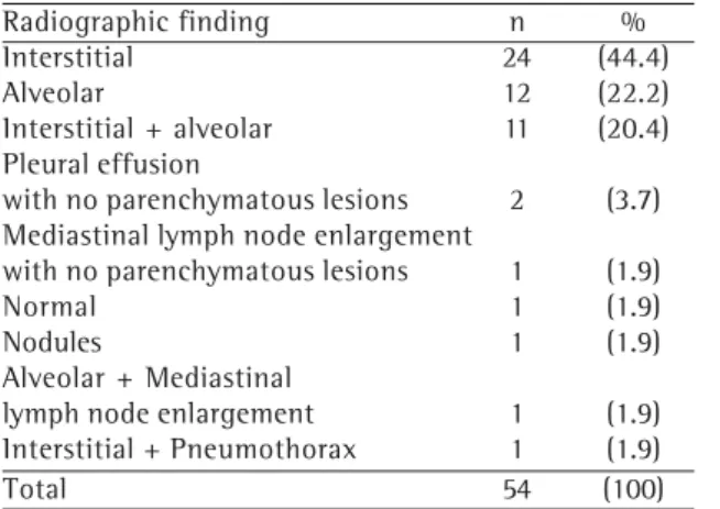
The most common radiological finding was interstitial pattern (44.4%), followed by alveolar pattern (22.2%). No radiological alterations were observed on the X-rays of 1.9% of the participants (Table 2).
The most common isolated symptom was dry cough (46.3%). The procedures for sample collection necessary for the diagnostic criteria (gold standard) are summarized in Table 3.
The most prevalent etiologic agent was P. jiroveci, followed by pyogenic bacteria. In 7 cases, the etiologic agent was not identified in any test. The etiologic agents identified are described in Table 4.
Nine patients (16.7%) presented spontaneous expectoration was seen in 9 patients (16.7%), 2 of which (1 infected with P. jiroveci and 1 infected
with Streptococcus viridans) presented results
concordant with the gold standard. No etiologic agent was isolated in any of the remaining samples. Among the cases in which M. tuberculosis was isolated, it was identified only through culture in 7 cases and only in induced sputum samples in 4
TABLE 1 Patient characteristics
n (%)
Age (years)* 35.7 (± 11.3)
Gender
Male 43 (79.6)
Female 11 (20.4)
Caucasian 46 (85.2)
Non-Caucasian 8 (14.8)
Duration of symptoms (days)* 24 ±
CD4+ T-lymphocyte count (/mm3) * 125 ±
*mean ± standard deviation n = 54
TABLE 2
Radiographic findings in 54 HIV-positive patients with lung disease
Radiographic finding n %
Interstitial 24 (44.4)
Alveolar 12 (22.2)
Interstitial + alveolar 11 (20.4)
Pleural effusion
with no parenchymatous lesions 2 (3.7)
Mediastinal lymph node enlargement
with no parenchymatous lesions 1 (1.9)
Normal 1 (1.9)
Nodules 1 (1.9)
Alveolar + Mediastinal
lymph node enlargement 1 (1.9)
Interstitial + Pneumothorax 1 (1.9)
Total 54 (100)
TABLE 3 Procedures adopted for
gold standard results
Procedure n %
Bronchoalveolar
lavage 32 (59.3)
Bronchoalveolar lavage and transbronchial
lung biopsy 12 (22.2)
Open lung biopsy 3 (5.6)
Pleural biopsy 2 (3.7)
Blood culture 2 (3.7)
Fiberoptic
bronchoscopy 2 (3.7)
Mediastinoscopy 1 (1.9)
cases. In no case was M. tuberculosis isolated through fiberoptic bronchoscopy with BAL and TBLB.
Induced sputum caused no complications. One patient presented post-TBLB pneumothorax and required thoracic drainage.
When compared with the gold standard, induced sputum presented 57.5% sensitivity, 42.9% specificity, 87.1% positive predictive value, 13% negative predictive value and 55.6% accuracy (Table 5).
In evaluating intra- and inter-observer agreement after the random selection of 20% of the cases (n = 11), a kappa value of 0.7 (which is considered good) was observed for both.
DISCUSSION
The difficulties encountered in diagnosing the etiologic agent of lower airway infections are multiplied in immunocompromised patients. These patients are susceptible to a great number of agents, and there is an unpredictable biological behavior that results from the interaction between the agent and the host. Consequently, it is extremely difficult to
make a diagnosis based solely on clinical and radiographic findings. Complicating matters further, there is no single preliminary method that presents a desirable diagnostic yield. It has been reported that techniques involving various concentrations of saline solutions and inhalation periods do not affect cellularity in samples(35). Chuard et al. stated that it is not completely impossible that hypertonic saline solutions might affect the viability of some pathogens(36). If this effect is real, we can infer that negative samples in patients with specific etiologic diagnoses might have been affected by this bias. However, additional studies are necessary in order to confirm or rule out this hypothesis. For processing of sputum samples, the highly desirable technique introduced by a member of our group (Petrillo), in which salivary contamination is eliminated, was
employed(18). Another way of reducing upper airway
contamination is through microscopic evaluation of sputum using well-established criteria for sample selection. High-quality samples increase the sensitivity of microscopy, as does the immediate delivery of
samples to the laboratory(37). The principal criticism
TABLE 4
Etiologic agents isolated in 54 HIV-positive patients with pulmonary disease
Gold Standard Induced Sputum
Etiologic agent n % n %
Pneumocystis jiroveci 28 (46.7) 16 (40.0)
Mycobacterium tuberculosis 10 (16.7) 9 (22.5)
Streptococcus pneumoniae 6 (10.0) 3 (7.5)
Streptococcus viridans 4 (6.7) 3 (7.5)
Pseudomonas aeruginosa 3 (5.0) 3 (7.5)
Klebsiella pneumoniae 2 (3.3) 2 (5.0)
Salmonella sp. 1 (1.7) 0 (0.0)
Citomegalovírus 1 (1.7) 0 (0.0)
Proteus vulgaris 1 (1.7) 1 (2.5)
Enterococcus sp. 1 (1.7) 1 (2.5)
Staphylococcus aureus 1 (1.7) 0 (0.0)
Cryptococcus neoformans var. grubii 1 (1.7) 0 (0.0)
Serratia liquefasciens 1 (1.7) 1 (2.5)
Total 60 (100) 40 (100)
TABLE 5
Induced sputum yield in 54 HIV-positive patients with lung disease
Induced Sputum Positive Gold Standard Negative Gold Standard Total
Positive 27 4 31
Negative 20 3 23
Total 47 7 54
of sputum examination as a diagnostic method for pulmonary infection is the potential contamination of samples. This is relevant since 45% of samples
sent to laboratories are contaminated by saliva(36).
This problem can be minimized with the combination of mechanical techniques for the removal of saliva from samples and microscopic selection of samples through the evaluation of alveolar macrophages, polymorphonuclear leukocytes and epithelial cells. Quantitative culture is a useful technique that facilitates the differentiation of colonization and
infection in cases of bacterial pneumonia(37,38).
Consequently, high concentration of microorganisms might be related to etiology, whereas low concentration of microorganisms might be related to contamination(39).
Of the 54 patients studied, 47 (87%) received a final diagnosis. Two final diagnoses were confirmed for 13 patients, and 3 final diagnoses were simultaneously defined for 1 patient. Rimland et al. reported that 13% of patients with pneumonia and HIV infection presented 2
etiologies, and 2% presented 3 etiologies(40). Jensen
et al. noted that co-infection has prognostic significance, resulting in higher short-term mortality and increasing the influence of the correct etiologic
diagnosis on the prognosis(41).
The most common etiologic agents were P. jiroveci (46.7%), pyogenic bacteria (33.5%) and M. tuberculosis (16.7%). Brazilian studies have shown a higher prevalence of pneumocystosis in this specific population. However, pyogenic bacteria
are not considered prevalent etiologic agents(42).
Danés et al. reported bacterial pneumonia as the most frequent diagnosis in HIV-positive patients
(63% of cases)(43). We confirmed the importance
of cultures for the diagnosis of pulmonary tuberculosis since 7 of the 10 diagnosed cases presented negative smear tests. Conde et al. reported higher sensitivity of cultures for the
diagnosis of pulmonary tuberculosis(44).
In a study comprising 40 patients diagnosed with AIDS and pneumonitis, Rolston et al. reported that induced sputum presented 45% overall
sensitivity(45). Miller et al. reported 13% sensitivity
in 82 patients with pneumocystosis(46). These
proportions are lower than those found in our study, which may be attributable to the fact that those studies did not include any invasive procedures other than fiberoptic bronchoscopy.
Our data showed that induced sputum is an effective technique for obtaining lower respiratory tract samples in this group of patients. Induced sputum proved particularly useful in populations in which spontaneous sputum production is infrequent, avoiding the use of traditional invasive procedures for sputum collection. Our results clearly showed that the best individual parameter of induced sputum as a diagnostic tool was the positive predictive value. This parameter, taken together with the other results, reinforces the fact that this diagnostic strategy is a promising technique since it reduces the necessity of invasive procedures for an appreciable number of patients. We conclude that induced sputum is an easily performed technique that does not require costly equipment and presents an alternative in the initial diagnosis of HIV-positive patients, especially in locales in which access to the more invasive methods is not practical.
REFERENCES
1. Suffredini AF, Masur H. Pulmonary dysfunction in patients infected with human immunodeficiency virus. In: Pennington JE, ed. Respiratory infections: diagnosis and management. 2nd ed. New York: Raven Press; 1988:241-63.
2. H a r a m a t i L B , J e n n y - A v i t a l E R . A p p r o a c h t o t h e diagnosis of pulmonary disease in patients infected with the human immunodeficiency virus. J Thorac Imaging 1998;13:247-60.
3. Stevens DA. Diagnosis of fungal infections: current status. J Antimicrob Chemother 2002;49:11-9. 4. Murray JF, Mills J. Pulmonary infectious complications
of human immunodeficiency virus infection (Part I). Am Rev Respir Dis 1990;141:1356-72.
5. Murray JF, Mills J. Pulmonary infectious complications of human immunodeficiency virus infection (Part II). Am Rev Respir Dis 1990;141:1582-98.
6. Masur H, Shelhamer J. Empiric outpatient management of HIV-related pneumonia: economical or unwise? Ann Intern Med 1996;124:451-3.
7. Smith PR. What diagnostic tests are needed for community-acquired pneumonia? Med Clin North Am;85:1381-96.
8. Santamauro JT, Mangino DA, Stover DE. The lung in the immunocompromised host: diagnostic methods. Respiration 1999;66:481-90.
9. Speich R. Diagnosis of pulmonary problems in HIV-infected patients. Monaldi Arch Chest Dis 1993;48:221-32. 1 0 . L u c e J M . S p u t u m i n d u c t i o n i n t h e a c q u i r e d
immunodeficiency syndrome. Am Rev Respir Dis 1986;133:513-4.
1 2 . Gracia JD, Miravitlles M, Mayordomo C, Ferrer A, Alvarez A, Bravo C, et al. Empiric treatments impair the diagnostic yield of BAL in HIV-positive patients. Chest 1997;111:1180-6.
1 3 . Barreto SM, Molinari JF. I Consenso brasileiro sobre pneumonias: pneumonias em portadores da Síndrome da Imunodeficiência Adquirida (SIDA/AIDS). J Pneumol 1998;24:95-100.
1 4 . Mendelson J. Principles of neoplasia. In: Harrison TR, e d . P r i n c i p l e s o f i n t e r n a l m e d i c i n e . 2a e d . N e w York:McGraw-Hill;1991.p.1576-87.
1 5 . O’Dell MW, Lubeck DP, O’Driscoll P, Matsuno S. Validity of the Karnofsky performance status in an HIV-infected sample. J Acquir Immune Defic Syndr Hum Retrovirol 1995;10:350-7.
1 6 . G u n d y K V, B oy l e n C T. F i b e ro p t i c b ro n c h o s c o py. I n d i c a t i o n s , c o m p l i c a t i o n s , c o n t r a i n d i c a t i o n s . Postgrad Med 1988;83:289-94.
1 7 . Efthimiadis A, Pizzichini E, Pizzichini MM, Hargreave F E . S p u t u m e x a m i n a t i o n f o r i n d i c e s o f a i r w a y inflamation:laboratory procedures. Canadian Thoracic Society 1997.
1 8 . Petrillo VF. A relevância clínica na preparação artesanal do esfregaço do escarro (judiciosamente selecionado) na bacteriologia dessa secreção: estudo comparativo com o aspirado de punção pulmonar transcutânea [Dissertação de Mestrado]. Porto Alegre:Universidade Federal do Rio Grande do Sul, 1987.
1 9 . M a r s h a l l J R . M a n u a l d e l a b o r a t ó r i o c l í n i c o – microbiologia. 1a ed. São Paulo: Santos Livraria Editora; 1995.
2 0 . Luna LG. Manual of histologic staining methods the armed forces institute of pathology. 3a ed. New York:McGraw-Hill Book Co New York; 1968.
2 1 . Behmer OA, Tolosa EMC, Neto AGF. Manual de técnicas para histologia normal e patológica. 1a ed. São Paulo:EDART;1976.
2 2 . Cordero E, Pachon J, Rivero A, Giron-Gonzalez JA, Gomez-Mateos J, Merino MD, et al. Usefulness of sputum culture for diagnosis of bacterial pneumonia in HIV-infected patients. Eur J Clin Microbiol Infect Dis 2002;21:362-7.
2 3 . Baughman RP, Dohn MN. Immunocompromised host. In:Baughman RP, ed. Bronchoalveolar lavage. St. Louis:Mosby Year Book Inc;1992:41-63.
2 4 . Skerret JS. Diagnostic testing for community-acquired pneumonia. Clin Chest Med 1999;20:531-48. 2 5 . Moreira JS. Valorização do exame de escarro. In:
Palombini BC, ed. Doenças das vias aéreas. Uma visão c l í n i c a i n t e g r a d o r a ( V i a e r o l o g i a ) . R i o d e Janeiro:Revinter;2001.p.187-90.
2 6 . Pisani RJ, Wright AJ. Clinical utility of bronchoalveolar lavage in immunocompromised hosts. Mayo Clin Proc 1992;67:221-7.
2 7 . S l o t a r D , Es c a l a n t e P, J o n e s B E . P u l m o n a r y manifestations of HIV/AIDS in the tropics. Clin Chest Med 2002;23:355-67.
2 8 . White DA, Matthay RA. Noninfectious pulmonary c o m p l i c a t i o n s o f i n f e c t i o n w i t h t h e h u m a n i m m u n o d e f i c i e n c y v i r u s . A m R e v R e s p i r D i s 1989;140:1763-87.
2 9 . Jeyapalan M, Steffenson S. Diagnosis of pulmonary Kaposi’s sarcoma in AIDS patients. AIDS Patient Care STDs 1997;11:9-12.
3 0 . Vander Els NJ, Stover DE. Approach to the patient with pulmonary disease. Clin Chest Med 1996;17:767-85. 31 . Trachiotis GD. Criteria for open lung biopsy. Focusing
on patients with AIDS. Chest 1995;108:293-4. 3 2 . Pereira CAC, Neder JA. Diretrizes para testes de função
pulmonar 2002. J Pneumol 2002;28:238.
3 3 . Fletcher RH, Fletcher SW, Wagner EH. Epidemiologia clínica. Bases científicas da conduta médica. 2a ed. Porto Alegre: Artes Médicas;1989.
3 4 . Menezes AMB, Santos IS. Curso de epidemiologia básica para pneumologistas. 4a parte – epidemiologia clínica. J Pneumol 1999;25:321-6.
3 5 . K i p s J C , P e l e m a n R A , P a u w e l s R A . M e t h o d s o f examining induced sputum: do differences matter? Eur Respir J 1998;11:529-33.
3 6 . Chuard C, Fracheboud D, Regamey C. Effect of sputum induction by hypertonic saline on specimen quality. Diagn Microbiol Infect Dis 2001;39:211-4.
3 7 . B a s e lsk i V, M a s o n K . P n e u m o n i a i n t h e immunocompromised host: the role of bronchoscopy and newer diagnostic techniques. Sem Respir Infect 2000; 15:144-61.
3 8 . Lode H, Schaberg T, Raffenberg M, Mauch H. Diagnostic problems in lower respiratory tract infections. J Antimicrob Chemoter 1993;32:29-37.
3 9 . Pirali F, Longo M, Gelmi M, Colombritta D, Ravizzola G, Pinsi G, et al. Diagnosis of bronchopulmonary infections by quantification of microflora. Eur J Epidemiol 1994;10:703-6.
4 0 . Rimland D, Navin TR, Lennox JL, Jernigan JA, Kaplan J, Erdman J, et al. Prospective study of etiologic agents of community-acquired pneumonia in patients with HIV infection. AIDS 2002;16:85-95.
41. Jensen BN, Gerstoft J, Skinhoj P. The prognosis in HIV-infected patients with pneumonia. Relation to microbiological diagnosis. Dan Med Bull 1991; 38:468-70. 4 2 . Weinberg A, Duarte MIS. Respiratory complications in b r a z i l i a n p a t i e n t s i n f e c t e d w i t h h u m a n immunodeficiency virus. Rev Inst Med Trop São Paulo 1993;35:129-39.
4 3 . Danés C, González-Martín J, Pumarola T, Raño A, Benito N , T o r r e s A , e t a l . P u l m o n a r y i n f i l t r a t e s i n immunosupressed patients:analysis of a diagnostic protocol. J Clin Microbiol 2002;40:2134-40. 4 4 . Conde MB, Soares SL, Mello FC, Rezende VW, Almeida
LL, Reingold AL, et al. Comparison of sputum induction with fiberoptic bronchoscopy in the diagnosis of tuberculosis: experience at an acquired immune deficiency syndrome reference center in Rio de Janeiro, Brazil. Am J Respir Crit Care Med 2000;162:2238-40. 4 5 . Rolston KV, Rodriguez S, McRory L, Uribe-Botero G,