Imaging of acute pulmonary thromboembolism*
C. ISABELA S. SILVA, NESTOR L. MÜLLER
The diagnosis of acute pulmonary thromboembolism is based on clinical probability, use of D- dimer (when available) and imaging. The main imaging modalities used in the diagnosis are ventilation- perfusion (V/Q), pulmonary angiography, and computed tomography (CT). In the last decade several studies have demonstrated that spiral CT has a high sensitivity and specificity in the diagnosis of acute pulmonary thromboembolism. The evaluation of the pulmonary arteries has further improved with the recent introduction of multidetector spiral CT scanners. Several groups of investigators have suggested that contrast enhanced spiral should replace scintigraphy in the assessment of patients whose symptoms are suggestive of acute PE. This article discusses the role of the various imaging modalities in the diagnosis of acute pulmonary thromboembolism with emphasis on spiral CT.
J Bras Pneum ol 2004; 30(5) 474- 9
Key words: Diagn ost ic imagin g. Pu lmon ary embolism./ diagn osis. An giography. Lu n g diseases/ radion u clide imagin g. Tomography, emission- computed single- photon/methods.
*St u d y carried o u t at Dep art m en t o f Rad io lo g y, Van co u ver Gen eral Ho sp it al an d Un iversit y o f Brit ish Co lu m b ia.
En d ereço p a ra co rresp o n d en cia : Nest o r L. Mü ller. Dep a rt m en t o f Ra d io lo g y, Va n co u ver Gen era l Ho sp it a l. 8 9 9 W. 1 2 t h Aven u e, Va n co u ver, BC V5 Z 1 M9 , Ca n a d a .Ph o n e : 1 - 6 0 4 - 8 7 5 – 4 3 5 5 – Fa x 1 - 6 0 4 - 8 7 5 4 7 2 3 - E- m a il: n m u lle r@ va n h o sp .b c.ca
INTRODUCTION
P u lm o n a r y t h r o m b o e m b o lis m (P TE) is a common clin ical en t it y t hat resu lt s in con siderable m o rb id it y a n d m o rt a lit y. Pro m p t a n d co rre ct d i a g n o s i s i s i m p o r t a n t b e c a u s e o f t h e co m p licat io n s o f PTE an d t h e co m p licat io n s fro m t reat m en t wit h an t ico ag u lan t s. Th e d iag n o sis o f acu t e PTE is b ased o n clin ical p ro b ab ilit y an d u se o f D- d im er (wh en availab le) b u t co n firm at io n o f d iag n o sis req u ires sp ecific im g in g m et h o d s (1).
Fo r m a n y yea rs, ven t ila t io n - p erfu sio n (V/ Q) scin t igraphy was t he main imagin g modalit y u sed
in t he evalu at ion of pat ien t s wit h su spect ed PTE (2).
A h ig h p ro b ab ilit y V/ Q scan p ro vid es su fficien t cert ain t y t o con firm t he diagn osis of PTE while a n ormal or n ear n ormal scan reliably exclu des t he diagn osis. However, on ly on e t hird of t he pat ien t s wit h clin ically su spect ed PTE fall in t o on e of t hese c a t e g o rie s; t w o t h ird s o f t h e p a t ie n t s h a ve
in con clu sive V/ Q scan resu lt s(2). It shou ld also be
n ot ed t here is limit ed availabilit y of scin t igraphy in Brazil t hu s fu rt her redu cin g it s u se.
Pu lm o n ary an g io g rap h y h as t rad it io n ally b een con sidered t o be t he gold st an dard for diagn osin g PTE (2 ,3 ). Pu lm o n ary an g io g rap h y h o wever is an in vasive m et h o d availab le in a sm all n u m b er o f cen t ers t h a t is p erfo rm ed less a n d less in t h e evalu at io n o f t h ese p at ien t s (4 , 5 ).
Th e i n t r o d u c t i o n o f s p i r a l c o m p u t e d t o m o g rap h y (CT) in t h e early 1 9 9 0 s h as m ad e it possible t o image t he en t ire chest in a short period o f t im e an d an alysis o f t h e p u lm o n ary art eries du rin g t he peak of con t rast en han cem en t . Several s t u d ie s h a ve s h o w n a h ig h s e n s it ivit y a n d sp ecificit y fo r sp iral CT in t h e d iag n o sis o f PTE (6, 7 ,8 ). Th e accu racy h as b een fu rt h er im p ro ved wit h
t he recen t in t rodu ct ion of m u lt idet ect or CT. In an in creasin g n u mber of cen t ers, spiral CT has become t h e im ag in g m o d alit y o f ch o ice in t h e d iag n o sis o f PTE.
Th e aim o f t h is m an u scrip t is t o review t h e in dicat ion s an d lim it at ion s of t he variou s im agin g t e c h n iq u e s u s e d in t h e d ia g n o s is o f a c u t e p u lm o n ary em b o lism , wit h em p h asis o n sp iral CT.
PULMONARY SCINTIGRAPHY
Th e d iag n o sis o f PTE o n scin t ig rap h y is b ased o n t h e p resen ce o f ven t ilat io n in t h e ab sen ce o f p erfu sio n , i.e., ven t ila t io n - p erfu sio n m ism a t ch , d ist al t o o b st ru ct in g em b o li. Th e fin d in g s o n t h e
ven t ilat ion an d perfu sion scin t igrams are classified in t erms of t he probabilit y of emboli bein g presen t is in t o h ig h p ro b ab ilit y, in t erm ed iat e p ro b ab ilit y, lo w p ro b ab ilit y, very lo w p ro b ab ilit y, an d n o rm al. A h ig h p ro b ab ilit y V/ Q scan p ro vid es su fficien t cert ain t y t o co n firm t h e d iag n o sis o f PTE wh ile a n o rm al o r n ear n o rm al scan reliab ly exclu d es t h e d iag n o sis. Ho wever, in t h e PIOPED (Pro sp ect ive In vest ig at io n o f Pu lm o n ary Em b o lism Diag n o sis) st u d y, in d et erm in at e scans, p resen t in 3 9 % (3 6 4 o f 9 31 ) o f p at ien t s, sh o wed a 3 0 % in cid en ce o f PTE an d lo w- p ro b ab ilit y scan s, seen in 3 4 % (31 2 o f 9 31 ) o f p at ien t s, a 1 4 % in cid en ce (2). Based o n t h e s e d a t a t h e a u t h o r s c o n c l u d e d t h a t in det erm in at e an d low probabilit y lu n g scan s (i.e., t wo- t hirds of V/ Q scan s in t he PIOPED st u dy) were n ot u sefu l in est ablishin g or exclu din g a diagn osis o f acu t e PTE. Fu rt h erm o re, alt h o u g h t h ere was good in t erobserver agreemen t for high- probabilit y a n d n o rm a l V/ Q sca n s, t h ere wa s a 2 5 % - 3 0 % d i s a g r e e m e n t b e t w e e n o b s e r v e r s i n t h e in t erpret at ion of in t ermediat e an d low- probabilit y scan s (2).
PULMONARY ANGIOGRAPHY
At p u lm o n a ry a n g io g ra p h y a c a t h e t e r is in t ro d u c e d t ra n sve n o u sly in t o t h e p ro xim a l p u lm o n a ry a rt e ry a n d co n t ra st m e d ia is ra p id ly in je ct e d . Th e t e ch n iq u e p ro vid e s h ig h sp a t ia l reso lu t io n a n d a llo ws d irect visu a liza t io n o f t h e a r t e r ia l lu m e n a n d d e t e c t io n o f e m b o li a s in t ra lu m in a l fillin g d efect s. Ho wever, p u lm o n a ry a n g io g ra p h y is a n in va sive m e t h o d a sso cia t e d w it h a 5 % r is k o f c a r d ia c a n d p u lm o n a r y co m p lica t io n s a n d 0 .3 % m o rt a lit y (3 ). Beca u se o f t h e s e p o t e n t ia l r is k s t h e r e is c o n s id e r a b le relu ct a n ce b y clin icia n s a n d ra d io lo g ist s in t h e p erfo rm a n ce o f p u lm o n a ry a n g io g ra p h y fo r PTE. Even in la rg e a ca d em ic cen t res in t h e Un it ed St a t es a n d Un it ed Kin g d o m it h a s b een est im a t ed t h at o n ly 5 t o 1 5 % o f p at ien t s wit h in d et erm in at e ve n t ila t io n - p e r f u s io n s c in t ig r a m s u n d e r g o p u lm o n a ry a n g io g ra p h y (4 , 5 ).
Abreviations used in this paper:
CT – Co m p u t ed t o m o g ra p h y PE – Pu lm o n a ry em b o lism V/ Q – Ven t ila t io n - p erfu sio n US – Ult ra so u n d
SPIRAL COMPUTED TOMOGRAPHY
Interpretation of Images:
Ch a ra ct erist ic fin d in g s o f a cu t e PTE a re: 1 ) part ial cen t ral or margin al fillin g defect su rrou n ded b y a t h in rim o f co n t rast m at erial (Fig . 1 ); o r 2 ) complet e fillin g defect wit h obst ru ct ion of an en t ire vessel sect io n (6 ,7 ,8 ). Pu lm o n ary art eries co m p let ely o b st ru ct ed b y an acu t e em b o lu s u su ally h ave an in creased d iam et er (Fig . 2 ). Diag n o sis o f acu t e PTE req u ires assessm en t o f b o t h t h e vascu lar an d
p aren ch ym al fin d in g s. Assessm en t o f t h e lu n g win d o ws is im p o rt a n t n o t o n ly t o id en t ify t h e p u lm o n a ry a rt e rie s b y t h e ir p ro xim it y t o t h e b ro n ch i, b u t also t o assess fo r t h e p resen ce o f an cillary sig n s t h at m ay b e h elp fu l in su g g est in g t h e p resen ce o f p u lm o n ary em b o lism (9, 10). Th e m o st h elp fu l an cillary sig n is t h e p resen ce o f a n o n - e n h a n c in g p le u ra l- b a se d w e d g e - sh a p e d p u lm o n a ry o p a cit y (Fig . 3 ). Lin ea r (p la t e- like) at elect asis is also seen wit h in creased freq u en cy o n CT in p at ien t s wit h acu t e PTE. Ot h er fin d in g s, su ch as areas of decreased at t en u at ion an d pleu ral effu sion , are n ot helpfu l in dist in gu ishin g pat ien t s wit h an d wit h o u t acu t e PTE (9, 10).
A n u mber of t echn ical, an at omical, an d pat ien t relat ed pit falls may lead t o misin t erpret at ion of t he CT im ag es. Tech n ical failu res o ccu r in 1 % t o 5 % o f scan s, an d are u su ally d u e t o m o t io n art ifact s in d ysp n e ic p a t ie n t s o r in su f f icie n t va scu la r en h a n cem en t . In p a t ien t s wit h severe d ysp n ea , m o t i o n a r t i f a c t s c a n p r o d u c e r e s p i r a t o r y m isreg ist rat io n an d in ad eq u at e sam p lin g o f t h e p u lm o n a ry ve sse ls re su lt in g in fo ca l a re a s o f d ecreased at t en u at io n t h at can m im ic a clo t .
Th e lym p h at ic an d co n n ect ive t issu e lo cat ed ad jacen t t o t h e p u lm o n ary art eries m ay m im ic t h e ap p earan ce o f p u lm o n ary em b o li. Th is p it fall can b e m in im ized b y carefu l review o f t h e im ag es an d t he u se of addit ion al imagin g ren derin g t ools su ch a s cin e- viewin g (wh ich we u se ro u t in ely) a n d m u lt iplan ar recon st ru ct ion s.
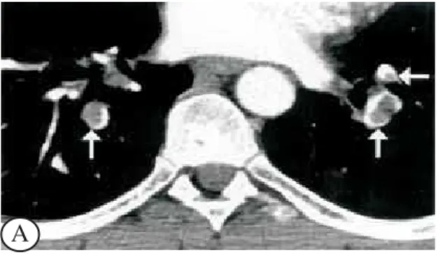
Figure 2A- Acute pulmonary thromboembolism in 15- year old female. The patient was paraplegic following a motor vehicle
accident. Spiral CT image performed on multidetector CT shows complete obliteration of the lumen of the left lower lobe pulmonary arterial branches by emboli. Note increased diameter of the occluded vessels. B - Lung windows demonstrate decreased attenuation and vascularity of the left lower lobe (Westermark sign) secondary to the occlusive emboli.
Diagnostic Accuracy of Spiral CT
Th e rep o rt ed d iag n o st ic accu racy o f sp iral CT h as varied d ep en d in g o n t h e t ech n iq u e u sed , t h e p at ien t p o p u lat io n , an d wh et h er t h e au t h o rs have lim it ed t he an alysis t o t he cen t ral pu lm on ary art eries down t he level of t he segm en t al vessels or have in clu ded su bsegmen t al art eries. Overall, t hese st u d ies h ave sh o wn a sen sit ivit y o f sp iral CT o f 90%, a specificit y of 90%, posit ive predict ive valu e o f 9 3 %, an d n eg at ive p red ict ive valu e o f 9 4 % fo r em b o li d o wn t o an d in clu d in g t h e level o f t h e segm en t al pu lm on ary art eries (11 ).
Th e resu lt s o f t h e vario u s st u d ies in t h e lit erat u re in d icat e t h at alt h o u g h sp iral CT h as a h ig h sen sit ivit y in t h e d et ect io n o f cen t ral em b o li (Fig . 4 ), it is o f lim it ed valu e in t h e d iag n o sis o f su bsegmen t al emboli. It shou ld be n ot ed, however, t h a t t h e c l i n i c a l s i g n i f i c a n c e o f i s o l a t e d su b seg m en t al em b o li, esp ecially in p at ien t s wit h n o u n d e r l y i n g d i s e a s e , i s c o n t r o v e r s i a l . Fu rt h erm o re, it h as b een sh o wn t h at even t h o u g h p u lm o n a ry a n g io g ra p h y is co n sid ered t h e g o ld st an dard for t he diagn osis of pu lmon ary embolism, t h e in t ero b server ag reem en t fo r t h e d iag n o sis o f su b seg m en t al em b o li o n an g io g rap h y is o n ly 6 6 %
(3). Exp erim en t al wo rk in a p o rcin e m o d el h as
sh o wn t h at sp iral CT is co m p arab le t o p u lm o n ary a n g io g ra p h y in t h e d ia g n o sis o f su b seg m en t a l p u lm o n a ry e m b o lism (1 2 ). Pre lim in a ry re su lt s in d ica t e t h a t t h e a ccu ra cy o f sp ira l CT in t h e
d iag n o sis o f su b seg m en t al em b o li is b e im p ro ved wit h t h e u se o f t h in n er sect io n s su ch as 1 o r 2 m m co llim at io n an d m u lt id et ect o r CT scan n ers (13, 1 4 ). Mu lt id et ect o r CT scan n ers allo w evalu at io n o f
t h e en t ire ch est wit h in a few seco n d s u sin g 1 m m t hick sect ion s, t hu s providin g bet t er depict ion t he seg m en t al an d su b seg m en t al p u lm o n ary art eries du rin g peak of con t rast opacificat ion (Fig. 5). These sca n n e rs a lso a llo w h ig h q u a lit y m u lt ip la n a r recon st ru ct ion s t hat fu rt her facilit at e diagn osis or exclu sio n o f PTE (Fig . 6 ) (1 3 , 1 4 ).
Becau se of t he limit at ion s of an giography as a g o ld st a n d a rd , a b et t er wa y t o d et erm in e t h e sen sit ivit y of spiral CT in t he det ect ion of acu t e p u lm o n a ry t h ro m b o em b o lism is t o lo o k a t t h e ou t come of pat ien t s in whom an t icoagu lat ion was wit hheld aft er a n egat ive spiral CT. The resu lt s of t he variou s st u dies performed so far have shown t hat t he ou t come of pat ien t s aft er a n egat ive CT is similar t o t hat report ed aft er a n egat ive an giogram
or negative V/Q scan (15, 16).Goodman et al compared
198 patien ts with n egative spiral CT fin din gs to 350 pat ien t s wit h a n egat ive V/ Q scan (n ormal or low p ro b a b ilit y) (1 5 ). Du rin g 3 - m o n t h f o llo w - u p , su bsequ en t PTE was observed in 1% of pat ien t s in t he spiral CT grou p compared t o 1.5% of pat ien t s in t h e V/ Q g ro u p (n o t st a t ist ica lly sig n ifica n t ). Swensen et al. reviewed 1512 consecu t ive pat ien t s who were referred for CT wit h clin ically su spect ed a cu t e p u lm o n a ry t h ro m b o e m b o lism (1 6 ). Nin e
Figure 3- Acu t e p u lm o n a ry t h ro m b o em b o lism in 3 7 - yea r o ld m a n . Sp ira l CT im a g e sh o ws t ria n g u la r p leu ra l b a sed o p a cit y in t h e p o st erio r seg m en t o f t h e rig h t u p p er lo b e, co n sist en t wit h p u lm o n a ry h em o rrh a g e d ist a l t o o cclu sive t h ro m b o em b o lism .
hu n dred an d n in et y t hree of t hese pat ien t s received n o an t icoagu lat ion an d had CT scan s in t erpret ed as n egat ive for acu t e pu lmon ary embolism. A 3-mon t h probabilit y of ven ou s t hromboembolism of 0.5% was iden t ified in t hese pat ien t s. The au t hors concluded that it is safe to withhold anticoagulation
in pat ien t s wit h a n egat ive spiral CT an d n o clin ical
eviden ce of deep vein t hrombosis (16)
.
Diagnostic Algorithm
Give n t h e d a t a in t h e lit e r a t u r e , t h e fo llo win g a lg o rit h m is re co m m e n d e d fo r t h e
Fig ure 6 A-Mu lt ip lan ar refo rm at io n s im ag es fro m m u lt id ect o r CT. A, Sag it t al refo rm at io n d em o n st rat es fillin g d efect s in t h e rig h t in t erlo b a r a n d lo wer lo b e p u lm o n a ry a rt eries. B. Cu rved a xia l refo rm a t io n d em o n st ra t es fillin g d efect s in t h e rig h t su p erio r seg m en t a l a n d in t h e lin g u la r p u lm o n a ry a rt eries (a rro ws). Also n o t e sm a ll rig h t p leu ra l effu sio n a n d su b ca rin a l lym p h a d en o p a t h y.
Fig ure 5 - Acu t e seg m en t a l a n d su b seg m en t a l p u lm o n a ry t h ro m b o em b o lism . A, Mu lt id et ect o r CT im a g e a t t h e level o f t h e lo wer lo b es d em o n st ra t es fillin g d efect s in t h e p ro xim a l se g m e n t a l p u lm o n a ry a rt e rie s (a rro ws). B, Mu lt id et ect o r CT im a g e a t a m o re ca u d a l level sh o ws fillin g d efect s in d ist a l seg m en t a l p u lm o n a ry a rt eries (a rro ws). C, Mu lt id et ect o r CT im a g e a t a lo wer level t h a n B re ve a ls f illin g d e f e ct s in su b se g m e n t a l p u lm o n a ry art eries (arro ws).
A
B
C
A
imagin g evalu at ion of pat ien t s su spect ed of havin g acu t e p u lm o n ary em b o lism (17):
1. All p at ien t s sh o u ld h ave a ch est rad io g rap h , t h e m a i n r o l e o f w h i c h i s t o e x c l u d e a b n o rm a lit ies su ch a s p n eu m o n ia t h a t m a y m im ic p u lm o n ary em b o lism clin ically.
2. Pat ien t s wit h sym p t o m s o r sig n s o f d eep vein t h ro m b o sis sh o u ld u n d erg o evalu at io n o f t h e leg vein s, t h e m o st co m m o n ly reco m m en d ed t echn iqu e bein g Doppler u lt rasou n d. If Doppler is p o sit ive, t h e p at ien t can b e co n sid ered t o h ave acu t e p u lm o n ary em b o lism an d u su ally d o es n o t req u ire fu rt h er in vest ig at io n .
3. Pat ien t s wit h clin ically su sp ect ed acu t e PTE a n d n o sig n s o r sym p t o m s o f DVT sh o u ld u n d erg o sp iral CT p u lm o n ary an g io g rap h y. It sh o u ld b e n o t ed t h at sp iral CT an g io g rap h y requ ires t he u se of iodin at ed con t rast m at erial. Pat ien t s wit h a co n t rain d icat io n t o t h e u se o f io d in a t ed co n t ra st m a t eria l sh o u ld u n d erg o ven t ilat ion - perfusion scin t igraphy. It shou ld be n o t ed t h at scin t ig rap h y rem ain s t h e im ag in g m et h o d o f ch o ice in cen t ers in wh ich sp iral CT is n o t availab le.
4. Pat ien t s in wh o m t h e CT scan s are su b o p t im al an d in wh o m t h e CT scan resu lt s are n eg at ive bu t who have a high clin ical in dex of su spicion for acu t e pu lmon ary embolism, shou ld u n dergo p u lm o n ary an g io g rap h y.
REFERENCES:
1 . Wells PS, Rodger M. Diagn osis of pu lmon ary embolism: when is imaging needed? Clin Chest Med. 2003;24:13- 28. 2 . The PIOPED In vest igat ors. Valu e of ven t ilat ion - perfu sion sc a n in a c u t e p u lm o n a ry e m b o lism . Re su lt s o f t h e p r o s p e c t ive in ve s t ig a t io n o f p u lm o n a r y e m b o lis m diagn osis (PIOPED). J AMA 1990; 263: 2753- 9. 3 . St ein PD, At h a n a so u lis C, Ala vi A. Co m p lica t io n s a n d
va lid it y o f p u lm o n a ry a n g io g ra p h y in a cu t e p u lm o n a ry em b o lism . Circu la t io n 1 9 9 2 ; 8 5 : 4 6 2 - 8 .
4 . Sc h l u g e r N , H e n s c h k e CI, Ki n g T. Di a g n o s is o f p u lm o n a ry em b o lism a t a la rg e t ea ch in g h o sp t it a l. J . Th o ra c Im a g 1 9 9 4 ; 9 : 1 8 0 - 4
5 . Co o p er TJ , Ha ywa rd MWJ , Ha rt o g M. Su rvery o n t h e u se o f p u lm o n a ry scin t ig ra p h y a n d a n g io g ra p h y fo r su p sp e ct e d p u lm o n a ry t h ro m b o e m b o lism in t h e UK. Clin Ra d io l 1 9 91 ; 4 3 : 2 4 3 - 5
6 . Re m y- J a rd in M, Re m y J , De sch ild re F. Dia g n o sis o f p u lm o n a ry em b o lism wit h sp ira l CT: Co m p a riso n wit h p u lm o n a ry a n g io g ra p h y a n d scin t ig ra p h y. Ra d io lo g y 1 9 9 6 ; 2 0 0 : 6 9 9 - 7 0 6 .
7 . M a yo J R, Re m y- J a r d in M , M ü lle r NL. P u lm o n a r y e m b o lism : p ro sp e ct ive co m p a riso n o f sp ira l CT wit h ve n t ila t io n - p e rf u sio n scin t ig ra p h y. Ra d io lo g y 1 9 9 7 ; 2 0 5 : 4 4 7 - 5 2 .
8 . Qa n a d li SD, El Ha jja m M, Me su ro lle B. P u lm o n a ry e m b o lism d e t e ct io n : p ro sp e ct ive e va lu a t io n o f d u a l-s e c t i o n h e l i c a l CT v e r l-s u l-s l-s e l e c t i v e p u l m o n a r y a r t e r i o g r a p h y i n 1 5 7 p a t i e n t s . Ra d i o l o g y 2 0 0 0 ; 2 1 7 : 4 4 7 - 5 5 .
9 . Co ch e EE, Mü ller NL, Kim W, Wig gs BR, Ma yo J R. Acu t e p u lm o n a ry e m b o lism : a n cilla ry fin d in g s a t sp ira l CT. Ra d io lo g y 1 9 9 8 ; 2 0 7 : 7 5 3 - 8 .
1 0 . Sh a h AA, Da vis SD, Ga m su G, In t riere L. Pa ren ch ym a l a n d p le u ra l f in d in g s in p a t ie n t s wit t h a n d p a t ie n t s wit h o u t a cu t e p u lm o n a ry em b o lism d et ect ed a t sp ira l CT. Ra d io lo g y 1 9 9 9 ; 2 11 : 1 4 7 - 5 3 .
11 . Ma ki DD, Ge f t e r WB, Ala vi A. Re c e n t a d va n c e s in p u lm o n a ry im a g in g . Ch e st 1 9 9 9 ; 11 6 : 1 3 8 8 - 4 0 2 . 1 2 . Ba ile EM, Ma yo J R, Kin g GG, Mü lle r NL, Co ch e EC,
Pa ré PD. Co n t ra st - e n h a n ce d sp ira l CT is co m p a ra b le t o p u l m o n a r y a n g i o g r a p h y f o r t h e d i a g n o s i s o f p u lm o n a ry em b o lism . Am J Resp Crit Ca re Med 2 0 0 0 ; 1 61 : 1 01 0 - 5 .
1 3 . Rem y- J a rd in M, Ma st o ra I, Rem y J . Pu lm o n a ry em b o lu s im a g in g wit h m u lt islice CT. Ra d io l Clin No rt h Am erica 2 0 0 3 ; 41 : 5 0 7 - 1 9
1 4 . P a t e l S, Ka z e r o o n i E A, Ca s c a d e P N . P u l m o n a r y e m b o lism : o p t im iz a t io n o f sm a ll p u lm o n a ry a rt e ry vis u a liz a t io n a t m u lt i- d e t e c t o r ro w CT. Ra d io lo g y 2 0 0 3 ; 2 2 7 : 4 5 5 - 6 0 .
1 5 . Go o d m a n LR, Lip ch ik RJ , Ku zo RS, Liu Y, McAu liffe TL, O’Brien DJ . Su b seq u en t p u lm o n a ry em b o lism : risk a ft er a n e g a t i v e h e l i c a l CT p u l m o n a r y a n g i o g r a m -p ro s-p e ct ive co m -p a riso n wit h scin t ig ra -p h y. Ra d io lo g y 2 0 0 0 ; 2 1 5 : 5 3 5 - 4 2 .
1 6 . Swen sen SJ , Sh eed y PF, Ryu J H, Picket a l. Ou t co m es a ft er wit h h o ld in g a n t ico a g u la t io n fro m p a t ien t s wit h su sp e ct e d a cu t e p u lm o n a ry e m b o lism a n d n e g a t ive co m p u t ed t o m o g ra p h ic fin d in gs: a co h o rt st u d y. Ma yo Clin ic Pro ceed in g s 2 0 0 2 ; 7 7 : 1 3 0 - 8 .