Smoke inhalation injury*
ROGÉRIO SOUZA(TE SBPT), CARLOS JARDIM, JOÃO MARCOS SALGE(TE SBPT), CARLOS ROBERTO RIBEIRO CARVALHO(TE SBPT)
J Bras Pneumol 2004; 30(5) 557-65.
*Study carried out in the Pulmonology Department of the Faculdade de Medicina da Universidade de São Paulo (FMUSP, University of São Paulo School of Medicine), São Paulo, SP.
Correspondence to: Rogério Souza. R. Afonso de Freitas 556, CEP 04006-052 – São Paulo, SP. Phone: 55-11-3889 7426. e-mail: [email protected]
Submitted: 28 April 2003. Accepted, after review: 20 February 2004.
Inhalation injury is the main cause of death in burn patients and has therefore, understandably, been the subject of numerous published studies. The pathogenesis of inhalation injury involves both local and systemic mechanisms, thereby increasing the repercussions of the injury. The search for tools that would allow earlier diagnosis of inhalation injury and for treatment strategies to lessen its deleterious effects is ongoing. In this review, we describe the physiopathological mechanisms of inhalation injury, as well as the current diagnostic tools and treatment strategies used in patients suffering from inhalation injury. We also attempt to put experimental therapeutic approaches into perspective.
INTRODUCTION
Inhalation injury results from the airway inflammatory response to inhalation of the products of incomplete combustion and is the leading cause of death (up to 77%) in burn patients(1,2). Approximately 33% of patients with extensive burns present inhalation injury, and the risk increases in proportion to the quantity of body surface area burned. The presence of inhalation injury per se is responsible for a 20% increase in mortality related to the extent of body surface area burned(3). Over the past few years, understanding of the physiopathological mechanisms of inhalation injury has grown, allowing more targeted approaches.
SMOKE PRODUCTION AND SMOKE
CONSTITUENTS
Smoke results from the burning of any fuel and is a mixture of gases and particles in suspension. Smoke production depends on two processes: pyrolysis and oxidation. Pyrolysis is the phenomenon in which fuel elements are liberated, through melting or boiling, by the heat alone. Oxidation is the process in which oxygen chemically reacts with fuel molecules, breaking them down into smaller compounds and producing light and heat. As examples of products resulting from oxidation, we can cite carbon monoxide (CO), nitrogen dioxide (NO2), and sulfur dioxide (SO2), as well as elemental carbon. The predominance of either process, as well as the temperature, ventilation, and type of material burned in the environment, may lead to the production of a large quantity of smoke elements, each with its peculiar level of toxicity and mechanism of injury(4,5).
Smoke products can be divided into two groups: particulate matter and gases. Both can cause airway injury, although they have different mechanisms of action and affect different areas.
Particulate matter may lead to airway obstruction (directly) through deposition or (indirectly) by inducing bronchospasm. Depending on the size of the particles, the area in which they are deposited varies. Particles greater than 5 micrometers tend to deposit in the upper airways, whereas particles smaller than 1 micrometer may reach the alveolar sacs. Increased airflow caused by tachypnea may lead to an increase in the particle deposition rate in more distal airways(6).
Depending on their mechanism of action, gases are divided into two categories: irritating and asphyxiating. Irritating gases cause mucosal injury through denaturation or oxidation reactions. They c a n c a u s e b r o n c h o s p a s m , c h e m i c a l tracheobronchitis, and even pulmonary edema. The site of action of irritating gases depends primarily on their solubility in water. More soluble gases such as ammonia and sulfur dioxide generally produce reactions in the upper airways, causing painful sensations in the mouth, nose or pharynx, or even in the eyes. On the other hand, less water-soluble gases are responsible for injury in more distal airways and, because they cause only slight irritation of the upper airways, may a l l o w a n o l i g o s y m p t o m a t i c p r e s e n t a t i o n , increasing the chances for, and the extent of, parenchymal injury. Asphyxiating gases are defined a s t h o s e t h a t r e m o v e o x y g e n f r o m t h e environment. Oxygen removal is caused both by the decrease of the fraction of inspired oxygen (FiO2) and by any other mechanisms that prevent o x y g e n c a p t u r e a n d d i s t r i b u t i o n i n t h e cardiovascular system. Consequently, both carbon dioxide (which decreases the oxygen fraction in the environment) and carbon monoxide (whose binding to hemoglobin decreases the oxygen supply to tissues) are considered asphyxiating gases(6,7).
MECHANISMS OF INJURY
The description of the components of smoke complete, we now focus on the objective of facilitating understanding, both in terms of clinical evidence and in terms of treatment. To that end, let us introduce the four mechanisms responsible for inhalation injury:
DIRECT THERMAL INJURY
HYPOXIC GAS INHALATION
During combustion, oxygen concentration drops progressively until the fire is extinguished as a result of the consumption generated by the combustion itself. Depending on the type of fuel, the duration of this process varies. In most cases of petroleum derivative combustion, the fire is extinguished when the oxygen fraction drops into the 13%-15% range. In the case of compounds that contain oxygen, combustion may be sustained at FiO2 values of less than 10%. This decrease in the FiO2 causes victims to experience dyspnea or dizziness, which can evolve to mental confusion, numbness, coma and, when the FiO2 falls to approximately 5% (considered incompatible with life), even death(11).
LOCAL TOXINS
Among the various smoke components, acrolein, formaldehyde, sulfur dioxide and nitrogen dioxide can cause direct airway injury. Such injury results from an acute inflammatory process, mediated by polymorphonuclear leukocytes, especially neutrophils. Symptoms related to this process may not appear until 24 hours after exposure and may include changes in capillary permeability, lymphatic flow or mucociliary clearance, as well as acute respiratory distress syndrome or secondary infections(12,13).
SYSTEMIC TOXINS
Among the substances inducing systemic effects, two gases are of particular importance: carbon monoxide and cyanide (both associated with high rates of morbidity and mortality).
Carbon monoxide intoxication is one of the most frequent causes of death in patients suffering from inhalation injury. Carbon monoxide has a high affinity for hemoglobin, which can be from 200 to 150 times higher than oxygen affinity with hemoglobin. The production of carboxyhemoglobin, an extremely stable complex, causes not only a decrease in oxyhemoglobin saturation, but also a shift of the dissociation curve to the left, reducing oxygen release to the tissues. In addition, competitive inhibition with cytochrome oxidase systems, especially cytochrome P450, impedes the use of oxygen for energy production. Carbon
monoxide also binds to myoglobin, impairing oxygen storage in muscles(14-16).
Cyanide toxicity is caused by the inhibition of cellular oxygenation, causing tissue anoxia through reversible inhibition of cytochrome oxidase enzymes (Fe3+). The inhibition of the aerobic glycolytic pathway forces the metabolism onto the alternative anaerobic pathway, causing an accumulation of acid byproducts(17,18).
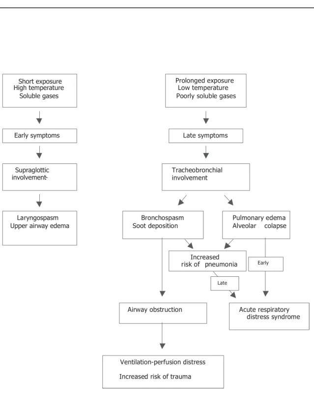
Type and length of exposure, or even the p r e d o m i n a n c e o f o n e o r m o r e o f t h e s e mechanisms, will determine the type of evolution seen in inhalation injury. Figure 1 shows the evolution of the phenomena related to inhalation injury in the various areas of the respiratory system resulting from the various types of exposure.
DIAGNOSIS OF INHALATION INJURY
Clinical presentation
In addition to patient reporting of smoke inhalation in a closed space, various signs and symptoms should arouse clinical suspicion of inhalation injury(19). The most significant signs and symptoms are listed in Table 1.
Certain findings should lead to suspicion of intoxication with particular substances. Carbon monoxide, for instance, tends to affect the central nervous system and the heart. Consequently, exposure to this agent may cause symptoms of cephalgia, visual alterations or mental confusion and may lead to tachycardia, angina, arrhythmia, or even convulsion and coma.
TABLE 1
Clinical characteristics of inhalation injury
Signs Symptoms Face or oral cavity burn Productive cough Singed/scorched vibrissae Hoarseness Sputum with smut or Dyspnea Profuse sputum
Conjunctivitis Wheezing sounds Disorientation/coma Tearing
Laryngeal stridor Respiratory distress
Imaging exams
In most patients, initial chest X-rays reveal normal results and therefore have low predictive value in the diagnosis of acute inhalation injury. However, when the chest X-ray reveals recent infiltrate, this is evidence of more severe inhalation injury, thereby indicating a worse prognosis. The main role of chest X-rays is to identify new infiltrate during the evolution of inhalation injury in the subacute and chronic stages, or even to reveal more diffuse evidence, such as acute respiratory distress syndrome. We must remember that the chronic stage of inhalation injury is characterized by secondary respiratory infections. Ongoing radiological examination of patients is therefore important in order to make an accurate diagnosis as early as possible.
In the initial treatment of severe burn patients, volume resuscitation and stabilization of airway patency are the main objectives. Computed tomography of the chest cannot be performed on such unstable patients. In view of these facts, it is not surprising that there have been few significant studies of the role of computed tomography in the early identification of inhalation injury(20).
Bronchoscopy
Inhalation injury is diagnosed through examination of the upper airways and trachea. The presence of edema or erythema, ulcerations in lower airways or even the presence of soot in more distal ramifications are suggestive of inhalation injury. The absence of these signs, however, should a l w a y s b e a n a l y z e d b e a r i n g i n m i n d t h e hemodynamic state of the patient since the initial
bronchoscopy may not reveal areas of edema or erythema in patients who have not yet undergone volume resuscitation. Apart from this exception, bronchoscopy has an accuracy of approximately 100% in the diagnosis of established inhalation injury(21,22).
Careful evaluation of upper airways, especially in patients who present no evidence of more distal injury, is extremely important during bronchoscopy. Severe edema in the supraglottic area, or a great quantity of upper-airway secretion, may indicate that these patients are more likely to present acute airway obstruction, and this is an indicator for the early intubation of patients suspected of having upper-airway injury. However, we must stress that examination of the upper airways does not preclude examination of the lower airways since involvement of the latter may occur independently of involvement of the former.
It is important to emphasize that anatomical alterations revealed by bronchoscopy precede gas exchange alterations and, more so, radiographic changes. Therefore, it is important that all patients under clinical suspicion of inhalation injury be submitted to bronchoscopy as soon as possible(20).
Arterial blood gas analysis
Radioisotopes
Ventilation-perfusion scans using 133 xenon
(Xe133) allow the identification of small-airway
o b s t r u c t i o n t h a t c a n n o t b e s e e n i n t h e bronchoscopic exam. Serial analyses are carried out after the administration of the radioisotope in order to determine the total retention time of the radiopharmacological agent in the lungs. Retention times longer than 90 seconds or significant heterogeneity in the distribution of the substance are considered predictive of inhalation injury(23). The rates of false-positive and false-negative results are 8% and 5%, respectively, and are usually due to hyperventilation (false-negative), atelectasis (false-positive) or chronic obstructive pulmonary disease (false-positive)(24).
T h e u s e o f X e1 3 3 i n h a l a t i o n - p e r f u s i o n scintigraphy is only indicated for patients in which there is high clinical suspicion of inhalation injury but normal chest X-ray and bronchoscopy results.
Pulmonary function tests
Although pulmonary function tests illustrate the physiopathological representation of the injury, they play only an adjuvant role in the diagnosis of inhalation injury, mainly because they are difficult to administer. Several factors contribute to this difficulty, including the presence of pain, poor patient cooperation, muscular weakness and the use of medications such as sedatives and opioids. These factors dramatically reduce the accuracy of pulmonary function tests(22).
In general, pulmonary function tests may characteristically reveal the following findings: static and dynamic compliance of normal respiratory system in the early stages of the injury, which progressively decreases as it develops, or even in chronic stages, when then reparation process may lead to restrictive profiles, evidenced by reduced respiratory system compliance and increased airway resistance, as well as decreases in forced expiratory volume in one second, the forced expiratory volume in one second/forced vital capacity ratio and peak expiratory flow, characteristic of airway obstruction and evidencing the flow-volume curve concavity typical of an obstructive pattern, which is caused as much by accumulation of soot and secretion as by edema of the airway mucosa.
T h e p r o g r e s s i v e a s p e c t o f m o n i t o r i n g p u l m o n a r y f u n c t i o n a l l o w s n o t o n l y t h e identification of the progress of the injury but also the evaluation of the response to the therapeutic measures adopted (ventilatory strategy, drug therapy, or even physical therapy).
INHALATION INJURY TREATMENT
Airway maintenance
Identifying patients at high risk for upper-airway obstruction, together with early intervention when injury has already been established is one of the principal points in the treatment of patients with i n h a l a t i o n i n j u r y, s i g n i f i c a n t l y re d u c i n g mortality(11,20). Clinical signs compatible with obstruction secondary to upper-airway injury, or even bronchoscopic evidence of this process, require early intervention.
The use of large-caliber tracheal tubes facilitates the bronchial hygiene necessary for the control of the considerable increase in the quantity of r e s p i r a t o r y s e c r e t i o n( 2 5 ). T r a c h e o s t o m y i s advantageous for patients, providing comfort and facilitating bronchial hygiene. However, according to recent studies, tracheostomy does not reduce the duration of mechanical ventilation, incidence of pneumonia, or even mortality due to inhalation injury. Consequently, tracheostomy is not indicated as a general therapeutic measure(26).
Oxygen
Patients with chronic obstructive pulmonary disease concomitant to carbon dioxide retention, together with comatose patients, compose a group in which immediate intubation is the most highly recommended means of increasing, as rapidly as possible, the inspired oxygen fraction without the potentially deleterious effects of greater carbon dioxide retention.
Ventilatory support
In relation to the treatment of burn patients, the subject of ventilatory support has, over the past few years, perhaps received the most attention in terms of the number of studies conducted.
W i t h t h e d e v e l o p m e n t o f n o n i n v a s i v e ventilation, the idea of avoiding intubation has become extremely attractive, especially in a population in which intubation is such a significant predictor of morbidity and mortality. The presence of facial injury in combination with the risk of decreased tissue perfusion at the points of support of the various types of masks used during noninvasive ventilation has limited the prolonged use of this technique. However, this has not prevented the performance of studies on its intermittent use, which have shown it capable of maintaining alveolar recruitment, thereby reducing the need for tracheal intubation. This fact alone makes the use of this technique attractive, even if only as an adjunct to physical therapy, keeping small airways and the alveolar space open. The development of new interfaces for noninvasive ventilation, with less involvement at the points of support, has presented even more alternatives for the prolonged use of noninvasive ventilation as an initial ventilatory support procedure in patients with inhalation injury(27).
If tracheal intubation becomes necessary, invasive ventilatory support must be based on strategies that keep the lungs open, providing better local clearing of secretion, as well as optimizing gas exchange.
Appropriate choice of ventilatory strategy is intimately related to the stage in which the patient shows respiratory insufficiency. This is because, in the initial stages, the principal physiopathological mechanism is direct airway injury, with edema and bleeding, combined with increased soot and secretion. In such cases, aggressive ventilatory
strategies, such as the use of high positive end-expiratory pressure, are seldom necessary.
In severe cases in the initial stages or in later-stage cases (in either of which patients clearly present acute respiratory distress syndrome), it is necessary to use alveolar recruitment strategies and low tidal volume(28) in combination with higher positive end-expiratory pressure. In relation to inhalation injury, the independent role of each of these strategies has yet to be established. However, clinical studies have shown the benefits of their use in acute respiratory distress syndrome related to other causes(29,30).
High-frequency ventilation and the use of nitric oxide are the current modalities for the use of invasive ventilatory support in patients with inhalation injury. Experimental studies have shown reduced rates of inhalation injury progression resulting from the use of nitric oxide, whereas clinical studies have only shown improved oxygenation from the use of the technique(31,32). Likewise, high-frequency ventilation has proven efficacious in improving oxygenation in patients with inhalation injury within the first hours after the use of the technique. However, there is insufficient evidence that, within this population of patients, this technique improves survival or lowers the rate of infection related to mechanical ventilation(33,34).
Although still preliminary, clinical studies attempting to prevent intubation, reduce the progression of inhalation injury and accelerate the response to the established injury are presently the major source of expectation that better treatments for severe burn patients will be found.
Antibiotic therapy
The early use of antibiotics when there was no clear evidence of infection has neither increased the survival of patients nor reduced the incidence of pneumonia, which is considered the most frequent infectious complication related to inhalation injury. Therefore, their use is not recommended(20,35).
profile, the more likely is infection caused by negative bacteria. In the more acute cases, gram-positive bacteria predominate. The role of bronchoscopy involving bronchial lavage and brushing in the identification of the etiologic agents, either in their early identification or in order to adjust empirical antibiotic regimens, still needs to be determined.
Specific measures according to the predominant physiopathology
Airway maintenance is the principal treatment for thermal injury. It is prudent to maintain the patient intubated until there is documented reversal of airway edema.
Regarding hypoxic gas inhalation, the strategy of removing the patient from the scene of the accident and using high inspired oxygen fractions interrupts the cascade effect of events secondary to hypoxemia(11,13,20).
As for carbon monoxide intoxication, the half-life of carboxyhemoglobin is 150 minutes in room air (inspired oxygen fraction of 0.21) and from 40 to 60 minutes in patients submitted to an inspired oxygen fraction of 1. Therefore, all patients should receive oxygen at 100% en route to the hospital. The use of hyperbaric therapy is still debatable. Some studies have shown no benefit(36), although there is evidence that its use may lead to a reduction in neurological damage(37). Another way to increase carbon monoxide elimination is through isocapnic hyperpnea, in which intubated patients are hyperventilated, providing them a supplementary source of carbon dioxide in order to avoid promoting respiratory alkalosis due to h y p o c a p n i a . H y p e r v e n t i l a t i o n r e d u c e s carboxyhemoglobin half-life, thereby minimizing the hazardous effects of the intoxication(38,39).
Some antidotes may be used in an attempt to limit the damage caused by cyanide intoxication: nitrates, which promote hemoglobin oxidation, transforming it into metahemoglobin, which competes with cytochrome oxidase for cyanide; sodium thiosulphate, which, in the presence of the mitochondrial enzyme rhodanase, transfers sulfur to the cyanide ion, forming thiocyanate (which is excreted in the urine), and hydroxocobalamin, a c h e l a t e t h a t r e a c t s w i t h c y a n i d e f o r m i n g cyanocobalamin(40,41).
Therapeutic perspectives
Advancements in the treatment of inhalation injury might have as significant an impact on burn patient outcomes as does the use of volume resuscitation, especially with the development of m e t h o d s t h a t a l l o w m o d u l a t i o n o f t h e inflammatory response of the respiratory system.
Various substances have shown potential benefits in the management of patients with inhalation injury. These include antiadherence agents (blocking neutrophil adherence in the p u l m o n a r y m i c r o v a s c u l a t u r e ) , l e u k o t r i e n e inhibitors, inhaled heparin, and antioxidants(42-44), although controlled clinical studies are still necessary before these new treatment alternatives can be adopted in clinical practice.
The focal point in the treatment of severe burn patients is the understanding that the intense inflammatory response and the consequent systemic and pulmonary involvement constitute a g l o b a l p h e n o m e n o n a n d a r e n o t i s o l a t e d complications.
REFERENCES
1. Ryan CM, Schoenfeld DA, Thorpe WP, Sheridan RL, Cassem EH, Tompkins RG. Objective estimates of the probability of death from burn injuries. N Engl J Med.1998;338:362-6.
2. Darling GE, Keresteci MA, Ibanez D, Pugash RA, Peters WJ, Neligan PC. Pulmonary complications in inhalation injuries with associated cutaneous burn. J Trauma. 1996;40:83-9.
3. Shirani KZ, Pruitt BA Jr, Mason AD Jr. The influence of inhalation injury and pneumonia on burn mortality. Ann Surg. 1987;205:82-7.
4. Birky MM, Clarke FB. Inhalation of toxic products from fires. Bull N Y Acad Med. 1981;57:997-1013. 5. Terrill JB, Montgomery RR, Reinhardt CF. Toxic gases
from fires. Science. 1978;200:1343-7.
6. Young CJ, Moss J. Smoke inhalation: diagnosis and treatment. J Clin Anesth. 1989;1:377-86.
7. Haponik EF. Clinical smoke inhalation injury: pulmonary effects. Occup Med. 1993;8:430-68.
8. Oldham KT, Guice KS, Till GO, Ward PA. Activation of complement by hydroxyl radical in thermal injury. Surgery. 1988;104:272-9.
9. Haponik EF, Crapo RO, Herndon DN, Traber DL, Hudson L, Moylan J. Smoke inhalation. Am Rev Respir Dis. 1988;138:1060-3.
1 0 . Ward PA, Till GO. Pathophysiologic events related to thermal injury of skin. J Trauma. 1990;30:75-9. 11 . Weiss SM, Lakshminarayan S. Acute inhalation injury.
Clin Chest Med. 1994;15:103-16.
1 3 . Demling RH. Smoke inhalation injury. New Horiz. 1993;1:422-34.
14. Piantadosi CA. Diagnosis and treatment of carbon monoxide poisoning. Respir Care Clin N Am. 1999;5:183-202. 1 5 . Ernst A, Zibrak JD. Carbon monoxide poisoning. N Engl
J Med. 1998;339:1603-8.
1 6 . Seger D, Welch L. Carbon monoxide controversies: neuropsychologic testing, mechanism of toxicity, and hyperbaric oxygen. Ann Emerg Med. 1994;24:242-8. 1 7 . Shusterman DJ. Clinical smoke inhalation injury:
systemic effects. Occup Med. 1993;8:469-503. 1 8 . Mayes RW. ACP Broadsheet No 142: November 1993.
Measurement of carbon monoxide and cyanide in blood. J Clin Pathol. 1993;46:982-8.
1 9 . Haponik EF. Clinical and functional assessment. In: Haponik EF, Munster AM, editors. Respiratory injury: smoke inhalation and burns. New York: McGraw-Hill; 1990. p.137-78.
2 0 . Lee-Chiong TL Jr. Smoke inhalation injury. Postgrad Med. 1999;105:55-62.
2 1 . B i n g h a m H G , G a l l a g h e r T J , P o w e l l M D . E a r l y bronchoscopy as a predictor of ventilatory support for burned patients. J Trauma. 1987;27:1286-8. 22. Haponik EF. Acute upper airway injury in burn patients.
Serial changes of flow-volume curves and nasopharyngoscopy. Am Rev Respir Dis. 1987;135:360-6. 2 3 . Agee RN. Use of 133 xenon in early diagnosis of
inhalation injury. J Trauma. 1976;16:218-24. 2 4 . Lull RJ, Anderson JH, Telepak RJ, Brown JM, and Utz
JA. Radionuclide imaging in the assessment of lung injury. Semin Nucl Med. 1980;10:302-10.
2 5 . Heimbach DM, Waeckerle JF. Inhalation injuries. Ann Emerg Med. 1988;17:1316-20.
2 6 . Saffle JR, Morris SE, Edelman L. Early tracheostomy does not improve outcome in burn patients. J Burn Care Rehabil. 2002;23:431-8.
2 7 . Smailes ST. Noninvasive positive pressure ventilation in burns. Burns. 2002;28:795-801.
2 8 . Sheridan RL. Permissive hypercapnia as a ventilatory strategy in burned children: effect on barotrauma, pneumonia, and mortality. J Trauma. 1995;39:854-9. 2 9 . Amato MB, Barbas CS, Medeiros DM, Magaldi RB, Schettino GP, Lorenzi-Filho G, et al. Effect of a protective-ventilation strategy on mortality in the acute r e s p i r a t o r y d i s t r e s s s y n d r o m e . N E n g l J M e d . 1998;338:347-54.
3 0 . Ventilation with lower tidal volumes as compared with traditional tidal volumes for acute lung injury and the a c u t e r e s p i r a t o r y d i s t r e s s s y n d r o m e . T h e a c u t e respiratory distress syndrome network. N Engl J Med. 2000;342:1301-8.
31. Sheridan RL, Hurford WE, Kacmarek RM, Ritz RH, Yin LM, Ryan CM, et al. Inhaled nitric oxide in burn p a t i e n t s w i t h r e s p i r a t o r y f a i l u r e . J T r a u m a . 1997;42:629-34.
3 2 . Musgrave MA, Fingland R, Gomez M, Fish J, Cartotto R. The use of inhaled nitric oxide as adjuvant therapy in patients with burn injuries and respiratory failure. J Burn Care Rehabil. 2000;21:551-7.
3 3 . Reper P, Wibaux O, Van Laeke P, Vandeenen D, Duinslaeger L, Vanderkelen A. High frequency percussive ventilation and conventional ventilation after smoke inhalation: a randomised study. Burns. 2002;28:503-8.
3 4 . Mehta S, MacDonald R, Hallett DC, Lapinsky SE, Aubin M, Stewart TE. Acute oxygenation response to inhaled nitric oxide when combined with high-frequency oscillatory ventilation in adults with acute respiratory distress syndrome. Crit Care Med. 2003;31:383-9. 3 5 . Bang RL, Sharma PN, Sanyal SC, Al Najjadah I. Septicemia
a f t e r b u r n i n j u r y : a c o m p a r a t i ve s t u d y. B u r ns. 2002;28:746-51.
3 6 . Scheinkestel CD. Hyperbaric or normobaric oxygen for acute carbon monoxide poisoning: a randomised controlled clinical trial. Med J Aust. 1999;170:203-1 0 .
3 7 . Weaver LK, Hopkins RO, Chan KJ, Churchill S, Elliott CG, Clemmer TP et al. Hyperbaric oxygen for acute c a r b o n m o n o x i d e p o i s o n i n g . N E n g l J M e d . 2002;347:1057-67.
3 8 . Fisher JA. Isocapnic hyperpnea accelerates carbon monoxide elimination. Am J Respir Crit Care Med. 1999;159:1289-92.
3 9 . Takeuchi A. A simple “new” method to accelerate clearance of carbon monoxide. Am J Respir Crit Care Med. 2000;161:1816-9.
4 0 . Langford RM, Armstrong RF. Algorithm for managing injury from smoke inhalation. BMJ. 1989;299:902-5. 41. Sauer SW, Keim ME. Hydroxocobalamin: improved public health readiness for cyanide disasters. Ann Emerg Med. 2001;37:635-41.
4 2 . Tasaki O. Effect of sulfo Lewis C on smoke inhalation injury in an ovine model. Crit Care Med. 1998;26:1238-4 3 .
4 3 . Demling R, LaLonde C, Ikegami K. Fluid resuscitation with deferoxamine hetastarch complex attenuates the lung and systemic response to smoke inhalation. Surgery. 1996;119:340-8.