Open Reduction and Internal Fixation of
Mandibular Fracture without Rigid
Maxillomandibular Fixation
Mohammad Waheed El-Anwar
1Magdy Abdalla Sayed El-Ahl
1Hazem Saed Amer
11Department of Otorhinolaryngology, Head and Neck Surgery,
Zagazig University, Zagazig, Egypt
Int Arch Otorhinolaryngol 2015;19:314–318.
Address for correspondence Mohammad Waheed El-Anwar, MD, Department of Otorhinolaryngology Head and Neck Surgery, Zagazig University, Zagazig 0020552309843, Egypt
(e-mail: [email protected]).
Introduction
With the exception of the nose, mandibular fractures occur twice as frequently as fracture of other facial bones. The importance of the mandible is not only cosmetic; it also functions in biting, chewing, and speaking.1
The purpose of the treatment of mandibular fractures is to restore proper dental occlusion and stable temporomandib-ular joint (TMJ) movement as well as the reduction of the displaced fracture.2
The ability to treat fracture with open reduction (OR) and internal fixation (IF) has dramatically revolutionized the Keywords
►
mandibular fractures
►
fracture
fi
xation
►
temporomandibular
joint
Abstract
Introduction
The ability to treat fracture with open reduction and internal
fi
xation
(OR/IF) has dramatically revolutionized the approach to mandible fracture. With OR/IF,
the postoperative role of rigid maxillomandibular
fi
xation (MMF) has declined, but it is
used to maintain proper occlusion until internal
fi
xation of the fracture is achieved.
Objective
To assess intraoperative manual MMF during OR/IF of selected cases of
mandibular fractures.
Methods
This prospective study was conducted on 80 patients with isolated
mandib-ular fractures managed by OR/IF using two titanium miniplates. The patients were
classi
fi
ed into two groups: a control group (40 patients) treated by OR/IF after
intraoperative rigid MMF followed by immediate MMF removal, and a study group
(40 patients) treated by rigid MMF, which was replaced by temporary intraoperative
manual MMF (3MF) until plate
fi
xation.
Results
There were no signi
fi
cant differences of the postoperative complication and
dental occlusion, although a highly signi
fi
cant reduction of operative time was achieved
in the 3MF group. Patient who received the 3MF technique had statistically signi
fi
cantly
better average intrinsic vertical mouth opening in the early postoperative period
(1 week after surgery), and normal mouth opening could be achieved in all cases in
both groups 8 weeks after surgery.
Conclusions
Intraoperative rigid MMF is not mandatory and can be replaced in
selected cases of fracture mandible by manual maintenance of proper dental occlusion
until hardware
fi
xation, gaining the advantages of shorter operative time and less risk of
blood-transmitted diseases to the surgical team and the patient in addition to the
bene
fi
ts of immediate postoperative mandible mobilization.
received
January 21, 2015
accepted
February 23, 2015
published online
March 30, 2015
DOI http://dx.doi.org/ 10.1055/s-0035-1549154.
ISSN 1809-9777.
Copyright © 2015 by Thieme Publicações Ltda, Rio de Janeiro, Brazil
approach to mandible fracture.3 Traditionally, OR/IF has required a period of postoperative mandibular immobiliza-tion by rigid maxillomandibular fixation (MMF) for up to 6 weeks for satisfactory healing. Difficulties associated with this period of immobilization include airway problems, poor nutrition, weight loss, poor mouth hygiene, phonation diffi -culties, insomnia, social inconvenience, patient discomfort, work loss, and difficult in recovering normal-range jaw function. Rigid fixation of mandible fractures allows early mobilization and restoration of jaw function and airway control; improves nutritional status, speech, oral hygiene, and patient comfort; and allows early return to the workplace.4,5
Many studies showed that immediate postoperative re-lease of rigid MMF after OR/IF using mini-titanium plates is as effective and safe as maintaining postoperative MMF.6,7 Therefore, the postoperative role of rigid MMF has declined, but it is essential for maintaining proper occlusion until internalfixation of the fracture.8
However, rigid intraoperative MMF lengthens the oper-ative time (for placement and removal of rigid MMF), carries a risk of teeth injury or vitality affection, and increases the risk of blood-transmitted diseases to patients and the surgical team.9These concerns encouraged us to assess temporary intraoperative manual MMF fixation (3MF) during OR/IF of selected cases of mandibular fractures.
Methods
Study Design
This prospective study was conducted from January 2008 to January 2014. The study was approved by the Institu-tional Review Board, and informed consent was signed by all enrolled subjects after explanation of the research purpose.
Study Subjects
Eighty patients with isolated traumatic mandibular fractures (symphyseal, parasymphyseal, and body) were included in this study. Inclusion criteria were age more than 12 years, isolated mandibular fracture (associated with no other facial fractures), and intact molar teeth in good condition. Exclusion criteria were condylar, subcondylar, or angle mandibular fractures; other associated maxillofacial fractures; history of pretrauma malocclusion or limited mouth opening; and edentulous patients.
The patients were stratified by gender then randomly assigned to control and study groups. The control group included 40 patients treated by OR/IF after intraoperative rigid MMF (using arch bars and wires) followed by imme-diate removal of MMF wires and release of the mandible after fracture stabilization. In the study group (40 patients), rigid MMF was not performed, and it was replaced by temporary intraoperative manual MMF (3MF) performed by the assistant surgeon to get and maintain proper dental occlusion until miniplate fixation of the fracture(s) (►Fig. 1).
Surgical Work
The clinicalfindings were correlated with diagnostic radio-graphic imaging including panoramic view of the mandible, computed tomography (CT) scans (coronal, axial cuts, and may three-dimensional CT). Panoramic view of the mandible was obtained postoperatively for follow-up.
The operation was performed under general anesthesia using nasal endotracheal intubation. Rigid MMF using arch bars and wires was performedfirst in the control group. Open reduction of the fracture (s) was conducted through a lower sublabial incision. In the study group, exposure, dissection, and disimpaction of the fracture were performed while the mandible was opened, then temporary intraoperative 3MF was performed by the assistant (maxillofacial surgeon) to get and maintain proper dental occlusion during IF.
Two titanium miniplates (1.5 mm thickness) with at least two holes on either side of each fracture line and bicortical screws were used (►Fig. 1). Care was taken during incision, dissection, and IF to avoid injury of the mental nerve. The sublabial incision was then closed, and in patients in the control group, rigid MMF was removed, with immediate mobilization of the mandible. Patients were kept on soft diet for 1 month. Prophylactic antibiotics and analgesics were prescribed for all patients. Neurotonics were prescribed for the cases of numbness of the chin, and the patients were discharged from the hospital the second day after surgery.
Postoperative follow-up was done every week for 1 month, then at 3 months and 6 months postoperatively, with monitoring of complications and functional results. Results were assessed by a surgeon blinded to the type of repair used according to the following criteria:
1. According to Angle’s classification of dental occlusion 2. According to the average intrinsic vertical mouth opening
(between the upper and lower central incisors)10 • Normal: intrinsic vertical mouth opening measures 40
to 50 mm
• Functional: intrinsic vertical mouth opening measures 25 to 35 mm
• Limited: intrinsic vertical mouth opening measures 10 to 24 mm
Fig. 1 Manual intraoperative maxillomandibularfixation allowing
Statistical Analysis
Statistical analysis was performed using SPSS 14.0 statistical software for Windows (SPSS Inc., Chicago, Illinois, United States). The significance level was set atp<0.05.
Results
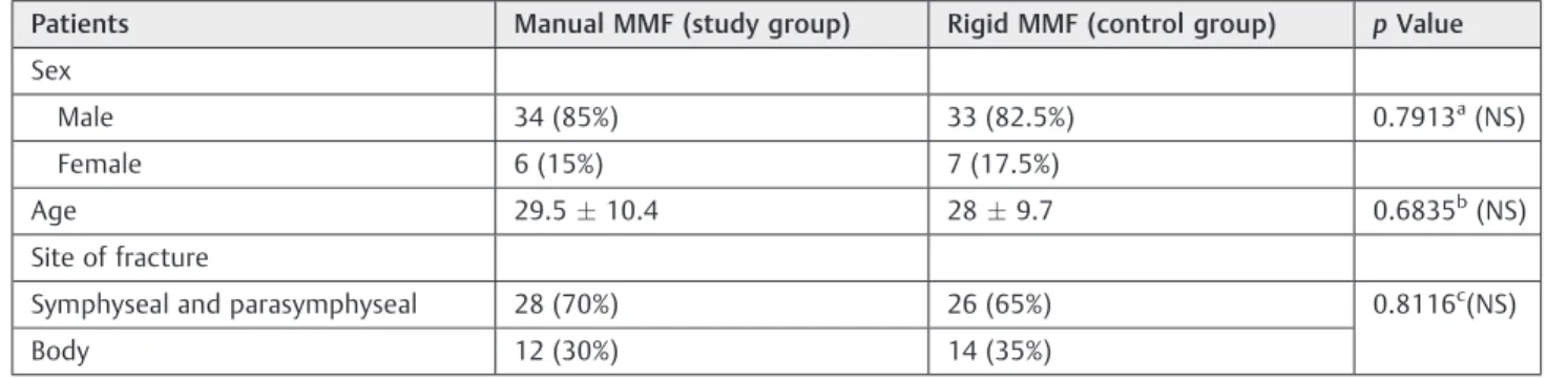
The 40 patients included in the study group and satisfying the selection criteria included 34 male (85%) and 6 female (15%) patients, with ages ranging from 16 to 63 years. The mean age and its standard deviation (SD) was 29.510.4 years. The site of mandibular fracture was symphyseal and parasym-physeal fracture in 28 patients (70%) and body fracture in 12 patients (30%).
The control group included 33 male (82.5%) and 7 female (17.5%) patients, with ages ranging from 17 to 59 years. The mean age (SD) was 28 (9.7) years. The site of mandibular fracture was symphyseal and parasymphyseal fracture in 33 patients (82.5%) and body fracture in 7 patients (17.5%). The differences between both groups regarding age, sex, and fracture site was not significant (►Table 1).
Postoperatively, all patients included in this study recov-ered easily from anesthesia; there was no need for intensive care unit admission. Normal (class I) dental occlusion was achieved in all cases.
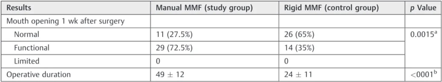
One week after surgery, mouth opening was normal in 26 patients (65%) and functional in 14 patients (35%) in the study group, and it was normal in 11 patients (27.5%) and functional in 29 patients (72.5%) in the control group. 3MF showed better early mouth opening, and the difference proved to be very statistically significant (►Table 2). Eight weeks after surgery, all patients in both groups achieved normal mouth opening.
Numbness occurred in the early postoperative period and was relieved in all cases but persisted in 1 (2.5%) patient in the study group and 3 (7.5%) patients in the control group; the difference was not significant (chi-square: 1.053; p¼0.3). Infection, malocclusion, delayed union, and nonunion were not encountered.
Actual operative duration was significantly shorter for the 3MF group (2411 minutes) compared with the control group (4912 minutes).
Exposure, dissection, and disimpaction of the fracture were easier and more rapid in the 3MF group, with the mandible opened and freely movable, than the rigid MMF group.
Discussion
The basic principle of fracture treatment is reduction,fi xa-tion, immobilizaxa-tion, prevention of infecxa-tion, and rehabilita-tion. The main goal in managing mandibular fracture is to restore preinjury form and function, with the least disability, smallest risk, and shortest recovery period.11The simplest method should be chosen whenever it is as effective as a more invasive method.12MMF is considered a mainstay of reduc-tion and stabilizareduc-tion of mandibular fracture.13
Recently, OR/IF has become the standard management of displaced fractures because it provides stable three-dimen-sional reconstruction, promotes bone healing, and shortens treatment time.14 OR/IF allows immediate jaw mobiliza-tion,7,15reduces dependence on MMF, and enables surgeons to reduce the period of MMF or immediately release the MMF after OR/IF with similar high success, efficiency, and safety as maintaining postoperative MMF.7,16,17Other than maintain-ing proper occlusion until internalfixation is achieved, rigid MMF has no postoperative role.8 However, intraoperative rigid MMF is a complex maneuver, lengthens the operative time, and increases the incidence of disease transmission and operative cost.
In the present study, rigid MMF was not performed in patients of the study group, depending on intraoperative proper manual reduction then maintaining proper dental occlusion by a trained assistant until the reduced fracture was
fixed by miniplates. After mandibular reconstruction, normal dental occlusion and mouth opening could be achieved in all cases. Comparing the results of 3MF with a control group treated by rigid MMF and immediate release, nonsignificant differences were reported, such as dental occlusion and mouth opening after 8 weeks of OR/IF.
Taking into consideration that both groups (manual MMF and rigid MMF) were matched for age, sex, and site of fracture, manual MMF is as effective as rigid MMF. Interestingly, 3MF
Table 1 Age, sex, and site of fracture of the study (manual MMF) and control (rigid MMF) groups
Patients Manual MMF (study group) Rigid MMF (control group) pValue
Sex
Male 34 (85%) 33 (82.5%) 0.7913a(NS)
Female 6 (15%) 7 (17.5%)
Age 29.510.4 289.7 0.6835b(NS)
Site of fracture
Symphyseal and parasymphyseal 28 (70%) 26 (65%) 0.8116c(NS)
Body 12 (30%) 14 (35%)
Abbreviation: MMF, maxillomandibularfixation; NS, not significant. aFisher exact test.
bttest
showed statistically significantly better early results in mouth opening than rigid MMF.
In addition, nonsignificant differences in mental nerve complication and absence of other complication in both groups proved that manual MMF is as safe as rigid MMF. It was obvious that 3MF reduced significantly the operative duration (p<0001).
This low incidence of postoperative complications and comparable functional results revealed that intraoperative rigid MMF is not mandatory, and manual MMF is preferred in parasymphyseal and isolated body mandibular fractures.
Many advantages were gained by using intraoperative manual occlusion instead of rigid MMF, including a significant reduction of the operative time and decreased length of general anesthesia. Owing to the absence of wire-based MMF, the operative liability of teeth, gum, or lid affection and the risk of percutaneous or mucosal wire punctures were minimized, reducing the incidence of blood-transmitted dis-eases to the surgical team or the patient caused by wires pricks. Wire pricks occurred more often in the rigid MMF group due to repeated manipulation of wires during release of the mandible as well asfixation of the wire-based MMF.
On the other hand, these results support the treatment option of immediate mandibular release following mandibular recon-struction by OR/IF; benefits gained include immediate mobili-zation as well as good oral hygiene, ease of feeding, avoidance of trismus and weight maintenance, better psychological impact on the patient, better anesthesia recovery, no need for intensive care unit admission and long postoperative care and hospitali-zation, and early return to work. There was also no need to keep the emergency quick release system available during recovery from anesthesia, as is necessary when rigid MMF is used.
The transoral approach, performed through an oral muco-sal incision, results in minimal external scarring or injury to the marginal mandibular nerve and allows direct visualiza-tion and confirmation of the desired occlusion during the placement of the miniplates.
Exposure, dissection, disimpaction, and reduction of the fracture were easier in the 3MF than the rigid MMF group because the mandible in 3MF was opened, giving space to work without limitation of the rigid MMF. In thefirst cases, manual MMF was maintained throughout all screwfixation. But in the later cases, two screws werefixed on one side of fracture while the mouth was opened, then manual MMF was
maintained duringfixation of the other two screws dimin-ishing period of manual MMF.
Based on the result obtained from this study, we can conclude that intraoperative manual MMF can replace rigid MMF in selected cases of mandibular fracture, and it is an effective alternative and gives a particular benefit and opti-mum solution when long general anesthesia is not recom-mended as in medically compromised patients or in patients with compromised pulmonary function.
Manual MMF represents an ideal modification on dealing with a mandible fracture in patients carrying a blood-borne virus such as hepatitis B or C that is a global public health problem. Thus, the current work provides a basis for future wider use of manual MMF. The financial benefits of this technique need to be studied.
Conclusion
Intraoperative rigid MMF is not mandatory and can be replaced in selected cases of mandible fracture by manual maintenance of proper dental occlusion until hardwarefi xa-tion, gaining the advantages of shorter operative time, lower cost, and less risk of blood-transmitted diseases to the surgi-cal team and the patient, in addition to the benefits of immediate postoperative TMJ release.
References
1 Gupta R, Surayana S, Pandya VK, et al. Traumatic mandibular fractures: pendulum swinging towards closed reduction? World Art Ear Nose Throat 2010;3:1
2 Imazawa T, Komuro Y, Inoue M, Yanai A. Mandibular fractures treated with maxillomandibularfixation screws (MMFS method). J Craniofac Surg 2006;17(3):544–549
3 Lazow SK. The mandible fracture: a treatment protocol. J Cranio-maxillofac Trauma 1996;2(2):24–30
4 Sorel B. Open versus closed reduction of mandible fractures. Oral Maxillofac Surg Clin North Am 1998;10:553
5 Schneidr M, Erasmus F, Gerlach K, et al. Open reduction and internalfixation versus closed treatment and mandibulomaxillary fixation of fracture of the mandible condylar process. J Oral Maxillofac Surg 2008;66(12):2537–2544
6 Kumar I, Singh V, Bhagol A, Goel M, Gandhi S. Supplemental maxillomandibular fixation with miniplate osteosynthesis-re-quired or not? Oral Maxillofac Surg 2011;15(1):27–30
Table 2 Difference between the study (manual MMF) and control (rigid MMF) groups in mouth opening and operative duration
Results Manual MMF (study group) Rigid MMF (control group) pValue
Mouth opening 1 wk after surgery
Normal 11 (27.5%) 26 (65%) 0.0015a
Functional 29 (72.5%) 14 (35%)
Limited 0 0
Operative duration 4912 2411 <0001b
Abbreviation: MMF, maxillomandibularfixation. aChi-square test, very statistically signi
ficant. bt-test, extremely statistically signi
7 Kaplan BA, Hoard MA, Park SS. Immediate mobilization following fixation of mandible fractures: a prospective, randomized study. Laryngoscope 2001;111(9):1520–1524
8 Smartt JM Jr, Low DW, Bartlett SP. The pediatric mandible: II. Management of traumatic injury or fracture. Plast Reconstr Surg 2005;116(2):28e–41e
9 Ayoub AF, Rowson J. Comparative assessment of two methods used for interdental immobilization. J Craniomaxillofac Surg 2003;31(3):159–161 10 Gallagher C, Gallagher V, Whelton H, Cronin M. The normal range of mouth opening in an Irish population. J Oral Rehabil 2004;31(2):110–116 11 Nacamuli RP, Longaker MT. Bone induction in craniofacial defects.
Orthod Craniofac Res 2005;8(4):259–266
12 Haug RH, Assael LA. Outcomes of open versus closed treatment of mandibular subcondylar fractures. J Oral Maxillofac Surg 2001; 59(4):370–375, discussion 375–376
13 Al-Belasy FA. A short period of maxillomandibularfixation for treatment of fractures of the mandibular tooth-bearing area. J Oral Maxillofac Surg 2005;63(7):953–956
14 Zimmermann CE, Troulis MJ, Kaban LB. Pediatric facial fractures: recent advances in prevention, diagnosis and management. Int J Oral Maxillofac Surg 2006;35(1):2–13
15 Yaman F, Atilgan S, Erol B. Malpractice in child with mandibular fracture: a case report. Biotechnol & Biotechnol Eq. 2006; 20:185–187
16 Singh V, Bhagol A, Kumar I. A new and easy technique for maxillomandibular fixation. Natl J Maxillofac Surg 2010;1(1): 24–25