Open and Endoscopic Management of Fourth
Branchial Pouch Sinus
–
Our Experience
Pavai Arunachalam
1Venkatraman Vaidyanathan
2Palaninathan Sengottan
21Department of Paediatric Surgery, PSG Institute of Medical Sciences & Research, Coimbatore, India
2Department of Otolaryngology and Head and Neck Surgery, PSG Institute of Medical Sciences & Research, Peelamedu,
Coimbatore, India
Int Arch Otorhinolaryngol 2015;19:309–313.
Address for correspondence Venkatraman Vaidyanathan, MBBS, MS (ENT), Department of Otolaryngology and Head and Neck Surgery, PSG Institute of Medical Sciences & Research, Peelamedu, Coimbatore 641004, India (e-mail: [email protected]).
Introduction
Children presenting with recurrent neck abscesses or thy-roiditis present a diagnostic dilemma. They are often treated symptomatically, which involves frequent incision and drain-age (I&D) procedures, adding to the child’s fear psychosis and parents’anxiety. An entity called congenital branchial pouch anomaly of the fourth arch has only been recently described as an underlying pathology in these children.1–3An infl
am-matory infiltration or abscess between the pyriform fossa and the thyroid bed in the lower left part of the neck may indicate
an infected third or fourth branchialfistula.1–16Acute
suppu-rative thyroiditis and thyroid abscess are extremely rare disorders. In this context, it is imperative to commence early diagnosis and treatment of the fourth pouch fistula. This article presents the author’s experience in treating patients with this rare anomaly.
Materials and Methods
A retrospective review and prospective follow-up offive cases of congenital fourth branchial pouch anomaly was done in our
Keywords
►
pyriform sinus
►
abscess
►
thyroiditis
Abstract
Introduction
Acute suppurative neck infections associated with third or fourth
branchial arch
fi
stulas are frequently recurrent. Third and fourth branchial arch
anomalies are much less common and usually present with recurrent left thyroid
lobe abscesses.
Objectives
The authors present their experience in treating such cases that were
observed exclusively in children.
Methods
The study involved performing a retrospective review of
fi
ve cases in PSG
Institute of Medical Sciences & Research. All cases were evaluated radiologically and
with Direct Rigid hypopharyngoscopy. De
fi
nitive surgery was performed, including
hemithyroidectomy.
Results
The patients consisted of
fi
ve children, two boys and three girls. All of them
presented with recurrent episodes of neck infection. Investigations performed included
computed tomography (CT)
fi
stulography, rigid hypopharyngoscopy and ultrasound,
which were useful in preoperatively delineating pyriform sinus
fi
stulous tract. All
patients underwent neck exploration with excision of the
fi
stulous tract and
hemi-thyroidectomy. Upon follow-up, all patients are asymptomatic.
Conclusions
Recurrent neck abscesses in a child should alert the clinician to the
possibility of a fourth branchial arch anomaly; therefore, children with this condition
require a complete evaluation so the anomaly can be ruled out.
received February 11, 2015 accepted May 17, 2015 published online July 13, 2015
DOI http://dx.doi.org/ 10.1055/s-0035-1556823. ISSN 1809-9777.
Copyright © 2015 by Thieme Publicações Ltda, Rio de Janeiro, Brazil
hospital, a tertiary care center. The study period included the years 2008–2013. The study was performed upon approval from the Institutional Human Ethics Committee (ref no.13/ 339). All the patients had presented to the pediatric surgery OPD with a history of recurrent neck abscesses with a past history of multiple I&D procedures. One patient had pre-sented with an acute suppurative inflammation in the neck. Once all the patients were admitted, they underwent a thorough evaluation, including complete blood counts with peripheral smear evaluation to document infection and to rule out neutrophil anomalies. The patients that had pre-sented with suppuration underwent a preliminary ultraso-nography of the neck to document the abscess. These patients were also started with broad spectrum antibiotics (Inj.Ceftri-axoneþInj.Flagyl). Pus was sent for culture and antibiotic
sensitivity while an I&D was done to relieve the acute symptoms.
Once all the symptoms of acute inflammation had subsid-ed, the children underwent a computerized tomography (CT) of the neck using contrast. Two children also underwent a magnetic resonance imaging (MRI) of the neck. The Otorhi-nolaryngologist performed a diagnostic hypopharyngoscopy/ endoscopy under general anesthesia on all the children. A
fistulous opening in the apex of the pyriform sinus was documented for allfive patients. There was no barium study done on any of the patients. The children were then planned for definitive management, which included an external ap-proach to delineate thefistulous tract and direct hypophar-yngoscopy to cauterize the internal opening in the pyriform sinus (►Figs. 1–5).
Procedure
External Approach
Only one patient had an external opening due to a previous failed drainage procedure. The remaining four patients had sinuses. A horizontal skin crease incision was done and a subplatysmal flap was raised. The authors identified the
fistulous tract and dissected it until it was free from other tissue. Surroundingfibrosis due to previous drainage proce-dures made these steps a challenge. Thefibrous tract was traced to the thyroid in all patients, although, on one patient, it only involved the left thyroid lobe. This patient underwent a left hemithyroidectomy. The tract was traced to the apex of the pyriform sinus in all patients, thus, proving that it was of fourth pouch origin. It was ligated at this level.
Endoscopic Approach
A direct hypopharyngoscopy is done and the internal opening was visualized. A 0° Hopkins rod endoscope with a camera is inserted into the hypopharyngoscope. This provided a mag-nified image of the finding, which enabled photographic
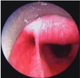
Fig. 1 Pus seen during hypopharyngoscopy at the internal opening in the pyriform sinus.
Fig. 2 Endoscopic view of the internal opening in the apex of left pyriform sinus.
documentation and was also used for teaching purposes. The opening is then cannulated by a guide wire from CV catheter and was cauterized using monopolar cautery.
The patients were extubated and shifted to the general ward. Oral feeds resumed 4 hours after surgery. Sutures were removed 5 days after surgery. The patients were subsequently on follow-up.
Results
All patients were children. There were 3 girls and 2 boys. The youngest was 3 years old while the oldest was 16. The lesions
occurred on the left side of the neck for all patients. In all cases, the internal opening of the sinus tract was confirmed by hypopharyngoscopy, and originated from the apex of the pyriform sinus, posterior to the fold made by the internal laryngeal nerve. In one case, surgical excision of the entire sinus tract was performed. In this patient, the sinus tract originated from the apex of the pyriform sinus, passed through the thyroid gland, and terminated in perithyroid tissue. This patient underwent a Left hemithyroidectomy with excision of the sinus tract.
The CT scans (n¼5) showed disease extending from the
pyriform sinus apex through the strap muscle layer to the thyroid or perithyroid tissue in all patients. In all patients, abnormal soft-tissue swelling and enhancement along the course of the disease were evident on the CT scans. In two patients, the lesions ended at the perithyroid level. There were no cases involving the mediastinums below the sternal notch. The involved pyriform sinus fossae were deformed by adjacent soft-tissue inflammation in all patients. One patient had cutaneous opening in the left anterior portion of the neck. One patient had thyroid gland involvement, including swell-ing of the thyroid gland, poor definition of the thyroid margin, and loss of high attenuation of the affected lobe on CT scans after contrast enhancement. MR images, obtained from two patients, showed the same disease course as did the CT scans. The patients continue to be under follow-up even after two years. There has been no evidence of recurrence in any of the patients (►Table 1).
Discussion
Fourth branchial pouch anomalies are rare, representing only 1–4% of branchial apparatus anomalies.6 They commonly occur as recurrent abscesses involving the neck and thyroid glands.1–3,12,13An anomaly can present as a sinus, a cyst or a
fistula. A sinus has an opening either in the pyriform sinus or the skin but not both; a cyst does not have an opening; a
fistula has an internal opening in the pyriform sinus and external opening in the skin, which makes it an epithelized tract.8Sinuses and be converted intofistulae by repeated I&D procedures.
The third and fourth branchial pouches are connected to the developing pharynx by the pharyngobranchial duct, which degenerates by the 7thweek of intrauterine life. Failure
to degenerate results in a 3rdor 4thbranchial pouch
anoma-ly.4,5 The course of the anomalies involving 3rd and 4th
branchial pouches is well described in literature. It is difficult to differentiate clinically between the two anomalies, though a definitive diagnosis can be established by radiological means and direct hypopharyngoscopy.
Thefistulous tract of a fourth branchial pouch originates at the apex of the pyriform sinus and descends to exit the pharynx inferior to the superior laryngeal nerve, cricothyroid muscle, and thyroid cartilage. The tract continues to descend lateral to the trachea and recurrent laryngeal nerve. On the left side, the tract curves forward, under the arch of the aorta, and then courses upward posterior to the internal carotid artery. On the right side, although rare, the tract circles
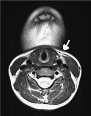
Fig. 5 MRI showing a neck abscess.
forward underneath the subclavian artery before ascending. The tract proceeds superiorly, coursing over the hypoglossal nerve and, possibly, open externally in the neck at the lower anterior portion of the sternocleidomastoid muscle.4,5There have only been a handful of cases in the literature claiming a true fourth pouchfistula10; none of our patients had a true fourth pouchfistula. All of them had blind sinuses which terminated in the neck and thyroid gland. The single patient with an external opening had it as a result of an I&D procedure.
Afistulous tract of a third branchial apparatus abnormality has a similar course to a fourth arch anomaly, albeit it exits the pharynx superior to the superior laryngeal nerve. The inter-nal opening in the pyriform sinus is also located higher up in the lateral wall rather than at the apex.4,5,9
The common symptoms of a fourth branchial pouch anomaly include recurring deep neck infections or abscesses, as well as soft fluctuant masses.2,11,14 Third and fourth branchial arch anomalies may also lead to acute suppurative thyroiditis. For this reason, some authors recommend inves-tigating the presence of a branchial arch anomaly in all cases of thyroiditis.1–3,12,13It is important to run diagnostic tests to
demonstrate a sinus or fistula originating in the pyriform sinus. A barium esophagogram can detect thesefindings but this procedure should only be done after the acute infection has resolved.12,15CT and magnetic resonance imaging (MRI) are the modalities of choice for displaying both location and extent of pyriform sinus anomalies, as well as thyroid in-volvement.11,14We performed direct hypopharyngoscopies in all our patients prior to open surgery to confirm our diagnosis and to cauterize the internal opening so as to abolish recurrence.17
All patients had been referred to the authors and had undergone multiple external drainage procedures. It was imperative to consider a diagnosis of fourth branchial pouch anomaly for these patients. The recommended treatment for fourth branchial anomalies is a complete surgical excision of the tract.8,11,14–16The most commonly used approach is the
external one, which involves complete exposure of the thyroid ala and carotid sheath on the affected side to expose the fistulous tract. The use of endoscopic cauterization limited to the sinus tract origin as a less-invasive procedure has been noted. Recently, use of sclerotherapy with OK-432 has been expanded to treat branchial cleft cysts.17,18 The
Table 1 Summary of patients
No. Age Year presented Sex Clinical presentation Radiology
1 3 2010 F Recurrent left-sided neck swelling I & D x 2
CT - Abscess extending from pyri-form apex to supraclavicular fossa
2 4 2011 F Recurrent left sided neck swelling I & D x 1
MRI - tract delineated to pyriform sinus
3 16 2011 M Recurrent neck left sided abscess7 years I & D x 6
CT- Left thyroid lobe abscessþ,
4 8 2010 F Recurrent left sided neck abscess I & D x 3
Exploration for lymphangioma done elsewhere
MRI- sinus tract seen communicat-ing with Left pyriform sinus
5 7 2009 M Recurrent left sided neck abscess I & D x 4
CT- abscess in high cervical region with possible communication with left pyriform sinus
No. Age Year presented Sex Hypophayngoscopy Treatment Status
1 3 2010 F Internal opening in apex of pyriform sinus with pus
External excision of tract with endoscopic diathermy of in-ternal opening
No recurrence
2 4 2011 F Positive Neck exploration with endo-scopic diathermy
No recurrence
3 16 2011 M Positive Neck exploration, left hemi-thyroidectomy, endoscopic diathermy
No recurrence
4 8 2010 F Positive Neck exploration with endo-scopic diathermy
No recurrence
5 7 2009 M Positive Neck exploration with endo-scopic diathermy
No recurrence
authors formulated a combined treatment modality for all our patients involving the pediatric surgeon and Otorhino-laryngologist. All the patients underwent external surgery with a concomitant therapeutic hypopharyngoscopy to en-sure complete excision of the anomalous tract. We did not use any chemical agents to cauterize the internal opening. The use of electro cautery was adequate. Due to the high incidence of secondary infection of these anomalies, early excision is recommended. A thorough examination is critical, and cannulation of the tract under direct visualization with a small catheter is very helpful in aiding a complete and safe dissection. Due to the intimacy of tracheal structures and
fibrosis, it is often ideal to remove a portion of the thyroid gland as well.
Conclusion
Fourth branchial pouch anomalies present a challenge to the clinician due to their rarity and ambiguous presentation. A detailed clinical history and examination should arouse suspicion. Radiology and diagnostic hypopharyngoscopy will confirm the diagnosis. Once diagnosed, early treatment is critical due to the high incidence of complications. The authors advocate a combined treatment involving external surgery with internal cauterization of the mucosal commu-nication to eradicate the disease process.
Key Points
• Fourth branchial pouch anomaly is rare
• Presents as recurrent neck abscess and thyroiditis mostly in children
• MRI and Direct hypopharyngoscopy are confirmatory tools
• Surgical exploration and excision of the tract is the treatment
• Hemithyroidectomy may be needed if the thyroid lobe is involved
• Cauterization of the internal opening prevents recurrence in all patients
Conflict of Interest
None.
Financial Disclosures
None.
References
1 Takai SI, Miyauchi A, Matsuzuka F, Kuma K, Kosaki G. Internal
fistula as a route of infection in acute suppurative thyroiditis. Lancet 1979;1(8119):751–752
2 Lucaya J, Berdon WE, Enriquez G, Regas J, Carreno JC. Congenital pyriform sinus fistula: a cause of acute left-sided suppurative thyroiditis and neck abscess in children. Pediatr Radiol 1990; 21(1):27–29
3 Tovi F, Gatot A, Bar-Ziv J, Yanay I. Recurrent suppurative thyroiditis due to fourth branchial pouch sinus. Int J Pediatr Otorhinolaryngol 1985;9(1):89–96
4 Williams PL. Gray’s Anatomy (38thed.). New York, NY: Churchill Livingstone; 1995
5 Benson MT, Dalen K, Mancuso AA, et al. Congenital anomalies of the branchial apparatus: embryology and pathologic. Radio-graphics 1992;12:943–960
6 Shrime M, Kacker A, Bent J, Ward RF. Fourth branchial complex anomalies: a case series. Int J Pediatr Otorhinolaryngol 2003; 67(11):1227–1233
7 Tucker HM, Skolnick ML. Fourth branchial cleft (pharyngeal pouch) remnant. Trans Am Acad Ophthalmol Otolaryngol 1973;77(5): ORL368–ORL371
8 Godin MS, Kearns DB, Pransky SM, Seid AB, Wilson DB. Fourth branchial pouch sinus: principles of diagnosis and management. Laryngoscope 1990;100(2 Pt 1):174–178
9 Nicoucar K, Giger R, Jaecklin T, Pope HG Jr, Dulguerov P. Management of congenital third branchial arch anomalies: a systematic review. Otolaryngol Head Neck Surg 2010;142(1): 21–28.e2
10 Patel AB, Hinni ML. The fourth branchial complex anomaly: a rare clinical entity. Case Rep Otolaryngol 2011;2011:958652 11 Yang C, Cohen J, Everts E, Smith J, Caro J, Andersen P. Fourth
branchial arch sinus: clinical presentation, diagnostic workup, and surgical treatment. Laryngoscope 1999;109(3):442–446 12 Cases JA, Wenig BM, Silver CE, Surks MI. Recurrent acute
suppura-tive thyroiditis in an adult due to a fourth branchial pouchfistula. J Clin Endocrinol Metab 2000;85(3):953–956
13 Tovi F, Gatot A, Bar-Ziv J, Yanay I. Recurrent suppurative thyroiditis due to fourth branchial pouch sinus. Int J Pediatr Otorhinolaryngol 1985;9(1):89–96
14 Liberman M, Kay S, Emil S, et al. Ten years of experience with third and fourth branchial remnants. J Pediatr Surg 2002;37(5):685–690 15 Nonomura N, Ikarashi F, Fujisaki T, Nakano Y. Surgical approach to
pyriform sinusfistula. Am J Otolaryngol 1993;14(2):111–115 16 Kubota M, Suita S, Kamimura T, Zaizen Y. Surgical strategy for the
treatment of pyriform sinusfistula. J Pediatr Surg 1997;32(1): 34–37
17 Kim MG, Lee NH, Ban JH, Lee KC, Jin SM, Lee SH. Sclerotherapy of branchial cleft cysts using OK-432. Otolaryngol Head Neck Surg 2009;141(3):329–334
18 Kim KH, Sung MW, Koh TY, Oh SH, Kim IS. Pyriform sinus