445
Laparoscopic Resection of Metastatic Liposarcoma Case Report
International Braz J Urol Vol. 32 (4): 445-447, July - August, 2006
Laparoscopic Resection of Metastatic Pelvic Liposarcoma
Hong B. Shim, Tae Y. Jung, Ja H. Ku
Department of Urology, Seoul Veterans Hospital, Seoul, Korea
ABSTRACT
We report a pelvic liposarcoma originating from the left spermatic cord that recurred following inadequate excision. In our case, the tumor was resected without performing orchiectomy previously. The patient was managed by laparoscopic resection, before undergoing radical orchiectomy in the left inguinal region. To our knowledge, no case of laparoscopic resection for the recurrent liposarcoma has been described. In addition, the present case serves to demonstrate that radi-cal orchiectomy with wide excision is needed for paratesticular tumor.
Key words: spermatic cord; liposarcoma; metastasis; pelvis; laparoscopy
Int Braz J Urol. 2006; 32: 445-7
INTRODUCTION
Paratesticular liposarcomas are rare malig-nant mesenchymal tumors that develop in fatty tis-sues. The choice of therapy for paratesticular liposa-rcoma is surgical, through orchiectomy and extensive local removal. We report here a pelvic liposarcoma originating from the recurrent paratesticular liposar-coma. We emphasize that the tumor should be treated with radical orchiectomy with high ligation of the cord and the patients must be followed periodically on a life-long basis.
CASE REPORT
A 73-year-old man presented at our hospital with a palpable mass at the left inguinal area. The patient had been treated for a paratesticular mass with resection via a high inguinal approach 7 years previ-ously at other hospital and had been told that histo-logical diagnosis at that time had been liposarcoma.
Physical examination revealed a firm non-tender im-mobile mass at the left inguinal area, which was the same region of the primary mass and an upward dis-placement of left testis. Hematologic examination and urinalysis were not remarkable.
446
Laparoscopic Resection of Metastatic Liposarcoma
after an additional 1 cm incision. Pathological exami-nation of resected specimens in both pelvis and left inguinal area revealed myxoid liposarcoma (Figure-2). Surgical margins were free of tumor. Without any postoperative adjuvant therapy, no evidence of recur-rence or metastasis was noted during the 6-month follow-up period.
COMMENTS
Liposarcoma is histologically subdivided into 3 types, including well differentiated, myxoid
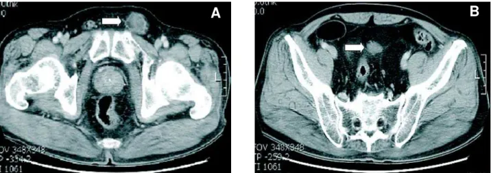
Figure 1 – Contrast-enhanced computed tomography of the pelvis. A) Partially fat containing soft tissue mass (arrow) at the left inguinal area. B) Homogenous enhanced soft tissue mass (arrow) adjacent to sigmoid mesocolon.
A
B
Figure 2 – Pathological findings of the tumor are indicative of myxoid liposarcoma. A) Paratesticular liposarcoma (HE, X40). B) Pelvic liposarcoma (HE, X200).
A
B
addi-447
Laparoscopic Resection of Metastatic Liposarcoma
tional therapeutic benefit, and the role of chemo-therapy is not well defined.
A case of port site metastasis after laparoscopy-assisted surgery for retroperitoneal li-posarcoma has been previously reported. A possible mechanism for port site cell implantation is a length operative procedure with high-pressure penumoperitoneum, which enables the tumor cell to implant locally or disseminate hematogeneously dur-ing the operation (1). Our case is the first description of pelvic liposarcoma metastasizing from the inguinal area, which was managed by laparoscopic procedure. In this case, there was no difficulty or special care during the laparoscopic procedure.
A case of liposarcoma has been described which was treated successfully by tumor excision without involvement of the testis (2). However, simple excision may be inadequate treatment for paratesticular sarcoma, since wide repeat excision may reveal microscopic residual disease in some of completely excised cases (3). Our finding emphasizes that in the treatment of spermatic cord liposarcoma complete mass resection and inguinal orchiectomy with high ligation of the spermatic cord must be
per-formed. However, as the histopathology of the ear-lier surgery was not available, the possibility of ma-lignant change in a preexisting lipoma could not be excluded. Regardless of initial therapy, the risk of recurrence of paratesticular liposarcoma always ne-cessitates long-term follow-up.
CONFLICT OF INTEREST
None declared.
REFERENCES
1. Martinez J, Targarona EM, Balague C, Pera M, Trias M: Port site metastasis. An unresolved problem in laparoscopic surgery. A review. Int Surg. 1995; 80: 315-21.
2. Crespo Atin V, Padilla Nieva J, Martin Bazaco J, Llarena Ibarguren R, Pertusa Pena C: Scrotal liposar-coma. Arch Esp Urol. 2001; 54: 729-32.
3. Catton C, Jewett M, O’Sullivan B, Kandel R: Paratesticular sarcoma: failure patterns after definitive local therapy. J Urol. 1999; 161: 1844-7.
Accepted after revision: March 6, 2006
Correspondence address: Dr. Ja Hyeon Ku
Dept Urology, Seoul Veterans Hosp 6-2, Doonchon Dong, Kangdong-Gu Seoul 134-791, Korea
Fax: + 82 2 4834260