DOI: 10.5935/2359-4802.20170079
CASE REPORT
Mailing Address: Joana Malheiro
Rua de Grijo, 100, 4E. Postal Code: 4150. Lordelo do Ouro, Porto – Portugal E-mail: [email protected]; [email protected]
Tachycardiomyopathy and Extracorporeal Membrane Oxygenation: A Case Report
Joana Malheiro, João Almeida, Daniel Caeiro, Adelaide Dias, Marlene Fonseca, Vasco Gama
Centro Hospitalar de Vila Nova de Gaia, Espinho – Portugal
Manuscript received October 06, 2016, revised manuscript February 08, 2017, accepted April 05, 2017. Cardiomyopathies; Tachycardia; Shock, Cardiogenic;
Extracorporeal Membrane Oxygenation; Atrial Fibrillation.
Keywords
Introduction
Tachycardiomyopathy (TCM) is a rare, but potentially reversible cause of cardiomyopathy and cardiogenic shock. It is defined by a global left ventricular systolic dysfunction secondary to persistent tachyarrhythmia, with partial (in patients with previous structural disease), or total recovery (in patients without previous structural disease) after cardiac rhythm normalization.1 The most
commonly implicated arrhythmia is atrial fibrillation. The hemodynamic changes that characterize the patient with TCM include: increase in ventricular telediastolic and telesystolic volumes, global hypokinesis, increase in ventricular and pulmonary artery filling pressures, and, finally, decrease in ejection fraction (EF).1,2
It manifests clinically by congestive heart failure, and, in some cases, it may develop into cardiogenic shock. There are no specific methods to identify the presence of TCM. The diagnosis is usually attained retrospectively with normalization or improvement of left ventricular dysfunction, through tachyarrhythmia reversion or control. The most frequent complications of TCM are embolic events, complications due to the evolution of arrhythmia severity with degeneration to Ventricular Tachycardia (VT)/Ventricular Fibrillation (VF) and cardiogenic shock.3
The treatment includes measures of hemodynamic support, frequency control, and reversion to sinus rhythm, when possible.2 Extracorporeal Membrane
Oxygenation (ECMO) is a mechanical device that ensures blood oxygenation and perfusion of the main organs for prolonged periods of time in patients with lung and/or
heart failure.4 The use of this device allows the patient
to remain alive and hemodynamically stable, working as a bridge to eventual recovery, transplantation, decision-making or even as a “bridge to another bridge” (for instance, as a bridge to left ventricular assist device – LVAD – as target therapeutics).
In cardiogenic shock of potentially reversible etiology, this measure can be lifesaving, as it temporarily assumes cardiac function during the refractory, intrinsic, and reversible phase of cardiogenic shock until patient recovery, minimizing the myocardial effort, improving organ perfusion, and preserving renal function.4 The
authors describe a case of a patient with cardiogenic shock secondary to TCM where the use of ECMO was very important in her evolution.
Clinical Case
The patient was a 54-year-old female patient with a history of hypertension, dyslipidemia, obesity, and rheumatic mitral stenosis. On May 5, 2015, she was submitted to a mitral mechanical valve implantation (St. Jude, n. 29) without complications and the echocardiogram at hospital discharge showed a normal-functioning prosthesis, in addition to preserved biventricular function. The patient came to the emergency department on July 6, 2015, due to history of approximately one month of palpitations and progressive worsening of the normal pattern of dyspnea, with complaints of dyspnea with minimal exertion (it usually was with moderate exertion), orthopnea and paroxysmal nocturnal dyspnea. She also reported antihypertensive drug discontinuation at the first postoperative month due to symptomatic hypotension (lipothymia). She denied chest pain and recent history of infection.
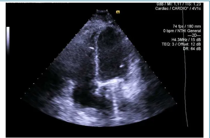
Figure 1 – Transthoracic echocardiography performed at admission disclosed global hypokinesis of the left and right ventricles, with severe decrease in biventricular systolic function.
Analytically, she also showed normocytic/normochromic anemia (Hgb 11.6 g/dL), troponin-T elevation (1.96 ng/mL) and supra-therapeutic International Normalized Ratio (INR) (6.0). The electrocardiogram disclosed atrial fibrillation with Rapid Ventricular Response (RVR) and the echocardiogram showed a mechanical prosthesis with normal function without regurgitation, moderate tricuspid regurgitation and global hypokinesis of the left ventricle and right ventricle, with severe depression of biventricular systolic function (Figure 1).
Due to suspected worsening of the systolic function associated with atrial fibrillation with RVR, an attempt was made to carry out chemical cardioversion with amiodarone and, subsequently, electrical cardioversion (100 J), which was successful. After the electrical cardioversion, she had several episodes of VT, one of which required cardioversion due to loss of consciousness and, subsequently, an episode of VF, which was successfully defibrillated. Due to the electrical instability and possible need for ventricular support, she was transferred to the Coronary Intensive Care Unit.
On admission to the unit, she was started on dobutamine, with good hemodynamic and gasometric response, with subsequent recurrence of electrical instability, and cardiopulmonary arrest in FV, which was successfully defibrillated. As she did not tolerate dobutamine, and given the context of refractory cardiogenic shock, it was decided to initiate hemodynamic support with Venoarterial ECMO (CARDIOHELP System® of Maquet), as well as invasive mechanical ventilation.
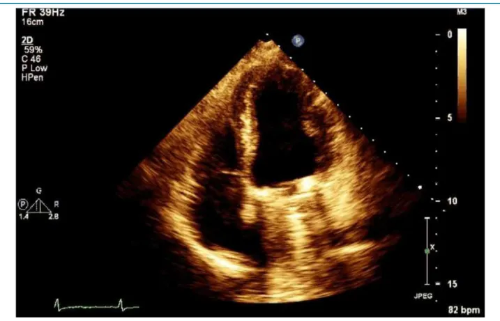
Figure 2 – Transthoracic echocardiogram performed on discharge showing moderate LVSF decrease with ejection fraction of 40% and preserved RVSF.
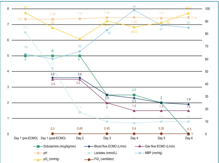
improvement of left ventricular systolic function in the echocardiographic control. Figure 3 summarizes the evolution of hemodynamic, and gasometric data, as well as the established parameters.
The venoarterial ECMO was removed on the 5th day,
the temporary pacemaker on the 6th day, and she was
extubated on the 8th day, maintaining clinical stability.
Beta-blocker and digoxin were initiated on the 11th day
of hospitalization, with good hemodynamic tolerance and good heart rate control. She underwent a coronary angiography, which excluded epicardial coronary disease, and a cardiac magnetic resonance, which showed no signs of myocardial edema or fibrosis. At discharge, with atrial fibrillation rhythm and controlled ventricular response, the transthoracic echocardiogram showed a normal-functioning mitral prosthesis, with mild prosthetic regurgitation, mild TI, moderate LVSF depression with 40% EF and preserved RVSF (Figure 2).
Discussion
TCM, being a rare and difficult-to-diagnose entity, may show an unfavorable evolution, if not diagnosed
early. Because there are no specific methods for its diagnosis, the latter is attained by a high index of clinical suspicion, based on clinical history and findings. Fenelon et al. proposed the following criteria for the diagnosis of TCM: dilated cardiomyopathy or heart failure; chronic or persistent cardiac arrhythmia, including incessant supraventricular tachycardia, atrial fibrillation, or flutter, and incessant VT.5
Generally, tachycardia that occurs in 10 to 15% of the day may result in cardiomyopathy.2 The ventricular rate
that causes TCM has not been defined, but any heart rate above 100 beats per minute may be relevant.2
8 7 6 5 4 3 2 1 0 0
Day 1 (pre-ECMO) Day 1 (post-ECMO) Day 2 Day 3 Day 4 Day 5 Day 6
10 20 30 40 50 60 70 80 90 100
0.3 0.45 0.45 0.4 0.35 0.3
0.8 0.8 0.8 1.4 4.2 6.5 0.7 3.5 3.5 2 2.5
2.3 2 1.9
1.5 1.5 1.5
3.6 3.6 5 5 5 2.5 0 64 66 83 99 85 60 97 85 90.4 85.5 87 89 96.6 7.35
7.34 7.37 7.44
7.44 7.4
7.45
Dobutamine (mcg/kg/min)
pH
pO2 (mmHg)
Blood flow ECMO (L/min)
Lactates (mmol/L)
FiO2 (ventilator)
Gas flow ECMO (L/min)
MBP (mmHg)
Figure 3 – Hemodynamic and gasometric data, and parameters established in extracorporeal membrane oxygenation (ECMO). pO2: oxygen partial pressure; FiO2: fraction of inspired oxygen; MBP: mean blood pressure.
As TCM is characterized by being reversible, the prognosis will be partially determined by the implemented hemodynamic support measures. Hemodynamic support with ECMO increasingly plays a key role in the approach of patients with cardiogenic shock. This technique is usually considered when hemodynamic instability does not respond to conventional pharmacological measures. There are few reports in the literature of patients with TCM who developed cardiogenic shock3,6,7
and, to the best of our knowledge, this is the first case of TCT with refractory shock in which ECMO was used as a hemodynamic support measure. In the reported case, given the context of electrical instability and the consequent intolerance to aminergic support, we chose the early onset with ECMO. The implementation of this
technique ensured tissue perfusion during the phase of greater electrical and hemodynamic instability, working as a bridge until heart rate control and recovery of left ventricular dysfunction were attained. After frequency control, progressive improvement of left ventricular systolic function was observed, which supported the diagnostic suspicion of TCM.
Author contributions
1. Araújo F, Ducla-Soares JL. Tachycardiomyopathies. Rev Port Cardiol. 2002;21(5):585-92.
2. Nam MC, Aravind A, Chan K, James S, Brown N. Tachycardia-induced
cardiomyopathy- a fully reversible phenomenon. Am Heart Hosp J. 2010;8(2):E115-7.
3. Gariglio L, Larraburu A, Nicolini M, Crespo M, Arrinda Calzetta J, Perrone SV, et al. Shock cardiogénico por taquicardiomiopatía: propranolol endovenoso en la etapa aguda. Insuf Card. 2013;8(2):95-101.
4. Lawler PR, Silver DA, Scirica BM, Couper GS, Weinhouse GL, Camp PC Jr. Extracorporeal membrane oxygenation in adults with cardiogenic shock. Circulation. 2015;131(7):676-80.
5. Nakazato Y. Tachycardiomyopathy. Indian Pacing Electrophysiol J. 2002;2(4):104-13.
6. Walker NL, Cobbe SM, Birnie DH. Tachycardiomyopathy: a diagnosis not to be missed. Heart. 2004;90(2):e7.
7. Zwicker C, Becker M, Lepper W, Koch KC, Westenfeld R. Cardiogenic shock due to tachycardiomyopathy after heart transplantation: successful treatment with ivabradine. Cardiology. 2010;116(3):174-7.
References
Potential Conflict of Interest
No potential conflict of interest relevant to this article was reported.
Sources of Funding
There were no external funding sources for this study.
Study Association