Métodos convencionais e moleculares para o diagnóstico da tuberculose pulmonar: um estudo comparativo
Stella Sala Soares Lima1, Wanessa Trindade Clemente2, Moisés Palaci3,
Reinaldo Vieira Rosa4, Carlos Maurício de Figueiredo Antunes5, José Carlos Serufo6
Abstract
Objective: To compare four laboratory methods in the diagnosis of pulmonary tuberculosis. Methods: Respiratory secretion specimens were collected from 160 patients suspected of having pulmonary tuberculosis. Direct testing for Mycobacterium tuberculosis was carried out using Ziehl-Neelsen staining, auramine staining, culture on Löwenstein-Jensen (LJ) medium and polymerase chain reaction (PCR). The strains isolated were identified by means of a radiometric method using p-nitro-alpha-acetylamino-beta-hydroxypropiophenone (NAP) and classical methods. The sensitivity of the methods was compared to the gold standard for the diagnosis of pulmonary tuberculosis, based on clinical, radiological and microbiological criteria. Results: Of the 160 patients, 142 were diagnosed with pulmonary tuberculosis according to the gold standard. The sensitivity of Ziehl-Neelsen staining, auramine staining, culture on LJ medium and PCR was 54.2%, 58.4%, 67.6% and 77.5%, respectively, when compared with the diagnostic criterion adopted. All four methods presented 100% specificity. In the identification of mycobacteria, there was high (96.8%) concordance between PCR and the radiometric method using NAP. The sensitivity of PCR was 50.8% in samples with negative sputum smear microscopy results and 98.8% in those with positive results. The sensitivity of PCR was lower in specimens with negative results in sputum smear microscopy and culture than in those with positive results (25.6% and 99.0%, respectively). Conclusions: We found PCR to be a promising method for the diagnosis of pulmonary tuberculosis, even in paucibacillary specimens. Simultaneous identification and faster results are additional advantages of this method.
Keywords: Tuberculosis, pulmonary/diagnosis; Culture media; Polymerase chain reaction; Sputum/microbiology.
Resumo
Objetivo: Comparar quatro métodos laboratoriais no diagnóstico de tuberculose pulmonar. Métodos: Foram realizadas pesquisa direta pelas colorações de Ziehl-Neelsen e auramina, cultura para micobactérias em meio Löwenstein-Jensen (LJ) e polymerase chain reaction (PCR, reação em cadeia da polimerase) para Mycobacterium tuberculosis em 160 amostras de secreção respiratória de pacientes com suspeita de tuberculose pulmonar. As cepas isoladas foram identificadas por método radiométrico utilizando-se p-nitro-alfa-acetilamino-beta-hidroxipropiofenona (NAP) e métodos clássicos. A sensibilidade dos métodos foi comparada com o padrão ouro para o diagnóstico da tuberculose pulmonar, definido por critérios clínicos, radiológicos e microbiológicos. Resultados: Dos 160 pacientes, 142 foram diagnosticados com tuberculose pulmonar de acordo com o padrão ouro. As técnicas de Ziehl-Neelsen e auramina, cultura em meio LJ e PCR apresentaram sensibilidade de 54,2%, 58,4%, 67,6% e 77,5%, respectivamente, quando comparados ao critério diagnóstico adotado. A especificidade dos quatro métodos foi de 100%. A concordância na identificação da micobactéria entre PCR e o método radiométrico utilizando NAP foi alta (96,8%). A sensibilidade da PCR foi de 50,8% nas amostras com baciloscopia negativa e de 98,8% naquelas com baciloscopia positiva. Nas amostras com resultados negativos na baciloscopia e cultura, a sensibilidade da PCR foi menor que nas com resultados positivos (25,6% e 99,0%, respectivamente). Conclusões: A PCR é método promissor no diagnóstico da tuberculose pulmonar, mesmo em amostras paucibacilares. Além disso, apresenta a vantagem da identificação simultânea e rapidez do resultado.
Descritores: Tuberculose pulmonar/diagnóstico; Meios de cultura; Reação em cadeia da polimerase; Escarro/microbiologia.
* Study carried out at the Júlia Kubitschek Hospital, Fundação Hospitalar do Estado de Minas Gerais – FHEMIG, Hospital Foundation of the State of Minas Gerais – Universidade Federal de Minas Gerais – UFMG, Federal University of Minas Gerais – School of Medicine, Belo Horizonte, Brazil; at the Micra Biotechnology Laboratory, Belo Horizonte, Brazil; and at the Universidade Federal do Espírito Santo – UFES, Federal University of Espírito Santo – Infectious Diseases Center, Vitória, Brazil.
1. Auditing Physician on the Committee for Nosocomial Infection Control. Universidade Federal de Minas Gerais – UFMG, Federal University of Minas Gerais – Hospital das Clínicas, Belo Horizonte, Brazil.
2. Assistant Professor in the Department of Complementary Assessment. Universidade Federal de Minas Gerais – UFMG, Federal University of Minas Gerais – School of Medicine, Belo Horizonte, Brazil.
3. Adjunct Professor. Universidade Federal do Espírito Santo – UFES, Federal University of Espírito Santo –Vitória, Brazil. 4. Head of the Department of Microbiology. Micra Biotechnology Laboratory, Belo Horizonte, Brazil.
5. Full Professor. Universidade Federal de Minas Gerais – UFMG, Federal University of Minas Gerais – Belo Horizonte, Brazil.
6. Adjunct Professor in the Department of Clinical Medicine. Universidade Federal de Minas Gerais – UFMG, Federal University of Minas Gerais – School of Medicine, Belo Horizonte, Brazil.
Correspondence to: Stella Sala Soares Lima. Rua Um, 108, Condomínio Veredas das Geraes, CEP 34000-000, Nova Lima, MG, Brasil. Tel 55 31 3541-7898. E-mail: [email protected]
Methods
Sputum samples collected from patients with presumed pulmonary TB who were treated at the Júlia Kubitschek Hospital, Belo Horizonte, Brazil, between January of 2001 and January of 2002, were analyzed. The patients underwent clinical evaluation, chest X-rays (anteroposterior and lateral) and HIV serology, as well as completing a form regarding epidemiological data. After a minimum of 18 months, the medical charts were reevaluated in order to confirm the diagnosis and determine the clinical evolution of the patients. Sample collec-tion and all laboratory procedures were performed following safety regulations, and standardized handling criteria were met.
The samples were decontaminated using monosodium phosphate and trisodium phosphate (modified Corper & Stoner method)(9) and were
concentrated by centrifugation. Subsequently, smears prepared from an aliquot of the sediment were submitted to staining (Ziehl-Neelsen and auramine),(10,11) after which they were examined
under microscopy. A quantity (0.1 mL) of the sedi-ment was added to at least two tubes containing Löwenstein-Jensen (LJ) medium, which were incu-bated at 37°C for up to eight weeks. The results were described using a semiquantitative scale.(12)
The strains isolated were identified by means of the growth inhibition test using p-nitro-alpha-acetylamino-beta-hydroxypropiophenone (NAP) in the BACTEC 460 system (Becton Dickinson Microbiology Systems, Sparks, MD, USA) and by means of other phenotyping tests ( morphological analysis of colonies and classical biochemical tests). (13) The Amplicor® PCR kit for M. tuberculosis
(Roche Molecular Systems, Branchburg, NJ, USA), with an internal control, was used according to the manufacturer specifications. (14)
The final criterion for the diagnosis of TB was defined using a combination of clinical, radiological and microbiological data, which were obtained by specialists at the Júlia Kubitschek Hospital, together with the response to the use of antituberculosis drugs.(5,7,8,15-18)
All patients included in the investigation gave written informed consent. The present study was approved by the Ethics in Research Committee of the Hospital Foundation of the State of Minas Gerais.
Introduction
Statistics regarding the occurrence of tuber-culosis (TB) worldwide reveal an alarming epidemiological situation, considering that approximately half of the world population is known to be infected with Mycobacterium tuberculosis, with 8.8 million new TB cases and 1.6 million deaths per year.(1) In Brazil, there
has been an evident resurgence of TB, which is related to the lack of an efficient public health care system, a paucity of TB control programs, successive economic crises, the growth of marginalized populations (urban and rural), increased migration and the AIDS epidemic.(2)
In 2005, the incidence rate of TB in Brazil was 43.78 cases/100,000 population, although it is believed that the number of patients infected is underreported.(3) Since TB is a disease for which
the use of vaccines has shown no efficacy, the best control mechanisms are early diagnosis and early treatment, which significantly reduce trans-mission rates. However, although conventional microbiology methods still constitute the prin-cipal tool for the diagnosis of TB, direct testing has low sensitivity, and culture requires long culturing time. Therefore, new diagnostic methods have been developed with the aim of replacing direct testing and culture. The ideal test would be one that has high sensitivity, high specificity, yields rapid results and is inexpensive. Molecular techniques, such as polymerase chain reaction (PCR), reduce the time necessary for detection and identification of M. tuberculosis. However, despite being promising for the diagnosis of TB, the sensitivity of molecular techniques is low in samples with negative sputum smear microscopy results and in extrapulmonary samples.(4-8) For
the Brazilian public health care system, PCR for M. tuberculosis remains costly, and it should be used only when presenting some advantage over the conventional methods currently available.
required to reject the null hypothesis was set at 5% (p ≤ 0.05) for all tests.
Results
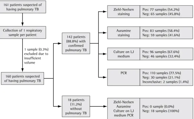
Between January of 2001 and January of 2002, 161 patients suspected of having pulmo-nary TB were evaluated, and one sputum sample was collected per patient. There was one sample loss due to insufficient volume. The mean age of the 160 patients included was 40.0 ± 12.8 years (range, 19-78 years). The general characteristics of the patients are described in Table 1.
Of the 160 patients included, 142 (88.8%) presented pulmonary TB according to the criteria adopted at the Júlia Kubitschek Hospital. Of those 142, 3 had silicotuberculosis and 12 had pulmo-nary multidrug-resistant TB (MDR-TB), according to the World Health Organization criteria.(15) After
the medical charts had been reevaluated, the clin-ical evolution of pulmonary TB was confirmed in 106 patients (74.6%). Of those, 35 (24.6%) improved after treatment with regimen I (rifampin + isoni-azid + pyrazinamide), 19 (13.4%) improved after treatment with regimen IR (regimen I + etham-butol), 10 (7.0%) improved after treatment with regimen III (streptomycin + ethionamide + etham-butol + pyrazinamide), and 10 (7.0%) improved after treatment with the MDR-TB treatment regimen (ethambutol + ofloxacin + clofazimine + teriz-idone + amikacin). Of the 106 patients, 18 (12.7%) abandoned treatment, 13 (9.1%) died, and 1 (0.7%) presented no improvement after treatment with the MDR-TB treatment regimen. Of the 142 patients with pulmonary TB, 36 (25.4%) were referred for treatment at referral centers close to their home, and it was not possible to ascertain their clinical evolution.
The results obtained using the laboratory methods evaluated are shown in Figure 1. The sensi-tivity of direct testing using Ziehl-Neelsen staining and auramine staining, as well as of culture on LJ medium and PCR, was 54.2%, 58.4%, 67.6% and 77.5%, respectively. All four methods presented 100% specificity. The kappa coefficients of the comparisons of the methods used are presented in Table 2.
The identification of M. tuberculosis, by means of a radiometric method using NAP, was performed in 95 of the 96 positive LJ medium cultures (one culture was contaminated, and it was not possible The data initially collected on the questionnaires
were entered into a database and analyzed using the Epi Info 2002 program, version 3.2.2. The kappa coefficient was used to compare the methods. Categorical variables were analyzed using the chi-square test with Yates’ correction. When there were fewer than five events or a null value, Fisher’s exact test was used. The level of statistical significance
Table 1 - Characteristics of the 160 patients suspected of having pulmonary tuberculosis.
Characteristic Patients
n %
Gender
Male 116 72.5
Female 44 27.5
History of tuberculosis contact
In the home 22 13.8
Occupational 6 3.8
Hospital 3 1.9
No contact 47 29.3
Unknown 82 51.2
Comorbidities
Alcoholism 86 53.8
Smoking 84 52.5
Positive HIV serology 20 12.5
Diabetes 12 7.5
Neoplasia 6 3.8
Silicosis 3 1.9
History of pulmonary tuberculosis
77 48.1
Abandonment of previous tuberculosis treatment
44 27.5
Symptoms
Cough 148 92.5
Weight loss 127 79.4
Fever 107 66.9
Night sweats 94 58.8
Dyspnea 78 48.8
Asthenia 72 45.0
Chest pain 61 38.1
Hemoptysis 34 21.3
Chest X-ray findings
Cavitations 85 53.1
Destroyed lung 20 12.5
Consolidation 38 23.8
Other alterations 6 3.8
Normal 2 1.3
In samples with negative results in direct testing and culture, the sensitivity of PCR was 15.6%, whereas, in those with positive results in direct testing and culture, the sensitivity of PCR was 99.0% (p < 0.0001).
In total, 77 patients reported a history of pulmo-nary TB (median time since previous diagnosis, 36 months), and, of those, 44 (57.1%) abandoned treatment. The sensitivity of PCR was 78.6% in patients with a history of pulmonary TB and 77.6% in those without such a history (p = 0.94). No false-negative results were obtained in patients with a history of TB but without active pulmonary TB at the time of the study.
Discussion
The present study was developed at a referral center for the treatment of pulmonary TB where there are high proportions of symptomatic patients, patients with comorbidities and patients with a history of TB who report treatment abandonment, as well as a considerable frequency of MDR-TB and death. In our to isolate the strain in successive subcultures). Of
the 95 strains isolated on LJ medium and identified by the BACTEC system, 92 (96.8%) were identified by PCR.
The sensitivity of PCR was 50.8% in samples with negative direct testing results, whereas, in those with positive sputum smear microscopy results, the sensitivity of PCR was 98.8% (p < 0.0001).
Figure 1 - Flowchart of the diagnosis and of the results obtained using the laboratory methods evaluated. TB: tuberculosis; LJ: Löwenstein-Jensen; PCR: polymerase chain reaction for Mycobacterium tuberculosis, Pos: positive; and Neg: negative.
1 sample (0.3%) excluded due to insufficient volume 161 patients suspected of
having pulmonary TB
Collection of 1 respiratory sample per patient
142 patients (88.8%) with confirmed pulmonary TB
160 patients suspected of having pulmonary TB
18 patients (11.2%) without pulmonary TB
Pos: 0 sample (0.0%) Neg: 18 samples (100%) Ziehl-Neelsen
Auramine Culture on LJ
medium PCR
Pos: 110 samples (77.5%) Neg: 30 samples (21.1%) Inconclusive: 2 samples (1.4%) PCR
Pos: 96 samples (67.6%) Neg: 46 samples (32.4%) Culture on LJ
medium
Pos: 83 samples (58.4%) Neg: 59 samples (41.6%) Auramine
staining
Pos: 77 samples (54.2%) Neg: 65 samples (45.8%) Ziehl-Neelsen
staining
Table 2 - Kappa coefficients obtained from the comparison of the diagnostic methods.
Diagnostic method Kappa (95% CI) Ziehl-Neelsen staining vs. auramine
staining
0.93 (0.87-0.98)
Ziehl-Neelsen staining vs. culture on LJ medium
0.62 (0.49-0.74)
Ziehl-Neelsen staining vs. PCR 0.54 (0.40-0.68) Auramine staining vs. culture on LJ
medium
0.66 (0.54-0.78)
In the present study, culture on LJ medium, which allows the definitive confirmation of the diagnosis of pulmonary TB, had a sensitivity of 67.6%, which is lower than the 80%-100% rate typically described,(19) and the influence of the
collection of a single sample should be considered. The main advantage of other culture media, as well as of automated and semi-automated detec-tion methods, is the shorter time required to detect mycobacteria (approximately 15 days rather than 3-8 weeks). However, LJ medium, which is approved by the World Health Organization, remains the most widely used in Brazil. Additional benefits include the fact that the strains can be stored for future studies, and that some strains grow only in this medium. For these reasons, the use of new methods does not dispense with the use of conventional culture.(8)
In relation to the other methods, PCR had higher sensitivity (77.5%), which is within the range (42%-90.9%) established in the literature, the variation depending principally on the characteris-tics of the patient sample.(4-6,8,15-17,22-24) The sensitivity
of PCR was found to be similar to that of culture (p = 0.09), as described in most of the studies comparing these two methods,(6,25) and there was
good concordance of results, which was expressed by a kappa coefficient of 0.78. Culture and PCR were superior to the direct testing methods (p = 0.048 and p = 0.00007, respectively), probably due to the greater capacity of culture and PCR for bacillus detection in paucibacillary samples. Direct testing detects bacilli in samples containing at least 5,000-10,000 bacilli, whereas culture requires only 10-100 bacilli, and PCR requires only 1-20 bacilli . (8,26,27) Ziehl-Neelsen
staining and auramine staining demonstrated good concordance of results with culture and PCR (kappa coefficient, 0.54-0.66; Table 2).
patient sample, 18 patients (11.2%) abandoned treat-ment, and this indicates the need for more efficient measures of treatment compliance, which is essential to control the dissemination of the disease.
The best method for the diagnosis of pulmonary TB is analysis based on the combination of clinical, radi-ological and microbiradi-ological characteristics . (5,7,8,16-18)
Based on the comparison with this criterion, the sensitivity of sputum smear microscopy using Ziehl-Neelsen staining and auramine staining was found to be 54.2% and 58.4%, respectively, results similar to those obtained in other studies, in which the sensitivity of sputum smear microscopy ranges from 50% to 80%.(8,19) The higher sensitivity
of direct testing using auramine staining in rela-tion to that of direct testing using Ziehl-Neelsen staining was not statistically significant. These two methods presented excellent concordance of results, which was expressed by a kappa coefficient of 0.93, as well as equivalence in clinical practice, although some authors suggest that the sensitivity of direct testing is higher when auramine staining is used.(20) Therefore, the choice of the method is
based on the characteristics and resources of the laboratory, since the time required to analyze the smears using auramine staining is shorter than that required to perform sputum smear microscopy using Ziehl-Neelsen (1-2 min vs. 10-15 min).(20,21)
The accuracy of auramine staining is similar to that of Ziehl-Neelsen staining, and the reading requires less time. However, auramine staining demands the use of expensive equipment and is therefore recommended only in laboratories where more than 100 slides are analyzed per day. In addition, the use of auramine staining does not dispense with the use of fuchsin-based stainings to study morphology of the bacillus.(8)
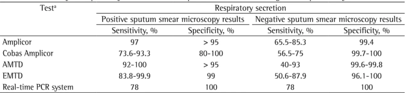
Table 3 - Sensitivity and specificity of nucleic acid amplification tests in the diagnosis of pulmonary tuberculosis.
Testa Respiratory secretion
Positive sputum smear microscopy results Negative sputum smear microscopy results Sensitivity, % Specificity, % Sensitivity, % Specificity, %
Amplicor 97 > 95 65.5-85.3 99.4
Cobas Amplicor 73.6-93.3 80-100 56.5-75 99.7-100
AMTD 92-100 > 95 40-93 99.6-99.8
EMTD 83.8-99.9 99 50.6-87.9 96.1-100
Real-time PCR system 78 100 78 100
aAmplicor and Cobas Amplicor: produced by Roche Molecular Systems, Branchburg, NJ, USA; Amplified Mycobacterium tuberculosis
time required to identify TB patients is important for controlling the dissemination of the disease, allowing the early institution of treatment, and this can have a positive impact on public health.
However, PCR results can remain positive for more than 12 months after diagnosis and initia-tion of treatment, even 6 months after conversion (direct testing and culture).(17,30) This precludes
the use of PCR in follow-up treatment.(8) In the
present study, there was no statistically signifi-cant difference between patients with a history of pulmonary TB and those without in terms of the sensitivity of PCR (78.6% and 77.6%, respectively). Among the patients with a history of TB, the rate of abandonment of previous treatment was high (44 of 77 patients; 57.1%), as was the incidence of active disease at the time of the study, which might have contributed to the absence of a difference between patients with a history of pulmonary TB and those without in terms of the sensitivity of PCR. In patients with a history of TB but without active disease, the median time since previous diagnosis was 36 months, longer than the 6- and 12-month intervals described in the literature as necessary for PCR detection of bacilli to persist.(16,29)
Due to the small number of patients without pulmonary TB and the absence of infection by other pathogens, the present study is not adequate to eval-uate specificity, although no false-positive results were observed, and other studies confirm the high specifi-city of nucleic acid amplification tests (Table 3).(8,30)
In the present study, direct testing was found to remain the method of choice for the initial evalua-tion of patients suspected of having TB, since it has high sensitivity (even in a single sample), is inex-pensive and is easily performed. The most sensitive method for the diagnosis of TB proved to be PCR, being equivalent to culture, with the advantages of faster results and simultaneous identification of M. tuberculosis, although having the disadvantage of being more costly. It has been said that, although the sensitivity of PCR in samples with negative sputum smear microscopy results is lower than desir-able, this method can still present an advantage, when compared with conventional methods, for the rapid diagnosis of paucibacillary pulmonary TB. Currently, there is no other, more effective method when the combined analysis of conventional clin-ical, radiological and microbiological findings does not establish the diagnosis.
The collection of more than one sample per patient would probably increase the sensitivity of all of the methods evaluated in the present study. However, the comparison of the methods using a single sample portrays common situations in clinical practice, as occurs in emergency care clinics.
The sensitivity of PCR was statistically lower in samples with negative sputum smear microscopy results than in those with positive results (50.8% and 98.8%, respectively), which is in accordance with the results of other studies (Table 3).(4,5,8,17,22,28) The
sensitivity of PCR in samples with negative results in direct testing and culture was even lower (25.6%), being statistically lower than that found in samples with positive conventional test results (99.0%). The decreased sensitivity in samples with negative sputum smear microscopy results is due to the presence of a reduced number of bacilli, the lack of homoge-neity of the patient sample and the use, in PCR, of a volume lower than that used in culture.(5,29,30) We
highlight the fact that, to date, the PCR technique used (Amplicor®) has not been approved by the Food and Drug Administration for use in samples with negative sputum smear microscopy results, since the sensitivity in this type of sample varies.(8) Although
the decreased sensitivity of PCR in samples with negative direct testing results is one of the greatest limitations of the method, it is of note that, in the present study, PCR detected bacilli in approximately half of the patients with negative sputum smear microscopy results and in approximately one fourth of the patients whose diagnosis would not have been clarified by direct testing or culture. In samples with negative direct testing results, the sensitivity of PCR was statistically higher than was that of culture (50.8% and 34.0%, respectively), considering that the sensitivity of culture in the present study was lower than that usually reported. Despite its higher cost, PCR yields faster results and has advantages such as simultaneous detection and identification of myco-bacteria, which is desirable in some situations, even in patients with positive direct testing results.(6)
17. Eing BR, Becker A, Sohns A, Ringelmann R. Comparison of Roche Cobas Amplicor Mycobacterium tuberculosis assay with in-house PCR and culture for detection of M. tuberculosis. J Clin Microbiol. 1998;36(7):2023-9.
18. Moore DF, Curry JI. Detection and identification of Mycobacterium tuberculosis directly from sputum sediments by Amplicor PCR. J Clin Microbiol. 1995;33(10):2686-91. 19. Diagnostic Standards and Classification of Tuberculosis in
Adults and Children. This official statement of the American Thoracic Society and the Centers for Disease Control and Prevention was adopted by the ATS Board of Directors, July 1999. This statement was endorsed by the Council of the Infectious Disease Society of America, September 1999. Am J Respir Crit Care Med. 2000;161(4 Pt 1):1376-95. 20. Salfinger M, Morris AJ. The role of the microbiology
laboratory in diagnosing mycobacterial diseases. Am J Clin Pathol. 1994;101(4 Suppl 1):S6-13.
21. Singh NP, Parija SC. The value of fluorescence microscopy of auramine stained sputum smears for the diagnosis of pulmonary tuberculosis. Southeast Asian J Trop Med Public Health. 1998;29(4):860-3.
22. Cohen RA, Muzaffar S, Schwartz D, Bashir S, Luke S, McGartland LP, et al. Diagnosis of pulmonary tuberculosis using PCR assays on sputum collected within 24 hours of hospital admission. Am J Respir Crit Care Med. 1998;157(1):156-61.
23. Schirm J, Oostendorp LA, Mulder JG. Comparison of Amplicor, in-house PCR, and conventional culture for detection of Mycobacterium tuberculosis in clinical samples. J Clin Microbiol. 1995;33(12):3221-4.
24. Al Zahrani K, Al Jahdali H, Poirier L, René P, Gennaro ML, Menzies D. Accuracy and utility of commercially available amplification and serologic tests for the diagnosis of minimal pulmonary tuberculosis. Am J Respir Crit Care Med. 2000;162(4 Pt 1):1323-9.
25. Chin DP, Yajko DM, Hadley WK, Sanders CA, Nassos PS, Madej JJ, et al. Clinical utility of a commercial test based on the polymerase chain reaction for detecting Mycobacterium tuberculosis in respiratory specimens. Am J Respir Crit Care Med. 1995;151(6):1872-7.
26. Hobby GL, Holman AP, Iseman MD, Jones JM. Enumeration of tubercle bacilli in sputum of patients with pulmonary tuberculosis. Antimicrob Agents Chemother. 1973;4(2):94-104.
27. Yeager H Jr, Lacy J, Smith LR, LeMaistre CA. Quantitative studies of mycobacterial populations in sputum and saliva. Am Rev Respir Dis. 1967;95(6):998-1004.
28. Yuen KY, Chan KS, Chan CM, Ho BS, Dai LK, Chau PY, et al. Use of PCR in routine diagnosis of treated and untreated pulmonary tuberculosis. J Clin Pathol. 1993;46(4):318-22. 29. Andersen AB, Thybo S, Godfrey-Faussett P, Stoker NG.
Polymerase chain reaction for detection of Mycobacterium tuberculosis in sputum. Eur J Clin Microbiol Infect Dis. 1993;12(12):922-7.
30. Ieven M, Goossens H. Relevance of nucleic acid amplification techniques for diagnosis of respiratory tract infections in the clinical laboratory. Clin Microbiol Rev. 1997;10(2):242-56.
References
1. World Health Organization. Global tuberculosis control: Surveillance, planning, financing. Geneva: World Health Organization; 2007.
2. Kritski AL, Conde MB, Souza GR. Tuberculose: Do Ambulatório à Enfermaria. 3rd ed. São Paulo: Editora Atheneu; 2005. 3. Departamento de Informática do SUS – DATASUS [homepage
on the Internet]. Brasília: Ministério da Saúde. [cited 2007 Dec 17]. Indicadores e dados básicos 2006. Taxa de incidência de tuberculose. Available from: http://tabnet.datasus.gov.br/ cgi/tabcgi.exe?idb2006/d0202.def
4. Rapid diagnostic tests for tuberculosis: what is the appropriate use? American Thoracic Society Workshop. Am J Respir Crit Care Med. 1997;155(5):1804-14.
5. Palomino JC. Nonconventional and new methods in the diagnosis of tuberculosis: feasibility and applicability in the field. Eur Respir J. 2005;26(2):339-50.
6. Iinuma Y, Ichiyama S, Yamori S, Oohama J, Takagi N, Hasegawa Y, et al. Diagnostic value of the Amplicor PCR assay for initial diagnosis and assessment of treatment response for pulmonary tuberculosis. Microbiol Immunol. 1998;42(4):281-7.
7. Sarmiento OL, Weigle KA, Alexander J, Weber DJ, Miller WC. Assessment by meta-analysis of PCR for diagnosis of smear-negative pulmonary tuberculosis. J Clin Microbiol. 2003;41(7):3233-40.
8. Sociedade Brasileira de Pneumologia e Tisiologia. II Consenso Brasileiro de Tuberculose. Diretrizes brasileiras para tuberculose 2004. J Pneumol. 2004;30(Supl 1):S2-S56. 9. Centro Panamericano de Zoonosis. Manual de normas
y procedimientos tecnicos para la bacteriologia de la tuberculosis: Parte I. La muestra, El examen microscopico. CEPANZO. Notas Tecnicas, no.26/Rev. 1. Buenos Aires: CEPANZO; 1988.
10. Smithwick RW. Laboratory manual acid-fast microscopy. Atlanta (GA): Public Health Service, Center for Disease Control, Bureau of Laboratories; 1976.
11. Union International Contra la Tuberculosis. Guia tecnico para recoleción, conservación y transporte de las muestras de esputo y examen por microscopia direct para la tuberculosis. Bol Un Int Tuberc. 1978;53(Supl 2):1-21
12. Fundação Nacional de Saúde. Centro de Referência Professor Helio Fraga. Manual de Bacteriologia da Tuberculose. 2nd ed. Rio de Janeiro: Ministério da Saúde; 1994.
13. Siddiqi SH. BACTEC 460 TB system. Product and procedure manual, revision D. Sparks (MD): Becton Dickinson Diagnostic Systems; 1995.
14. Roche Molecular Diagnostics. [homepage on the Internet], Basel, Switzerland: AMPLICOR® Mycobacterium tuberculosis (MTB) Test. 2007 [cited 2007 Dec 17]. Available from: http:// molecular.roche.com/diagnostics/mycobacteria/products_ mycobacteria_2.html
15. Espinal MA. The global situation of MDR-TB. Tuberculosis (Edinb). 2003;83(1-3):44-51.