large sample of patients with respiratory symptoms*
Esofagomanometria e pHmetria esofágica de 24 h em uma amplaamostra de pacientes com manifestações respiratórias
Mirna da Mota Machado1, Paulo Francisco Guerreiro Cardoso2,
Iana Oliveira e Silva Ribeiro3, Idílio Zamin Júnior1, Rene Jacobsen Eilers1
Abstract
Objective: To determine the prevalence of gastroesophageal reflux disease (GERD) and to evaluate the esophageal motor profile of patients with respiratory symptoms referred to a digestive motility referral center for esophageal function testing. Methods: The results of esophageal manometry and 24-h esophageal pH-metry were analyzed. The inclusion criterion was presenting respiratory symptoms, with or without accompanying digestive symptoms. Results: Of the 1,170 patients included in the study, 602 (51.5%) reported having digestive and respira-tory symptoms (DRS group), and 568 (48.5%) reported having only respirarespira-tory symptoms (RS group). Asthma was diagnosed in 142 patients in the RS group (RS-A subgroup) and in 201 of those in the DRS group (DRS-A subgroup). Of the 346 cases of esophageal dysmotility, hypomotility was found in 175 (14.3% and 15.6% in the DRS and RS groups, respectively), and lower esophageal sphincter (LES) hypotonia was found in 411 (40.3% and 30.2%, respectively). Hypotonia correlated with GERD. Exposure of the distal esophagus to acid was markedly abnormal in the supine position. The prevalence of GERD in the sample as a whole, the RS-A/DRS-A subgroups and the RS-A subgroup alone was 39.8%, 44.0% and 35.2%, respectively. Conclusions: Hypotonic LES was the most common abnormality and correlated with GERD. Although GERD was more evident in the DRS group, approximately one third of the patients in the RS group also presented GERD (silent GERD). The findings suggest that GERD can be an extrapulmonary cause of chronic respiratory symptoms unresponsive to conventional therapy.
Keywords: Gastroesophageal reflux; Signs and symptoms, respiratory; Asthma; Esophageal pH monitoring; Manometry.
Resumo
Objetivo: Determinar a prevalência da doença do refluxo gastroesofágico (DRGE) e avaliar o perfil motor esofágico de portadores de mani-festações respiratórias encaminhados para avaliação funcional esofágica em um serviço de referência em motilidade digestiva. Métodos: Foram analisados os resultados de esofagomanometria e de pHmetria esofágica de 24 h. O critério de inclusão foi a presença de sintomas respi-ratórios, acompanhados ou não de sintomas digestivos. Resultados: Dos 1.170 pacientes incluídos no estudo, 602 (51,5%) relataram manifestações digestivas associadas às respiratórias (grupo MRD) e 568 (48,5%), apenas respiratórias (grupo MR). A asma foi diagnosticada em 142 indivíduos no grupo MR (subgrupo MR-A) e em 201 no grupo MRD (subgrupo MRD-A). Dentre os 346 casos de dismotilidade do corpo esofágico, a hipomotilidade esteve presente em 175 (14,3% e 15,6%, respectivamente, no grupos MRD e MR) e hipotonia do esfíncter esofágico inferior (EEI) em 411 (40.3% e 30,2% nos mesmos grupos, respectivamente). A hipotonia se correlacionou com DRGE. A exposição do esôfago distal ao ácido foi marcadamente anormal no período de decúbito. A prevalência de DRGE na amostra total, nos subgrupos MR-A/MRD-A e somente no subgrupo MR-A foi de 39,8%, 44,0% e 35,2%, respectivamente. Conclusões: A hipotonia do EEI foi a alter-ação manométrica preponderante, correlacionando-se com DRGE. Embora a DRGE foi mais evidente no grupo MRD, aproximadamente um terço dos pacientes do grupo MR apresentou DRGE (DRGE silencioso). Os achados sugerem a DRGE como possível causa extrapulmonar de sintomas respiratórios crônicos não responsivos à terapêutica convencional.
Descritores: Refluxo gastroesofágico; Sinais e sintomas respiratórios; Asma; Monitoramento do pH esofágico; Manometria.
* Study conducted in the Laboratory of Digestive Motility. Pereira Filho Ward, Porto Alegre Santa Casa Hospital, Porto Alegre, Brazil. 1. Physician in the Laboratory of Digestive Motility. Pereira Filho Ward, Porto Alegre Santa Casa Hospital, Porto Alegre, Brazil. 2. Associated Professor of Thoracic Surgery. Federal University of Health Sciences of Porto Alegre, Porto Alegre, Brazil
3. Professor in the Department of Clinical Medicine. Health Sciences Center, Federal University of Rio Grande do Norte, Natal, Brazil.
Correspondence to: Paulo Francisco Guerreiro Cardoso. Pavilhão Pereira Filho, Santa Casa de Porto Alegre, Rua Professor Annes Dias, 285, 1 PPF, Centro, CEP 90020-090, Porto Alegre, RS, Brasil.
Tel 55 51 3221-2232. E-mail: [email protected] Financial support: None.
disorder.(8,9) The prevalence of GERD in asthma can
vary from 34% to 89%.(9)
In a previous study carried out in our labora-tory, we found that a significant number of patients presented respiratory symptoms and GERD. This fact was particularly relevant in patients with asthma referred for esophageal function testing.(10)
The aim of the present study was to determine the prevalence of GERD and to evaluate the motor esophageal profile of patients with respiratory symptoms referred for esophageal function testing at a referral center for digestive motility.
Methods
Retrospective study of the results of esophageal manometry and 24-h esophageal pH-metry in adult patients referred to the Laboratory of Digestive Motility of the Pereira Filho Ward, Porto Alegre Santa Casa Hospital, located in the city of Porto Alegre, Brazil. In the patients referred to stationary computed esophageal manometry and 24-h esophageal pH-metry, the inclusion criterion was presenting respiratory symptoms, with or without accompanying digestive symptoms.
In the specific case of asthma, we defined asthma patients as those definitively diagnosed as specified in the request provided by the attending physicians, without classification of severity or spirometry-based stratification. Patients who under-went pH-metry while under the effect of antacid medications or who were submitted to previous esophageal-gastric surgery were excluded from the study, as were pregnant patients, patients in whom pH-metry monitoring time was shorter than 22 h,
Introduction
Gastroesophageal reflux disease (GERD) is a common disease, and data regarding its prevalence in the general population remains imprecise, GERD prevalence rates typically being underestimated. It is characterized as a chronic disorder resulting from the retrograde flow of the gastroduodenal content into the esophagus,(1,2) as well as into adjacent
organs, such as the mouth and larynx, and into the tracheobronchial tree,(3) causing a variable spectrum
of esophageal/extraesophageal signs and symp-toms, with or without tissue damage.(4) The clinical
symptoms of GERD can include typical symptoms (heartburn and regurgitation), atypical symptoms or extraesophageal symptoms, as well as complications (ulcers, stenosis and Barrett’s esophagus). Atypical symptoms of GERD comprise noncardiogenic chest pain, asthma, chronic cough, laryngitis, dysphonia, posterior chronic pharyngitis, pharyngeal globus sensation, sinusitis, erosion of the dental enamel and recurrent pneumonia, among others.(1,2) The
respiratory symptoms are among the most common extraesophageal manifestations, representing a diagnostic dilemma, since they can appear without digestive symptoms of reflux. Few studies in Brazil have addressed the question of reflux and chronic cough.(5,6)
The high prevalence of GERD in patients with asthma and cough has been the object of numerous studies, and it is known that this combination involves nervous reflexes, cytokines, neuroendo-crine and inflammatory cells, as well as tracheal aspiration of the gastric content.(7) This disease
has been singled out as the third cause of chronic cough, affecting up to 40% of individuals with this
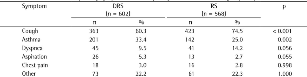
Table 1 - Prevalence of the respiratory symptoms most frequently observed in the two groups of patients studied.
Symptom DRS
(n = 602)
RS (n = 568)
p
n % n %
Cough 363 60.3 423 74.5 < 0.001
Asthma 201 33.4 142 25.0 0.002
Dyspnea 45 9.5 41 14.2 0.056
Aspiration 26 5.3 13 2.7 0.055
Chest pain 18 3.0 16 2.8 0.998
Other 73 22.2 61 22.3 1.000
relaxation were studied during 5 swallows of 5 mL of water. The esophageal body was evaluated in terms of the morphology, amplitude, duration and velocity of the esophageal contractions generated by the sequence of 10 swallows of 5 mL of water at 30-s intervals, with sensors placed at 3, 8, 13 and 18 cm above than the LES. The amplitude ranged between 40 and 180 mmHg, considered normal for the contractions in the distal esophagus. The study of the UES encompassed the tonus (mid-expiratory pressure), location/extension, relaxation and coordi-nation with the pharyngeal contractions. The results were based on the criteria of normality obtained in our laboratory,(11) which are similar to those found
in the literature.(12) The length of the esophagus
was defined as the distance between the proximal border of the LES and the distal border of the UES. The criteria for the definition of the primary and secondary esophageal disorders were based on the literature.(13)
After the esophageal manometry, pH-metry was performed. We used an external reference electrode connected to the partially disposable catheter of a sensor (Zinectics; Medtronic-Synetics, Skorlunde, Denmark) or two antimony sensors (Alacer Biomédica, São Paulo, Brazil) connected to a portable computed recorder (Digitrapper MK III; Synectics Medical) capable of registering one pH measurement every 4 s during a 24-h period. The distal pH-metry electrode was placed 5 cm above the proximal border of the LES, which had been previ-ously identified in the esophageal manometry. When a dual-sensor catheter was used, there was a 15-cm distance between the two sensors. Prior to each test, the electrodes were calibrated in buffer solutions as well as those in whose cases there was breakage
or dislocation of the electrode which hindered the complete acquisition of data.
The outcome measure of this cross-sectional study was the profile of the esophageal manometry and of the pH-metry. The protocol was approved by the Ethics in Research Committee of the Santa Casa Sisters of Mercy Hospital in Porto Alegre (Process no. 1420/06).
The esophageal manometry testing was carried out using a computed stationary manometry system, composed of a 4.5-mm diameter polyvinyl catheter, with 8 perforations, with 4 distal radial and 4 longitudinal channels axially distributed at intervals of 5 cm (Synectics Medical, Stockholm, Sweden). We used a system of perfusion involving a low-compliance capillary pneumohydraulic pump (Mui Scientific, Mississauga, Ontario, Canada) at a constant flow of 0.5 mL/min. The openings of the perfusion catheter were connected to external pressure transducers. The pressures detected were registered by a digital computed polygraph (PC Polygraf HR; Synetics Medical). The analysis was made using specific software (Polygram; Synectics Medical). All procedures were carried out following a fast of at least 4 h. For the esophageal manometry, after topical nasal anesthesia (2% lidocaine gel), nasoesophageal intubation into the stomach was carried out with the patient in the supine posi-tion. The catheter was then retracted in increments of 1 cm (slow removal technique). During this process, the lower and upper esophageal sphinc-ters (LES and UES, respectively) were analyzed, as was the esophageal body. In the LES, the tonus (mid-expiratory pressure), location / extension and
Table 2 - Correlations between hypotonia of the lower esophageal sphincter and abnormal 24-h esophageal pH-metry findings in the groups and subgroups.
Parameters with abnormal results Hypotonia of the lower esophageal sphincter
RS, p DRS, p RS-A, p DRS-A, p
%TT 0.033 < 0.001 0.022 0.005
%TO 0.039 0.001 0.085 0.028
%TSU 0.014 < 0.001 0.047 0.053
Acid reflux episodes 0.030 0.002 0.343 0.051
DeMeester score 0.019 < 0.001 0.015 0.025
Results
Between January of 1995 and December of 2005, we conducted 4,020 esophageal manom-etry tests and 3,486 pH-mmanom-etry tests. For this study, 1,170 patients met the inclusion criteria. Of those, 568 (48.5%) presented respiratory symptoms without digestive symptoms (RS group), whereas 602 (51.5%) reported digestive symptoms associ-ated with respiratory symptoms (DRS group). Of the 309 male patients, 152 (26.4%) were in the RS group and 157 (26.8%) were in the DRS group. Of the 861 (73.6%) female patients, 416 (73.2%) were in the RS group and 445 (73.9%) were in the DRS group. The distribution by gender was homogeneous between the two groups. The clinical symptoms that led the patients to be referred for functional evalu-ation of the esophagus are described in Table 1. Cough and asthma were the most common respira-tory symptoms among the patients in both groups. Other symptoms included recurrent pneumonia, lung abscess, bronchiectasis, chronic obstructive pulmonary disease, pulmonary fibrosis, and sleep apnea.
In the DRS group, the most frequently reported digestive symptom was heartburn (in 91.9% of the patients), followed by regurgitation (in 10.6%), dysphagia (in 3.0%), vomiting (in 2.5%) and other symptoms, including a sensation of gastric full-ness, epigastric pain, odynophagia or retrosternal discomfort, pharyngeal globus sensation, halitosis and choking (in 18.0%).
(Alacer Biomédica) at pH 7 and pH 1. The external reference electrode was fixed to the skin in the anterosuperior region of the chest. Nasoesophageal intubation, positioning and fixation of the electrode were then carried out, with subsequent triggering of the chronometer, initiating the 24 h registering. Patients were instructed to discontinue the use of antacids and prokinetics 7 days prior to the test, as well as to fast for 4 h before the test. Medications used for the control of respiratory symptoms were maintained. A log was provided for registration of the times at which meals began and ended, as well as periods in decubitus and any symptoms. At the end of the 24-h period, the patients returned to the laboratory for removal of the catheter and analysis of the data recorded (Esophogram; Synectics Medical), and a graphic, descriptive report was issued in order to show the results of the test. The analysis was based on the data provided by the distal electrode, using the table devised by Johnson and the scoring system developed by DeMeester.(14,15) Tests in which
the DeMeester score was higher than 14.7 were considered abnormal.
Fisher’s exact test and the chi-square test were used for the analysis of the qualitative variables, whereas the Student’s t test was used for the analysis of quan-titative variables. Data are presented as frequency and percentage for the categorical variables and as mean and standard deviation for the quantitative variables. A significance level of 5% was adopted. Data were analyzed using the Statistical Package for the Social Sciences, version 13.0 (SPSS Inc., Chicago, IL, USA).
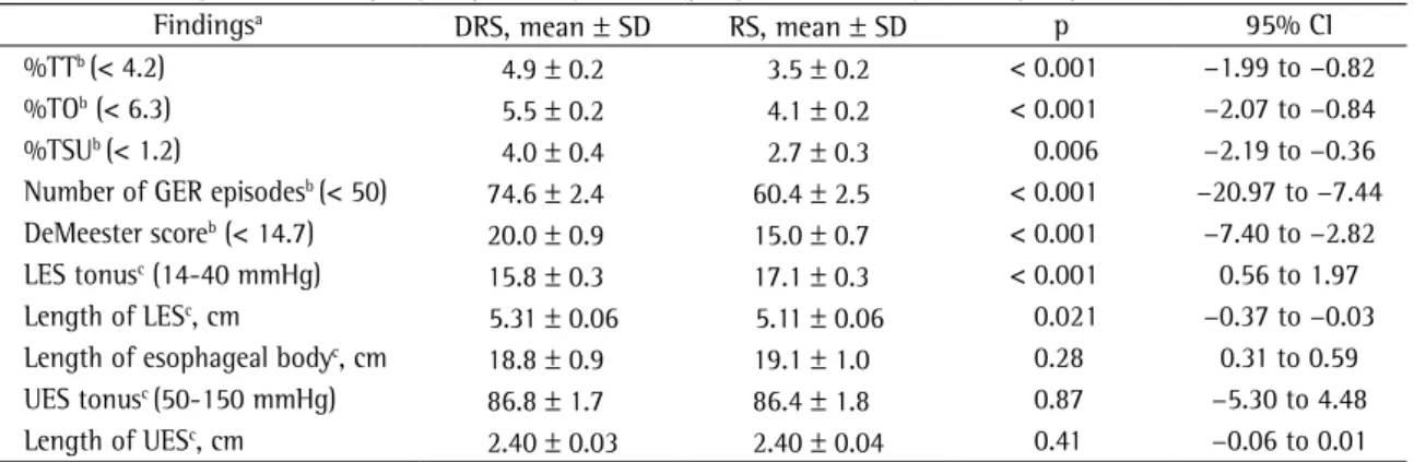
Table 3 - Findings of 24-h esophageal pH-metry and esophageal manometry in the groups studied.
Findingsa DRS, mean ± SD RS, mean ± SD p 95% CI
%TTb (< 4.2) 4.9 ± 0.2 3.5 ± 0.2 < 0.001 −1.99 to −0.82
%TOb (< 6.3) 5.5 ± 0.2 4.1 ± 0.2 < 0.001 −2.07 to −0.84
%TSUb (< 1.2) 4.0 ± 0.4 2.7 ± 0.3 0.006 −2.19 to −0.36
Number of GER episodesb (< 50) 74.6 ± 2.4 60.4 ± 2.5 < 0.001 −20.97 to −7.44
DeMeester scoreb (< 14.7) 20.0 ± 0.9 15.0 ± 0.7 < 0.001 −7.40 to −2.82
LES tonusc (14-40 mmHg) 15.8 ± 0.3 17.1 ± 0.3 < 0.001 0.56 to 1.97
Length of LESc, cm 5.31 ± 0.06 5.11 ± 0.06 0.021 −0.37 to −0.03
Length of esophageal bodyc, cm 18.8 ± 0.9 19.1 ± 1.0 0.28 0.31 to 0.59
UES tonusc (50-150 mmHg) 86.8 ± 1.7 86.4 ± 1.8 0.87 −5.30 to 4.48
Length of UESc, cm 2.40 ± 0.03 2.40 ± 0.04 0.41 −0.06 to 0.01
Of the 1,170 patients included, 343 reported asthma. Of these, 142 reported no digestive symptoms (subgroup RS-A). In the 201 patients with asthma accompanied by digestive symptoms (subgroup DRS-A), heartburn was the most common complaint (reported in 94.0%).
The comparison between the RS-A and DRS-A subgroups revealed that the abnormalities detected by the esophageal manometry were irrelevant, except for the tonus of the LES, which, although significantly different between the two subgroups (p = 0.001), remained within normality. However, LES hypotonia was present in 26.8% of the RS-A subgroup patients and in 40.3% of those in the DRS-A subgroup.
The pH-metry profile revealed that esophageal exposure to acid was greater, and that the number of abnormal parameters was higher, in the patients with accompanying digestive symptoms (DRS group and DRS-A subgroup) than in those without diges-tive symptoms (Tables 3 and 4).
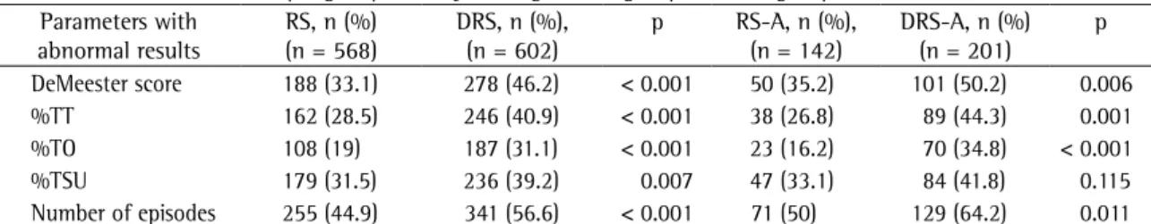
Markedly abnormal exposure of the distal esophagus to acid in the period of decubitus was observed in all of the groups, occurring in approxi-mately one third of the individuals without digestive symptoms (Table 5).
The prevalence of GERD in the patients studied was pronounced, being identified in 39.8% of the sample as a whole and in 188 (33.1%) of those who had reported no digestive symptoms. Among the patients in the asthma subgroups, the pH-metry test The manometric diagnosis of 1,170 patients
revealed normal test results in 481 (41.1%): 262 RS group patients (22.4% of the sample as a whole) and 219 DRS group patients (18.7% of the sample as a whole); (p = 0.001).
Among the manometric alterations, motor disor-ders were found in 1.9% of the cases (nutcracker esophagus in 15 cases and diffuse esophageal spasm in 7) and nonspecific disorders were found in 4.4% (52 cases). Other findings abnormal to the study occurred in 474 tests (40.5%): hypomotility of the esophageal body; hypotonia, hypertonia and relaxation disorder in the LES and UES; unco-ordinated UES; and alterations suggestive of collagenosis. Hypotonia in the LES was the altera-tion most frequently found and the only alteraaltera-tion for which there was significant difference between the two groups (30.2% in the RS group and 40.0% in the DRS group; p < 0,001), although both means were representative of normal tonus. Among the 346 cases of esophageal dysmotility, hypomotility was present in 175 (14.3% of the DRS group patients and 15.6% of the RS group patients). Hypotonia of the LES, present in 411 of the 1,170 patients, was the only esophageal manometry finding that corre-lated with the abnormal pH-metry parameters, as shown in Table 2.
The pH-metry profile revealed that exposure of the distal esophagus to acid was greater in the DRS group than in the RS group, whether or not the indices were within the parameters of normality (Table 3).
Table 4 - Findings of 24-h esophageal pH-metry and esophageal manometry in the subgroups of patients diagnosed with asthma.
Findingsa DRS-A, mean ± sd RS-A, mean ± sd p 95% CI
%TTb (< 4.2) 5.6 ± 0.5 3.8 ± 0.4 0.01 −3.11 to −0.36
%TOb (< 6.3) 5.9 ± 0.5 4.0 ± 0.3 0.001 −3.09 to −0.85
%TSUb (< 1.2) 4.6 ± 0.8 3.6 ± 0.8 0.36 −3.26 to 1.19
Number of GER episodesb (< 50) 87.0 ± 4.8 71.0 ± 6.4 0.03 −31.81 to −1.16
DeMeester scoreb (< 14.7) 22.5 ± 2.0 17.0 ± 1.7 0.45 −10.92 to −0.13
LES tonusc (14-40 mmHg) 15.7 ± 0.4 17.8 ± 0.5 0.001 0.84 to 3.47
Length of LESc, cm 5.1 ± 0.1 5.1 ± 0.1 0.91 −0.30 to 0.33
Length of esophageal bodyc, cm 19.0 ± 0.2 19.1 ± 0.2 0.78 −0.41 to 0.55
UES tonusc (50-150 mmHg) 84.4 ± 2.7 86.1 ± 3.8 0.71 −7.32 to 10.69
Length of UESc, cm 2.36 ± 0.04 2.40 ± 0.05 0.59 −0.09 to 0.17
DRS-A: subgroup composed of DRS group patients diagnosed with asthma; RS-A: subgroup composed of RS group patients diagnosed with asthma; %TT: percentage of the total study time at pH < 4.2; %TO: percentage of the total study time spent in the orthostatic position at pH < 4.2; and %TSU: percentage of the total study time spent in the supine position at pH < 4.2; GER: gastroesophageal reflux; LES: lower sphincter esophageal; and UES: upper sphincter esophageal. aValues in parentheses are
prevalence of GERD in asthma might be due to the inclusion of cases of asthma of different complexi-ties, as well as to the creation of study-specific parameters of normality that differ from the criteria stipulated by DeMeester.(14,15) This can lead to an
underestimation of the proportion of individuals presenting asthma and GERD.
In the evaluation of patients with GERD-related respiratory symptoms, the esophageal manom-etry test results are often normal. However, in the present study, GERD was most often observed in the patients with LES hypotonia (30.2% in the RS group and 40.0% in the DRS group). This finding corroborates the findings of one author(20) and is in
contrast with those of other authors,(22) who found
inefficacious esophageal motility to be the prin-cipal GERD-related manometric alteration, being present in 20% of the patients with typical GERD (heartburn) and in more than 50% of the patients with GERD and respiratory symptoms.(2) In fact, LES
hypotension and esophageal dysmotility were the manometric alterations most frequently observed in the present study. The distribution of hypomotility of the esophageal body was similar between the RS and DRS groups (14.3% and 15.6%, respectively; p = 0.51). Recent studies of patients with idiopathic pulmonary fibrosis and who were lung transplant candidates have shown that reflux is related to both alterations.(23) This dual manometric alteration might
be related to the severity of the pulmonary profile, which is much greater in this type of terminal pulmonary disease patient than in those included in other studies.(20,22) In this regard, the failure of
two of the principal mechanisms of the antireflux barrier (LES and peristalsis), allowing the ascension of the refluxed material and its permanence in the esophagus for a long time, can, together, contribute results were abnormal in 44%, including 50 (35.2%)
of the asthma patients who presented no digestive symptoms.
Discussion
The respiratory manifestations of gastroesopha-geal reflux are frequently unaccompanied by typical symptoms, and the diagnosis is therefore rarely suspected in such cases. For instance, GERD can stimulate the cough reflex through irritation of the upper respiratory tract without aspiration, through irritation of the lower respiratory tract due to micro-aspiration or macromicro-aspiration, as well as through stimulation of an esophageal-bronchial neural mechanism, in which the simple presence of the content refluxed to the distal esophagus would be sufficient to trigger cough. In this context, the radi-ological and endoscopic test results can be normal. Although GERD causes cough by irritating the larynx, with alterations detectable by bronchoscopy and chest imaging studies, the presence of these alterations should not be unquestioningly imputed to the reflux, since the inflammation and edema of the larynx and the lower airways can result from the trauma of cough caused by other diseases. (16)
Similarly, asthma can be exacerbated, or even trig-gered, by GERD, which leads some authors to use the term “gastric asthma”.(17,18) In the literature,
the prevalence of GERD in asthma patients varies between 32% and 82%,(8,19,20) and up to 50% of
asthma patients present “silent GERD”.(3) In a study
in which groups with asthma and without associ-ated digestive symptoms were compared,(21) reflux
was as severe in patients with asthma as in those with silent GERD, and the prevalence of GERD was 62% in patients presenting stable asthma without digestive symptoms. The great variability in the
Table 5 - Abnormal 24-h esophageal pH-metry findings in the groups and subgroups studied. Parameters with
abnormal results
RS, n (%) (n = 568)
DRS, n (%), (n = 602)
p RS-A, n (%), (n = 142)
DRS-A, n (%) (n = 201)
p
DeMeester score 188 (33.1) 278 (46.2) < 0.001 50 (35.2) 101 (50.2) 0.006 %TT 162 (28.5) 246 (40.9) < 0.001 38 (26.8) 89 (44.3) 0.001 %TO 108 (19) 187 (31.1) < 0.001 23 (16.2) 70 (34.8) < 0.001
%TSU 179 (31.5) 236 (39.2) 0.007 47 (33.1) 84 (41.8) 0.115
the analysis prevented us from providing these data with greater reliability.
When evaluating the means of the parameters evaluated by esophageal manometry, we observed that their values were normal in the RS and in the DRS groups, as well as in the two subgroups with asthma symptoms. However, normal esophageal manometry results do not exclude GERD. Therefore, in patients with respiratory symptoms, the func-tional investigation of the esophagus should be carried forward even if the esophageal manometry results are normal. In addition, although the corner-stone of the functional esophageal investigation of these patients is the pH-metry, esophageal manom-etry is essential for the appropriate intra-esophageal positioning of the pH-metry electrode.(26-28)
Hypotonia of the LES figured as the most common manometric abnormality and also as the only such abnormality for which there was a statis-tically significant difference between the groups (p < 0.001). Therefore, the correlation between LES hypotonia and abnormal pH-metry test results was analyzed. In the RS and DRS groups, significant correlations were found for all such parameters. In the same comparison carried out among those who reported asthma, the correlation between hypotonia and time of exposure to acid in the supine position was found to be significant in the RS-A subgroup, which elevated to equally significant levels the percentage of the total time spent in exposure, as well as the DeMeester score. In the DRS-A group, reflux in the orthostatic position contributed significantly to elevating the total reflux time and, consequently, to elevating the final DeMeester score (Table 4). The use of bronchodilators apparently plays no role in these findings, since it has been demonstrated that there is no appreciable modification in the tonus of the LES,(29) and the that presence of the reflux does
not depend on the use of bronchodilators.(30)
A quantitative increase in the episodes of reflux and in the exposure during the period in decubitus was observed through pH-metry in the RS group and RS-A subgroup. In the DRS group and DRS-A subgroup, there was also an increase in the time of exposure of the distal esophagus to acid over 24 h.
The comparison between the DRS and RS groups showed highly significant differences in all aspects of the pH-metry. However, the comparison between the DRS-A and RS-A subgroups showed significant differences only in the percentages of time with to its aspiration, generating or aggravating the
pulmonary profile. However, whether esophageal dysmotility results from the reflux or vice-versa has yet to be established.(16)
The most precise methods for the diagnosis of GERD are conventional pH-metry and impedanciom-etry. The former is able to detect episodes of reflux, regardless of their nature, whereas the latter identi-fies acid reflux episodes, presenting, in prospective studies for the investigation of cough, sensitivity and specificity values ranging from 66% to 100%.(16)
In addition to correlating the episodes of reflux with the digestive or respiratory symptoms reported by the patients, pH-metry is able to quantify the reflux, as well as to establish its pattern and periodicity. In cases of GERD-related cough under treatment with antireflux therapy and presenting no improvement in the symptoms, pH-metry can also be employed in order to verify the therapeutic efficacy.(16)
It has been demonstrated that up to 75% of patients with chronic cough without GERD symp-toms and 24% of patients with difficult-to-control asthma can present GERD through the pH-metry. In such patients, antireflux therapy can result in pronounced improvement or even in the remission of the respiratory symptoms.(9)
In the present study, the prevalence of GERD was high (39.8%). The pH-metry test results were abnormal in 44% of the patients with asthma, including 50 (35.2%) of the asthma patients who presented no digestive symptoms. These percent-ages are similar to those found in one study (36% and 25%, respectively)(19) but are inferior to those of
another study (72% and 62%, respectively).(20)
In all the groups analyzed, the period of greatest pathologic exposure of the distal esophagus to acid was always the time spent in decubitus. Our find-ings confirm those of a study in which exposure of the distal esophagus to acid was also found to be greater in the patients who presented asthma and typical symptoms of GERD.(24)
The decision to classify asthma as a subgroup of respiratory symptoms was made based on the fact that it is highly prevalent in patients in Brazil and represents a line of research pursued in previous studies conducted in our laboratory.(25) We
3. Richter JE. Ambulatory esophageal pH monitoring. Am J Med. 1997;103(5A):130S-4S.
4. Vakil N, van Zanten SV, Kahrilas P, Dent J, Jones R; Globale Konsensusgruppe. The Montreal definition and classification of gastroesophageal reflux disease: a global, evidence-based consensus paper [Article in German]. Z Gastroenterol. 2007;45(11):1125-40.
5. Gastal OL, Castell JA, Castell DO. Frequency and site of gastroesophageal reflux in patients with chest symptoms. Studies using proximal and distal pH monitoring. Chest. 1994;106(6):1793-6.
6. Palombini BC, Villanova CA, Araújo E, Gastal OL, Alt DC, Stolz DP, et al. A pathogenic triad in chronic cough: asthma, postnasal drip syndrome, and gastroesophageal reflux disease. Chest. 1999;116(2):279-84.
7. Theodoropoulos DS, Pecoraro DL, Efstratiadis SE. The association of gastroesophageal reflux disease with asthma and chronic cough in the adult. Am J Respir Med. 2002;1(2):133-46.
8. Poelmans J, Tack J. Extraoesophageal manifestations of gastro-oesophageal reflux. Gut. 2005;54(10):1492-9. 9. Harding SM, Richter JE. The role of gastroesophageal reflux in
chronic cough and asthma. Chest. 1997;111(5):1389-402. 10. Ribeiro IOS. Manifestações respiratórias e doença do refluxo
gastro-esofágico: perfil da esofagomanometria e pHmetria esofágica de 24 horas [dissertation]. Porto Alegre (RS): UFRGS; 2002.
11. Barros I, Felicetti J, Camargo J, Cardoso PFG. Parâmetros de normalidade para esofagomanometria. J Pneumol. 1995;21(supl 1):19.
12. Richter JE, Wu WC, Johns DN, Blackwell JN, Nelson JL 3rd, Castell JA, et al. Esophageal manometry in 95 healthy adult volunteers. Variability of pressures with age and frequency of “abnormal” contractions. Dig Dis Sci. 1987;32(6):583-92. 13. Nasi R, Michelson N, editores. Manometria e pHmetria
esofágicas. 1st Ed. São Paulo: Roca; 2001.
14. Johnson LF, Demeester TR. Twenty-four-hour pH monitoring of the distal esophagus. A quantitative measure of gastroesophageal reflux. Am J Gastroenterol. 1974;62(4):325-32.
15. Johnson LF, DeMeester TR. Development of the 24-hour intraesophageal pH monitoring composite scoring system. J Clin Gastroenterol. 1986;8 Suppl 1:52-8.
16. Irwin RS. Chronic cough due to gastroesophageal reflux disease: ACCP evidence-based clinical practice guidelines. Chest. 2006;129(1 Suppl):80S-94S.
17. Makkar RP, Sachdev GK. Gastric asthma: a clinical update for the general practitioner. MedGenMed. 2003;5(3):4. 18. Peters FT, Kleibeuker JH, Postma DS. Gastric asthma: a
pathophysiological entity? Scand J Gastroenterol Suppl. 1998;225:19-23.
19. Kiljander TO, Laitinen JO. The prevalence of gastroesophageal reflux disease in adult asthmatics. Chest. 2004;126(5):1490-4.
20. Harding SM. Recent clinical investigations examining the association of asthma and gastroesophageal reflux. Am J Med. 2003;115 Suppl 3A:39S-44S.
21. Harding SM. Gastroesophageal reflux, asthma, and mechanisms of interaction. Am J Med. 2001;111 Suppl 8A:8S-12S.
22. Fouad YM, Katz PO, Hatlebakk JG, Castell DO. Ineffective esophageal motility: the most common motility abnormality
reflux in the orthostatic position, in the total time of exposure of the distal esophagus to acid and in the number of episodes of reflux in 24 h. Nevertheless, it is important to observe that all the means of the pH-metry parameters of the patients with associ-ated digestive symptoms, either in the DRS group or in the DRS-A subgroup, presented higher values than those of the RS group, regardless of whether they were within the range of normality or not. The individuals with accompanying digestive symptoms presented greater exposure of the distal esophagus to acid.
In conclusion, the manometric profile of the patients with respiratory manifestations revealed that LES hypotonia was the most common altera-tion in both groups, although it was more frequently observed in the patients with digestive symptoms accompanied by respiratory symptoms, correlating to GERD. The pH-metry profile revealed a greater frequency of GERD and more altered parameters in the patients with associated digestive complaints. In all groups, the greatest exposure of the distal esophagus to refluxed acid occurred during the period in decubitus. Approximately one third of the patients with respiratory complaints and without digestive symptoms presented GERD (silent GERD). This confirmation is particularly important since it permits the suggestion of GERD as an extrapul-monary cause of chronic respiratory symptoms of undetermined origin that are unresponsive to conventional treatment. The findings of the present study have limitations imposed by its retrospective design, and future prospective studies are needed in order to clarify this issue further.
Acknowledgments
The authors would like to thank Edna da Mota Machado for the grammar editing, Mathias Bressel for the statistical analysis and Professor Doutor José da Silva Moreira from the Research and Post-graduation in Pulmonology Program of the Federal University of Rio Grande do Sul for the support.
References
1. Chinzon D, Neto M. Doença do Refluxo Gastroesofagiano: avaliação clínica e diagnóstico. In: Domingues G, editor. Esôfago. 1st Ed. Rio de Janeiro: Rubio; 2005. p. 245-59. 2. Katz PO. Gastroesophageal reflux disease--state of the art.
27. Kahrilas PJ, Quigley EM. Clinical esophageal pH recording: a technical review for practice guideline development. Gastroenterology. 1996;110(6):1982-96.
28. Pandolfino JE, Kahrilas PJ; American Gastroenterological Association. AGA technical review on the clinical use of esophageal manometry. Gastroenterology. 2005;128(1):209-24.
29. Sontag S. The prevalence of gastroesophageal reflux disease in asthma. In: Stein M, editor. Gastroesophageal reflux disease and airway disease. New York: Marcel Dekker; 1999. p. 115-38.
30. Sontag SJ, O’Connell S, Khandelwal S, Miller T, Nemchausky B, Schnell TG, et al. Most asthmatics have gastroesophageal reflux with or without bronchodilator therapy. Gastroenterology. 1990;99(3):613-20.
in patients with GERD-associated respiratory symptoms. Am J Gastroenterol. 1999;94(6):1464-7.
23. Sweet MP, Patti MG, Leard LE, Golden JA, Hays SR, Hoopes C, et al. Gastroesophageal reflux in patients with idiopathic pulmonary fibrosis referred for lung transplantation. J Thorac Cardiovasc Surg. 2007;133(4):1078-84.
24. Harding SM, Guzzo MR, Richter JE. The prevalence of gastroesophageal reflux in asthma patients without reflux symptoms. Am J Respir Crit Care Med. 2000;162(1):34-9. 25. Santos LH, Ribeiro IO, Sánchez PG, Hetzel JL, Felicetti JC,
Cardoso PF. Evaluation of pantoprazol treatment response of patients with asthma and gastroesophageal reflux: a randomized prospective double-blind placebo-controlled study. J Bras Pneumol. 2007;33(2):119-27.