Stability comparison of two different dentoalveolar
expansion treatment protocols
Ezgi Atik1, Tülin Taner2
Objective: The aim of this study was to compare the longitudinal stability of the conventional straight-wire system after the use of a quad-helix appliance with Damon self-ligating system in patients with Class I malocclusion. Methods: 27 adolescent patients were evaluated at three different periods: pre-treatment (T1), post-treatment (T2) and three years post-treatment (T3). Group 1 included 12 patients (with a mean age of 14.65 year) treated with Damon 3MX bracket system; and Group 2 included 15 patients (with a mean age of 14.8 year) who underwent orthodontic treatment with Roth prescribed brackets after expansion with Quad-Helix appliance. Relapse was evaluated with dental cast examination and cephalometric radiograph tracings. Statistical analysis was performed with IBM-SPSS for Windows software, version 21 (SPSS Inc., Chicago, IL). A p-value smaller than 0.05 was considered statistically signifi-cant. Results: There were significant increases in all transverse dental and postero-anterior measurements (except for UL6-ML mm in Group 1) with active treatment. There was some significant relapse in the long-term in inter-canine width in both groups and in the inter-first premolar width in Group 2 (p < 0.05). Significant decrease in all frontal measurements from T2 to T3 was seen for both groups. Upper and lower incisors significantly proclined in T1-T2 (p < 0.05), however no relapse was found for both groups. When two systems were compared, there was no significant difference for the long-term follow-up period. Conclusion: Conventional (quad-helix appliance with conventional brackets) and Damon systems were found similar with regard to the long-term incisor positions and transverse dimension changes of maxillary arch.
Keywords:Conventional brackets. Self-ligating brackets. Stability.
1 Assistant Professor in Orthodontics, School of Dentistry, University of
Hacettepe (Ankara, Turkey).
2 Professor in Orthodontics, School of Dentistry, University of Hacettepe
(Ankara, Turkey).
» The authors report no commercial, proprietary or financial interest in the products or companies described in this article.
DOI: https://doi.org/10.1590/2177-6709.22.5.075-082.oar
How to cite: Atik E, Taner T. Stability comparison of two different dento-alveolar expansion treatment protocols. Dental Press J Orthod. 2017 Sept-Oct;22(5):75-82. doi: https://doi.org/10.1590/2177-6709.22.5.075-082.oar
Submitted: September 28, 2016 - Revised and accepted: March 12, 2017
Contact address: Ezgi Atik – Assistant Professor, Department of Orthodontics, Faculty of Dentistry, Hacettepe University, Sihhiye, 06100, Ankara, Turkey E-mail: [email protected]
Objetivo: comparar a estabilidade longitudinal após o tratamento de pacientes com má oclusão de Classe I usando o sistema Straight--wire convencional — depois da expansão com aparelho quadri-hélice — e o sistema autoligável Damon. Métodos: 27 pacientes adolescentes foram avaliados em três períodos distintos: pré-tratamento (T1), pós-tratamento imediato (T2) e três anos pós--tratamento (T3). O Grupo 1 incluiu 12 pacientes (com idade média de 14,65 anos) tratados com o sistema de braquetes Damon 3MX e o Grupo 2 incluiu 15 pacientes (com idade média de 14,8 anos), submetidos a tratamento ortodôntico com braquetes prescrição Roth após expansão com aparelho quadri-hélice. A recidiva foi avaliada por meio de exame dos modelos de estudo e traçados cefalométricos. A análise estatística foi realizada com o software IBM-SPSS para Windows, versão 21 (SPSS Inc., Chicago, IL). Valores de p < 0,05 foram considerados estatisticamente significativos. Resultados: após o tratamento ativo, ocor-reu aumento significativo em todas as medidas transversais dentárias e posteroanteriores (exceto para a UL6-ML mm, no Grupo 1). Em longo prazo, ocorreu recidiva significativa (p < 0,05) na distância intercaninos em ambos os grupos, e na distância interprimeiros pré-molares no Grupo 2. De T2 para T3, observou-se diminuição significativa em todas as medidas frontais, para ambos os grupos. De T1 para T2, os incisivos superiores e inferiores sofreram vestibularização significativa (p < 0,05); porém, nenhuma recidiva ocorreu em qualquer um dos dois grupos. Ao se comparar os dois sistemas, não foi encontrada qualquer diferença significativa no período de acompanhamento em longo prazo. Conclusão: o sistema convencional (aparelho quadri-hélice e braquetes convencionais) e o sistema Damon apresentaram desempenho semelhante, em longo prazo, em termos das posições dos incisivos e das mudanças ocorridas na dimensão transversal da arcada superior.
INTRODUCTION
One of the important aspects of orthodontic treat-ment is to maintain arch form and prevent the possi-bility of relapse. However, increasing the arch perim-eter in non-extraction orthodontic treatment results in both transverse expansion of the arches and proclina-tion of the incisors.1,2 On the other hand, it is known
that arch dimensional changes most probably inluence prolonged stability. Both widening the inter-canine dimension width and tipping incisors labially results in unstable post-treatment results.3,4
Posterior expansion in the maxillary and mandibular arches is one of the comparison issues of self-ligating and conventional brackets. Transverse expansion with self-ligating systems is explained by low friction between the brackets and the archwires.5 In the literature, some
studies have indicated greater arch width increases with self-ligating brackets,6-8 while other studies have shown
no diferences between self-ligating and conventional appliances.9-11 It has been purported that the lower force
produced by self-ligating brackets might lead to more stable treatment results.12 It has also been claimed that
passive self-ligating brackets can introduce stable arch dimensional changes.13 However, few studies14,15 in the
literature have evaluated the stability of treatment re-sults associated with self-ligating bracket systems.
Since there is a lack of studies comparing the long-term stability of conventional and self-ligating systems, the present study aimed to comparatively evaluate the long-term post-treatment efects of self-ligating and conventional systems on the transverse dimensions of maxillary arches, and identify the dentoalveolar cepha-lometric changes. The null hypothesis assumed that there was not signiicant diferences regarding the long-term stability between both systems.
MATERIAL AND METHODS
The subjects in this study were derived from a sam-ple of 33 patients who had been previously treated in Hacettepe University Department of Orthodontics. The treatment results were originally presented in other article.9 The following inclusion criteria were used:
pa-tients were 13-17 years of age at the start of treatment; they had Class I malocclusion (ANB angle between 2° and 4°); moderate maxillary and mandibular crowding (between 3 mm and 6 mm); maxillary constriction caused by dental transverse discrepancy, characterized by palatal tipping of
the upper premolar and/or molar teeth; had undergone the non-extraction treatment protocol using the same archwire sequence with self-ligating brackets (Damon 3MX, Ormco/A Company, San Diego, CA, USA) or quad-helix expansion followed by conventional brackets (Forestadent, Pforzheim, Germany); had not undergone any adjunctive method, such as stripping, power chain, or intermaxillary elastics so as not to constrict the maxillary arch; and had undergone the same retention protocol in both the upper and lower dental arch for approximately one year. All patients had available records from before treatment (T1), ater treatment (T2), and three years af-ter treatment (T3). In this retrospective clinical study, no sample size calculation was done because all the available records were included. The power analysis was done and the power of the efect of diferent treatment protocols according to the obtained results and hypothesis was found to be 99.47%.
Ethical approval was obtained from the University of Hacettepe Research Committee (No. GO 16/573-13). A total of 33 patients were recalled to the Orthodontic Department of Hacettepe University, for a follow-up investigation three years post-treatment. Previous-ly, the patients were randomly allocated to one of the two treatment systems. Six patients did not return for long-term post-treatment records. Therefore, the inal number of follow-up patients was 27; 12 subjects were assigned to the Damon bracket group (Group 1) and 15 subjects were assigned to the conventional bracket group (Group 2), as shown in Table 1. In the present study the pre-treatment and post-treatment variables were not obtained from the aforementioned previous study9, being re-evaluated, since the sample size was
smaller in this study.
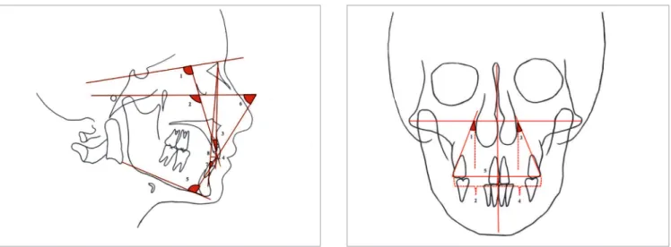
Figure 1 - Lateral cephalometric dental angular and linear measurements:
1) U1-SN (degrees); 2) U1-FH (degrees); 3) U1-NA (degrees); 4) U1-NA (mm);
5) IMPA (degrees); 6) FMIA (degrees); 7) L1-NB (degrees); 8) L1-NB (mm).
Figure 2 - Posteroanterior cephalometric measurements: 1) UR6-ML (degrees);
2) UR6-ML (mm); 3) UL6-ML (degrees); 4) UL6-ML (mm); 5) UR6-UL6 (mm).
The conventional bracket group (15 female pa-tients with a mean age of 14.8 years and an age range of 12.1-15.9 years) was previously bonded with the 0.022-in Roth prescription bracket (Forestadent, Pforzheim, Germany). At the beginning of the treat-ment, the maxillary arch was expanded using a quad-he-lix appliance until the lingual cusps of the maxillary irst molars were in contact with the buccal cusps of the man-dibular irst molars. Ater the desired expansion of the maxillary arch was achieved, the quad-helix appliance was removed and the transpalatal arch, with two arms behind the upper right and let premolar teeth, was put in place for retention until the SS archwires were applied to the maxillary arch. The following sequence of arch-wires was used for leveling and aligning: 0.014, 0.018-in CuNiTi; 0.014 × 0.025, 0.017 × 0.025-in CuNiTi, fol-lowed by 0.017 × 0.025 and 0.019 × 0.025-in SS. In the Damon bracket group, the Damon CuNiTi archwires were in uniform archwire form, and they were not co-ordinated to the original dental arch form. In the con-ventional bracket group, standard CuNiTi archwires were used. When these two types of archwires were compared, they displayed the same shape in the front region; however, the Damon CuNiTi archwires were wider in the region distal to the canines. In the Da-mon bracket group, DaDa-mon SS archwires were used; in the conventional bracket group, medium arch form SS archwires were used. The Damon SS archwires were broader than the standard SS archwires used in the con-ventional bracket group.
When the treatment was completed, upper and lower Hawley retainers were applied to all patients, for the reten-tion protocol. The devices were made with Orthocryl®
(Dentaurum, Ispringen, Germany). The patients were instructed to wear the Hawley retainers full time for six months, except when eating and brushing their teeth and thereater 6 months for every night. The total reten-tion period was one year. The patients were evaluated at three-month intervals until the completion of the reten-tion period (one year) to determine their motivareten-tion, ad-dress hygiene issues, and assess breakage.
Statistical analysis
Descriptive and analytical statistical analyses were performed with IBM-SPSS for Windows sotware, version 21 (SPSS Inc., Chicago, IL, USA). The Sha-piro–Wilk test was used to determine if the continu-ous data were normally distributed. Data were shown as mean ± standard deviation or median (min-max), where applicable. Degrees of reliability were calculated by intra-class correlation coeicients and 95% coni-dence interval for each clinical parameter: the degree of concordance observed was classiied as satisfactory and excellent, respectively.
Mann-Whitney test was used to compare the demo-graphic variables among the groups. Two-way repeated ANOVA measure test was used to compare the difer-ences among the groups. The non-parametric Friedman test was used to evaluate the statistical signiicance of the mean diferences between the pre-treatment, post-treatment, and post-retention measurements within the groups. A p-value smaller than 0.05 was considered to be statistically signiicant.
RESULTS
All the dental arch width measurements were found to have signiicantly increased from the T1 period to T2 period in both groups (p < 0.05). From T2 to T3, there was some signiicant relapse in inter-canine width in both groups (p < 0.05) and in the inter-irst premo-lar width in Group 2, the conventional bracket group (p = 0,019) (Table 2).
Statistically signiicant increases were found in the pos-tero-anterior measurements (except for UL6-ML mm in the Damon bracket group, Group 1) in the T1-T2 period for both groups. Moreover, there was a signiicant de-crease (p < 0.05) in all the frontal measurements in the T2-T3 period for both groups (Table 3).
In the T1-T2 period, U1-SN (degrees), U1-FH (degrees), FMIA (degrees), L1-NB (degrees), and L1-NB (mm) signiicantly changed in Group 1; and U1-NA (degrees), IMPA (degrees), FMIA (degrees), L1-NB (degrees), and L1-NB (mm) signiicantly changed in Group 2. From T2 to T3, no signiicant relapse was found in the lateral cephalometric measurements for both groups (Table 4).
The inter-group comparison results showed that there was no statistically signiicant diference between the groups for all measurements (Table 2, 3 and 4).
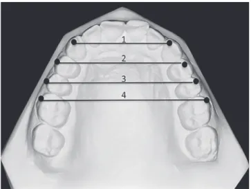
Figure 3 - 1) Inter-canine width (3-3), which is the distance between the right and left maxillary canine cusp tips. 2) Inter-first premolar width (4-4), which is the distance between the buccal cusp tips of the right and left maxillary first premolars. 3) Inter-second premolar width (5-5), which is the distance be-tween the buccal cusp tips of the right and left maxillary second premolars.
4) Inter-molar width (6-6), which is the distance between the mesiobuccal cusp tips of the right and left maxillary first molars.
Variables Group 1 (Damon) Group 2 (Conventional) p-value
Number of subjects 12 15
Age (year) 14.65 (13.10-16.70) 14.80 (12.10-15.90) 0.905a
Mand. crowding (mm) 3.75±0.94 3.31±0.74 0.256a
Max. crowding (mm) 4.24±1.14 3.66±0.90 0.236a
Table 1 - Demographic and clinical characteristics of the sample.
Table 2 - Dental Model Meausurements of the Groups at T1 (pre-treatment) ,T2 (post-treatment) and T3 (3 years post-treatment) Periods
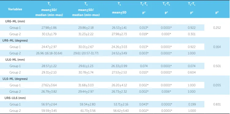
Table 3 - Postero-anterior cephalometric measurements of the groups at T1 (pre-treatment), T2 (post-treatment) and T3 (3 years post-treatment) periods. *Statistically significant (p < 0,05).
a: Friedman test, comparison of pre-treatment, post-treatment and 3 years post-treatment measurements within the groups. b: Two-way repeated ANOVA measure test, comparison of groups.
*Statistically significant (p < 0,05).
a: Friedman test, comparison of pre-treatment, post-treatment and 3 years post-treatment measurements within the groups. b: Two-way repeated ANOVA measure test, comparison of groups.
Variables T1 T2 T3 T2-T1 T3-T2 T3-T1
mean±SD mean±SD mean±SD pa pa pa pb
3-3
0,647 Group 1 (Damon) 33.83±3.54 35.99±2.41 34.81±2.34 0.001* 0.007* 1.000
Group 2 (Conventional) 32.861±3.15 34.94±1.93 34.08±1.20 0.0001* 0.019* 0.301
4-4
0.438 Group 1 (Damon) 38.16±2.95 43.58±2.13 42.35±1.98 0.0001* 0.662 0.007*
Group 2 (Conventional) 38.09±2.32 44.03±2.06 42.24±2.20 0.0001* 0.019* 0.019*
5-5
0.462 Group 1 (Damon) 42.82±3.49 48.28±2.76 47.16±2.30 0.0001* 0.662 0.007*
Group 2 (Conventional) 43.79±3.38 49.12±2.26 47.47±2.49 0.0001* 0.053 0.010*
6-6
0.976 Group 1 (Damon) 48.85±3.73 52.70±3.34 51.75±2.89 0.0001* 0.662 0.007*
Group 2 (Conventional) 49.90±3.90 53.77±2.91 52.73±3.18 0.0001* 0.134 0.006*
Variables
T1 T2 T3 T2-T1 T3-T2 T3-T1
mean±SD/
median (min-max)
mean±SD/
median (min-max) mean±SD p
a pa pa pb
UR6-ML (mm)
0,252 Group 1 27.98±1.86 29.86±2.18 26.53±1.41 0.013* 0.0001* 0.922
Group 2 30.13±1.79 31.23±2.22 27.98±2.73 0.019* 0.000* 0.301
UR6-ML (degrees)
0.164 Group 1 24.47±2.97 30.01±2.67 24.26±3.03 0.013* 0.0001* 0.922
Group 2 26.96 (16.18-30.64) 29.61 (20.57-31.77) 24.52±3.49 0.003* 0.0001* 1.000
UL6-ML (mm)
0.501 Group 1 28.57±1.22 29.61±1.23 26.33±0.99 0.074 0.0001* 0.074
Group 2 29.31±2.10 30.78±1.74 27.53±2.53 0.010* 0.0001* 0.604
UL6-ML (degrees)
0.055 Group 1 27.62±3.64 31.68±3.03 26.20±4.12 0.002* 0.0001* 1.000
Group 2 26.79±3.82 29.44±2.97 26.73±2.32 0.002* 0.006* 1.000
UR6-UL6 (mm)
0.831 Group 1 56.97±2.64 59.34±2.80 53.71±2.16 0.043* 0.0001* 0.199
Table 4 - Lateral cephalometric measurements of the groups at T1 (pre-treatment), T2 (post-treatment) and T3 (3 years post-treatment) periods.
*Statistically significant (p < 0,05).
a: Friedman test, comparison of pre-treatment, post-treatment and 3 years post-treatment measurements within the groups. b: Two-way repeated ANOVA measure test, comparison of groups.
Variables T1 T2 T3 T2-T1 T3-T2 T3-T1
mean±SD mean±SD mean±SD pa pa pa pb
U1-SN (degrees)
0,173 Group 1 102.47±8.41 107.07±7.35 106.92±5.70 0.007* 1.000 0.043*
Group 2 102.00±7.17 103.48±5.73 103.40±7.50 0.448 1.000 0.08
U1-FH (degrees)
0.342 Group 1 112.76±7.20 116.99±6.09 116.80±5.48 0.024* 1.000 0.074
Group 2 111.86±6.70 113.09±5.54 114.12±6.37 0.432 0.706 0.024*
U1-NA (degrees)
0.386 Group 1 22.08±7.80 27.00±5.30 27.42±6.10 0.074 1 0.124
Group 2 22.86±6.65 25.16±5.16 25.16±7.29 0.018* 0.523 0.083
U1-NA (mm)
0.45 Group 1 4.64±2.59 6.06±1.61 6.15±1.46 0.074 1.000 0.024*
Group 2 5.13±2.18 5.86±2.45 5.64±2.18 0.053 1 0.249
IMPA (degrees)
0.867 Group 1 95.70±3.58 101.41±6.24 100.50±5.30 0.074 0.922 0.003*
Group 2 92.95±6.43 97.56±7.70 97.35±7.92 0.019* 1.000 0.019*
FMIA (degrees)
0.764 Group 1 61.27±6.97 54.05±6.78 55.91±8.31 0.024* 1 0.013*
Group 2 62.47±6.24 56.56±6.71 57.66±7.66 0.002* 1 0.002*
L1-NB (degrees)
0.702 Group 1 26.08±4.72 32.55±5.14 30.90±6.11 0.002* 1.000 0.003*
Group 2 23.84±5.90 28.77±6.58 27.98±7.18 0.002* 1.000 0.006*
L1-NB (mm)
0.128 Group 1 5.40±2.65 7.77±2.58 7.41±3.01 0.001* 1.000 0.007*
Group 2 5.17±2.22 6.76±2.37 6.17±2.50 0.000* 0.134 0.067
DISCUSSION
In orthodontic treatment, it is diicult to sustain the patient’s dental arches in the position attained by active treatment. Several reasons might cause the tendency toward relapse, such as inter-canine width, mandibu-lar growth rotation, third momandibu-lar eruption, and diferent treatment patterns.16-19
While evaluating the long-term stability of an orth-odontic treatment, the pattern and magnitude of the dentoalveolar arch dimensional changes must be taken into consideration. The increase in the inter-canine arch width and proclination of the incisors are the main causes of unstable results.3,20 Therefore, it is important to
maintain the arch form during orthodontic treatment,
and doing so is highly recommended. There seems to be little basis for the claim that self-ligating brackets in-duce stable dental arch expansion.13
of the teeth with straight-wire appliances, including conventional brackets. However, in the Damon brack-et group, due to the expansion feature of the Damon CuNiTi archwires,21 the expansion appliance was not
used before the leveling and alignment stages.
In both treatment systems, the arch widths were signiicantly larger post-treatment. Although an expan-sion appliance was not used in the Damon bracket group (Group 1), the increase in the arch widths in that group can be attributed to the larger CuNiTi and SS archwires in the region distal to the canines. In the three-year post-treatment period (from T2 to T3), signiicant relapse in the inter-canine width was observed in both groups, and relapse was observed in the inter-irst premolar width in the conventional bracket group (Group 2) (Table 3). The inter-canine width relapse amounts were 1.18 mm and 0.86 mm for Group 1 and Group 2, respectively; the inter-irst premolar width decrease was 1.79 mm for Group 2. Several studies have shown an inter-canine and inter-molar width decrease during the post-retention period when it had been expanded during active treat-ment.22-24 In the present study, the signiicant relapse in
the inter-canine width in both groups ater the post-treatment phase may be due to the constriction of the expanded inter-canine dimension.25 This relapse could
also be explained by the disuse of the Hawley appliances ater one year, since the use of removable appliances was under the control of the patients. In addition, when the two groups were compared, the arch width decrease in the self-ligating brackets group (Group 1) did not dif-fer from the arch width decrease in the conventional bracket group (Group 2) ater the post-treatment period. Yu et al14 compared the long-term stability of treatment
with self-ligating brackets and conventional brackets with a mean follow-up period of 7.68 years. Diferent from the present study, these authors14 found a greater increase in
the inter-molar width with self-ligating brackets than with conventional appliances. This diferent result can be attributed to diferent treatment modalities, such as the expansion appliance used with the conventional brackets in the present study. Similar to the results of the present study, they14 did not ind a signiicant
diference in the inter-canine and inter-molar width change between the bracket systems over the long-term follow-up period. This result is concurrent with our study, and indicates that the bracket type do not afect the stability associated with dental arch width.
The proclined maxillary and mandibular incisors were similar from T1 period to T2 period in both groups, which is consistent with the indings reported in other studies.6,26,27 In this present study, the cephalometric
evaluations showed that the relief of the maxillary and mandibular anterior crowding mainly occurred as a result of labial inclination, independent of the type of bracket. Nevertheless, the studies in the literature6,26,27
did not evaluate long-term incisor position changes with self-ligating brackets. The results of the present study showed that the changes in the maxillary and mandibular dental measurements during the three-year follow-up period were insigniicant in the conventional and self-ligating bracket groups. Similar to the pres-ent study, only Bascitci et al15 evaluated the long-term
dentoalveolar efects of self-ligating brackets. However, these authors15 did not compare the experimental group
to a control group. In keeping with the indings of the present study, the maxillary and mandibular incisor po-sition changes were not signiicant from the immediate post-treatment period to the two-year follow-up peri-od.15 However, diferent from the present study, they15
reported that the upper inter-canine width remained stable in the self-ligating bracket patients during all the retention periods. In the present study, some signiicant relapse was observed in the inter-canine width in both the self-ligating bracket group (1.18 mm) and the con-ventional bracket group (0.86 mm). This diference may be due to the retention protocol we used, since we did not apply upper and lower lingual retainers as Bascitci et al15 did in their study.
In the present study, the postero-anterior cepha-lometric evaluation measurements indicated signii-cant buccal tipping of the upper molars in both treat-ment groups at the end of the treattreat-ment. In accordance with this inding, Yu et al.28 showed buccal tipping of
the molars when using rapid palatal expansion (RPE) and the Damon technique with a non-extraction treat-ment approach. Cattaneo et al.29 also indicated buccal
tipping in the self-ligating bracket group. It is known that increased tipping of the maxillary molars would put a patient at risk of future relapse. Therefore, diferent from other studies14,15 that investigated the stability of
sig-niicant relapse. The relapse amounts were not statisti-cally diferent from each other when both groups were compared. However, the p value (0.055) of UL6-ML degrees (Table 3), which indicated a more clinically-signiicant relapse of the upper let molar teeth inclina-tion in the Damon bracket group, must be taken into consideration. This result is probably due to the greater buccal tipping of the molars at the end of the treatment in the Damon bracket group, as found in a previous study.9 In the Damon bracket group, the low buccal
root torque and the increased tipping of the maxillary molars could be considered as relapse risk factors.
The limitation of this study might be its retrospec-tive design. However, to reduce the disadvantage of
po-tential selection bias,30 the same treatment protocol (the
non-extraction treatment included the same archwire sequence with no other appliances) and the same reten-tion protocol were applied by only one practireten-tioner in the same clinic, while creating the samples.
CONCLUSION
In this study, no signiicant diferences were found in terms of long-term stability between the self-ligating (Damon brackets) and the conventional (quad-helix ap-pliance with conventional brackets) treatment systems. However, further long-term follow-up, randomized controlled trials are needed to precisely know how us-ing self-ligatus-ing brackets impacts stability.
1. Weinberg M, Sadowsky C. Resolution of mandibular arch crowding in growing patients with Class I malocclusions treated nonextraction. Am J Orthod Dentofacial Orthop. 1996 Oct;110(4):359-64.
2. Fleming PS, DiBiase AT, Sarri G, Lee RT. Comparison of mandibular arch changes during alignment and leveling with 2 preadjusted edgewise appliances. Am J Orthod Dentofacial Orthop. 2009 Sept;136(3):340-7.
3. Burke SP, Silveira AM, Goldsmith LJ, Yancey JM, Van Stewart A, Scarfe WC. A meta-analysis of mandibular intercanine width in treatment and postretention. Angle Orthod. 1998 Feb;68(1):53-60.
4. Little RM. Stability and relapse of mandibular anterior alignment: University of Washington studies. Semin Orthod. 1999 Sept;5(3):191-204.
5. Capistrano A, Cordeiro A, Siqueira DF, Capelozza Filho L, Cardoso MA, Almeida-Pedrin RR. From conventional to self-ligating bracket systems: is it possible to aggregate the experience with the former to the use of the latter? Dental Press J Orthod. 2014 May-June;19(3):139-57.
6. Vajaria R, BeGole E, Kusnoto B, Galang MT, Obrez A. Evaluation of incisor position and dental transverse dimensional changes using the Damon system. Angle Orthod. 2011 July;81(4):647-52.
7. Pandis N, Polychronopoulou A, Eliades T. Self-ligating vs conventional brackets in the treatment of mandibular crowding: a prospective clinical trial of treatment duration and dental efects. Am J Orthod Dentofacial Orthop. 2007 Aug;132(2):208-15.
8. Pandis N, Polychronopoulou A, Makou M, Eliades T. Mandibular dental arch changes associated with treatment of crowding using self-ligating and conventional brackets. Eur J Orthod. 2010 June;32(3):248-53.
9. Atik E, Ciğer S. An assessment of conventional and self-ligating brackets in Class I maxillary constriction patients. Angle Orthod. 2014 July;84(4):615-22.
10. Scott P, DiBiase AT, Sherrif M, Cobourne MT. Alignment eiciency of Damon3 self-ligating and conventional orthodontic bracket systems: a randomized clinical trial. Am J Orthod Dentofacial Orthop. 2008 Oct;134(4):470.e1-8.
11. Fleming PS, Lee RT, Mcdonald T, Pandis N, Johal A. The timing of signiicant arch dimensional changes with ixed orthodontic appliances: data from a multicenter randomised controlled trial. J Dent. 2014 Jan;42(1):1-6.
12. Chen SS, Greenlee GM, Kim JE, Smith CL, Huang GJ. Systematic review of self-ligating brackets. Am J Orthod Dentofacial Orthop. 2010 June;137(6):726.e1-18; discussion 726-7.
13. Damon D. Damon System: the workbook. Orange: Calif; 2004.
14. Yu Z, Jiaqiang L, Weiting C, Wang Y, Zhen M, Ni Z. Stability of treatment with self-ligating brackets and conventional brackets in adolescents: a long-term follow-up retrospective study. Head Face Med. 2014;10:41.
15. Basciftci FA, Akin M, Ileri Z, Bayram S. Long-term stability of dentoalveolar, skeletal, and soft tissue changes after non-extraction treatment with a self-ligating system. Korean J Orthod. 2014 May;44(3):119-27.
REFERENCES
16. Rossouw PE, Preston CB, Lombard CJ, Truter JW. A longitudinal evaluation of the anterior border of the dentition. Am J Orthod Dentofacial Orthop. 1993 Aug;104(2):146-52.
17. Fudalej P, Artun J. Mandibular growth rotation efects on postretention stability of mandibular incisor alignment. Angle Orthod. 2007 Mar;77(2):199-205. 18. Harradine NW, Pearson MH, Toth B. The efect of extraction of third molars on
late lower incisor crowding: a randomized controlled trial. Br J Orthod. 1998 May;25(2):117-22.
19. Little RM, Riedel RA, Engst ED. Serial extraction of irst premolars--postretention evaluation of stability and relapse. Angle Orthod. 1990 Winter;60(4):255-62. 20. Mills JR. The long-term results of the proclination of lower incisors. Br Dent J.
1966 Apr 19;120(8):355-63.
21. Birnie DJ. The Damon passive self-ligating appliance system. Semin Orthod. 2008;14(1):19-35.
22. Kahl-Nieke B, Fischbach H, Schwarze CW. Post-retention crowding and incisor irregularity: a long-term follow-up evaluation of stability and relapse. Br J Orthod. 1995 Aug;22(3):249-57.
23. Amott RD. A serial study of dental arch measurements on orthodontic subjects: 55 cases at least 4 years postretention [MSD thesis]. Chicago (IL): Northwestern University Dental School; 1962.
24. Arnold ML. A study of the changes of the mandibular intercanine and intermolar widths during orthodontic treatment and following postretention period of ive or more years [MSD Thesis]. Seattle (WA): University of Washington; 1963. 25. Little RM. Stability and relapse of dental arch alignment. Br J Orthod. 1990
Aug;17(3):235-41.
26. Jiang RP, Fu MK. [Non-extraction treatment with self-ligating and conventional brackets]. Zhonghua Kou Qiang Yi Xue Za Zhi. 2008 Aug;43(8):459-63. 27. Pandis N, Strigou S, Eliades T. Maxillary incisor torque with conventional and
self-ligating brackets: a prospective clinical trial. Orthod Craniofac Res. 2006 Nov;9(4):193-8.
28. Yu YL, Tang GH, Gong FF, Chen LL, Qian YF. [A comparison of rapid palatal expansion and Damon appliance on non-extraction correction of dental crowding]. Shanghai Kou Qiang Yi Xue. 2008 June;17(3):237-42.
29. Cattaneo PM, Treccani M, Carlsson K, Thorgeirsson T, Myrda A, Cevidanes LH, et al. Transversal maxillary dento-alveolar changes in patients treated with active and passive self-ligating brackets: a randomized clinical trial using CBCT-scans and digital models. Orthod Craniofac Res. 2011 Nov;14(4):222-33.