Kathy Saraiva-Pava,Faculdade de Engenharia, Universidade Católica Portuguesa, Campus de Sintra, 2635-631 Rio de Mouro, Portugal
Nazanin Navabi, Emma C Skoog, Sara K Lindén, Department of Medical Biochemistry and Cell Biology, Institute of Biomedicine, University of Gothenburg, SE-40530 Gothenburg, Sweden
Mónica Oleastro, Departamento de Doenças Infeciosas, Instituto Nacional Saúde Dr. Ricardo Jorge, 1649-016 Lisboa, Portugal Mónica Roxo-Rosa,Chronic Diseases Research Center, CEDOC, Nova Medical School, Campo Mártires da Pátria, 1169-056 Lisboa, Portugal
Author contributions:Lindén SK, Oleastro M and Roxo-Rosa M designed the research; Saraiva-Pava K, Navabi N and Skoog EC performed the research; Roxo-Rosa M, Lindén SK and Oleastro M analyzed the data; Saraiva-Pava K, Lindén SK and Roxo-Rosa M wrote the paper.
Supported by Grants from the Fundação para a Ciência e a Tecnologia (FCT, Portugal), No. PPCDT/SAL-IMI/57297/2004 and No. PTDC/BIM-MEC/1051/2012; The Swedish Cancer foundation; The Swedish Research Council, No. K2010-79X-21372-01-3; Forska utan djurförsök, Animal Free Research; and by Research fellowship 2011 from the Sociedade Portuguesa de Gastrenterologia (Portugal).
Conflict-of-interest: The authors have no competing interests. Data sharing: Technical appendix, statistical code, and dataset available from the corresponding author at monica.oleastro@ insa.min-saude.pt. Participants gave informed consent for data sharing.
Open-Access: This article is an open-access article which was selected by an in-house editor and fully peer-reviewed by external reviewers. It is distributed in accordance with the Creative Commons Attribution Non Commercial (CC BY-NC 4.0) license, which permits others to distribute, remix, adapt, build upon this work non-commercially, and license their derivative works on different terms, provided the original work is properly cited and the use is non-commercial. See: http://creativecommons.org/ licenses/by-nc/4.0/
Correspondence to: Mónica Oleastro, PhD, Departamento de Doenças Infeciosas, Instituto Nacional Saúde Dr. Ricardo Jorge,
Av. Padre Cruz, 1649-016 Lisboa,
Portugal. [email protected] Telephone: +351-217519200
Fax: +351-217526400
Received: October 14, 2014
Peer-review started: October 15, 2014 First decision: January 8, 2015 Revised: February 7, 2015 Accepted: March 31, 2015 Article in press: March 31, 2015 Published online: June 7, 2015
Abstract
AIM: To establish a cellular model correctly mimicking the gastric epithelium to overcome the limitation in the study of
Helicobacter pylori
(H. pylori
) infection.METHODS: Aiming to overcome this limitation, clones of the heterogenic cancer-derived NCI-N87 cell line were isolated, by stably-transducing it with the human telomerase reverse-transcriptase (hTERT) catalytic subunit gene. The clones were first characterized regarding their cell growth pattern and phenotype. For that we measured the clones’ adherence properties, expression of cell-cell junctions’ markers (ZO-1 and E-cadherin) and ability to generate a sustained transepithelial electrical resistance. The gastric properties of the clones, concerning expression of mucins, zymogens and glycan contents, were then evaluated by haematoxylin and eosin staining, Periodic acid Schiff (PAS) and PAS/Alcian Blue-staining, immunocytochemistry and Western blot. In addition, we assessed the usefulness of the hTERT-expressing gastric cell line for
H. pylori
research, by performing co-culture assays and measuring the IL-8 secretion, by ELISA, upon infection with twoH.
pylori
strains differing in virulence.RESULTS: Compared with the parental cell line, the DOI: 10.3748/wjg.v21.i21.6526 © 2015 Baishideng Publishing Group Inc. All rights reserved.
ORIGINAL ARTICLE
New NCI-N87-derived human gastric epithelial line after
human telomerase catalytic subunit over-expression
Basic Study
most promising NCI-hTERT-derived clones (CL5 and CL6) were composed of cells with homogenous phenotype, presented higher relative telomerase activities, better adhesion properties, ability to be maintained in culture for longer periods after confluency, and were more efficient in PAS-reactive mucins secretion. Both clones were shown to produce high amounts of MUC1, MUC2 and MUC13. NCI-hTERT-CL5 mucins were shown to be decorated with blood group H type 2 (BG-H), Lewis-x (Lex), Ley and Lea and, in a less extent, with BG-A antigens, but the former two antigens were not detected in the NCI-hTERT-CL6. None of the clones exhibited detectable levels of MUC6 nor sialylated Lex and Lea glycans. Entailing good gastric properties, both NCI-hTERT-clones were found to produce pepsinogen-5 and human gastric lipase. The progenitor-like phenotype of NCI-hTERT-CL6 cells was highlighted by large nuclei and by the apical vesicular-like distribution of mucin 5AC and Pg5, supporting the accumulation of mucus-secreting and zymogens-chief mature cells functions.
CONCLUSION: These traits, in addition to resistance to microaerobic conditions and good responsiveness to
H. pylori
co-culture, in a strain virulence-dependent manner, make the NCI-hTERT-CL6 a promising model for futurein vitro
studies.Key words:
Helicobacter pylori
infection; Pathogenesis; Human gastric epithelium; Cellular model; NCI-N87 cells© The Author(s) 2015. Published by Baishideng Publishing Group Inc. All rights reserved.
Core tip: In this study, we aimed to establish and characterize novel human gastric epithelial cell lines derived from NCI-N87 cells after over-expression of human telomerase catalytic activity. The two most promising NCI-N87-derived clones were shown to be composed of cells with homogenous phenotype, to produce gastric zymogens and to produce and secrete neutral mucins. In addition, these clones showed very good
in vitro
growth properties, resistance to microaerobic conditions and good responsiveness toHelicobacter pylori
. Due to their improved properties, compared to the heterogeneous parental line, these NCI-N87-derived clones are promising models of the human gastric epithelium.Saraiva-Pava K, Navabi N, Skoog EC, Lindén SK, Oleastro M, Roxo-Rosa M.New NCI-N87-derived human gastric epithelial line after human telomerase catalytic subunit over-expression. World J Gastroenterol 2015; 21(21): 6526-6542 Available from: URL: http://www.wjgnet.com/1007-9327/full/v21/i21/6526.htm DOI: http://dx.doi.org/10.3748/wjg.v21.i21.6526
INTRODUCTION
The normal physiological functions of the human
stomach rely on the distinct populations of specialized epithelial cells present at the gastric mucosa. Early in foetal life, this simple columnar epithelium forms invaginations into the inner layers of the mucosa, which originate the gastric pit glands. In the continuous renewing of this tissue, its organization is maintained through a bidirectional migration of the gastric stem and progenitor cells, primarily located at the isthmus of the glands (reviewed in). In oxyntic glands (glands from fundus and corpus), the stem cells migrating upward into the pit and surface give rise to mucin (MUC) 5AC (MUC5AC)-secreting cells, while those that migrate down differentiate into MUC6-secreting and parietal (HCl-secreting) cells at the neck, and mature chief (zymogens-secreting) cells at the base of the gland. In pyloric glands (glands from the antrum), instead of parietal and chief cells, the downward migrating stem cells give rise to cells specialized in the production of gastrin (G cells) and of somatostatin (D cells). These hormones ensure the endocrine regulation of the acid production by oxyntic parietal cells[1-4].
Despite our knowledge on the importance of this organization, the research on the identification of
specific regulators of the development and maintenance
of human gastric functions, and their underlying mechanisms, requires the establishment of human gastric cell lines closely resembling the natural features of the gastric epithelium. Such in vitro model is also urgently needed for the study of the still poorly understood molecular mechanisms involved in the pathogenesis of severe gastric diseases associated with the Gram-negative bacterium Helicobacter pylori (H. pylori).Having the human gastric mucosa as ecological niche, this spiral-shaped, microaerophilic bacterium survives the extreme conditions of the human stomach and, constantly evading (or interacting with) the human immune system, persists throughout the patient life. Indeed, almost invariably, this pathogen/host interaction starts in patient’s childhood, always eliciting an acute
host immune response, which is however inefficient for
bacteria clearance[5,6]
. Although often asymptomatic, most patients experience dyspeptic symptoms (non-ulcer dyspepsia) because of the resulting chronic gastritis[7] and, later in the adulthood, 15%-20% of them end up developing peptic ulcer disease and 2%-5% gastric cancer[8,9]
. Despite all the spent efforts, there is still a lack of knowledge on the events that occur in the time lag that goes from the colonization in the childhood to peptic ulceration and/or gastric carcinogenesis, late in the patients’ adult life. We have shown that these events are dependent on the virulence of the strain itself[10-13]
, but also on its interaction with the human host, changing the expression pattern of mucins of the patient’s gastric mucosa[14-17]
. Indeed, sustained infection is known to depend on the bacteria adhesion to host mucins of the epithelial cells’ surface and mucus layer lining the gastric epithelium[5,6]
gastric biopsy samples[1,4,18]
. Albeit being more representative of the human gastric epithelium, such primary cultures present important limitations regarding the availability of human donors, restricting the number of experiments that can be performed. Furthermore, primary cultures can only be cultured for a few days before they become apoptotic, limiting the type of experiments that can be performed. Similarly, for many studies gastric primary cultures of rodents are not a suitable option due to important distinctive features of their physiology compared to the human’s[19]
. In fact, only human chief cells produce/secrete not only pepsinogen, but simultaneously an acid-tolerant gastric lipase, ensuring gastric digestion of triglycerides, a prerequisite for optimal intestinal lipolysis. Again, these are not a suitable option for long-term studies because, beyond the limitations of their availability, these cells also proliferate for only a small number of doublings before entering into senescence[1,4]
. An extensively used alternative are cancer cell lines derived from poorly differentiated [e.g., KATO-III [American type culture collection (ATCC) HBT-103] and Hs746t (ATCC HBT-135) cells], or moderately [e.g., AGS (ATCC CRL-1739) cells] and well differentiated [e.g., NCI-N87 (ATCC CRL-5822) and MKN-7 (Riken Cell Bank, Japan) cells] human gastric carcinomas[1,20,21]
. Lacking important epithelial and/or glandular properties, these in vitro cellular models are limited in resembling the native tissue. For instance, AGS cells harbour a mutated E-cadherin encoding gene that results in a non-functional truncated form of this protein, thus these cells form monolayers that do not polarize and eventually lose their integrity after reaching confluency[1,21-23]
. Furthermore, despite reaching a good polarization status upon transfection with E-cadherin cDNA, they do not secrete mucus, acid or zymogens[22,24]
. The MKN7 cell line, polarize and present the mucin MUC1 apically, however, again the problem is that it does not produce an adherent mucus layer[21,25]
. As far as we can ascertain, the properties of this cell line regarding the acid/zymogens production/ secretion have never been described.
The NCI-N87 cell line is known by its distinctive features, including colony-forming growth pattern, maintenance at post-confluency, expression of increased levels of gastric zymogens [namely, human gastric lipase (HGL) and fundic-type pepsinogen-5 (Pg5)], as well as expression of MUC6, zonula adherens marker E-cadherin and zonula occludens marker ZO-1[1]
. Compared to the widely use AGS cells (ATCC, CRL-1739), the NCI-N87 cell line responds to H. pylori
infection in a closer manner to that of primary gastric epithelial cell preparations[26]
. However, the expression of these epithelial/gastric markers are confined only to some cell sub-populations[23]
. Indeed, this is a heterogenic cell line composed of several phenotypic variants, also including non-epithelial cells. Homotypic epithelial phenotype was, interestingly, achieved by isolating non-transfected clones (using the limit-dilution approach) of those cell sub-populations, allowing the
establishment of two NCI-N87-derived clones: the HGE-17 (human gastric epithelial-17 cell line), exhibiting features reminiscent of the granule-free stem cell type found in the isthmus of the glands; and the HGE-20, possessing a more differentiated, pre-zymogenic-like
status (simultaneous synthesis and efficient secretion of
MUC6 and zymogens)[23] .
The ectopic expression of human telomerase reverse-transcriptase catalytic subunit gene (hTERT) in differentiated normal human cells is sufficient to restore telomerase activity, thus preventing telomeres’ shortening and inducing continuous cell proliferation[27]
. Indeed, telomerase activity is only dependent on the presence of its hTERT subunit, since its template RNA component, used for the addition of telomere repeats at the chromosome tips at each cell cycle, is constitutively synthesized in normal cells[28]
. Based on these findings, the induction of hTERT has been intensively used in the last decade in the establishment of novel immortalized cell lines from primary cultures of a variety of human cells and tissues (e.g., skin fibroblasts[28]
, T lymphocytes[29]
, airway epithelial cells[30]
, proximal renal tubules epithelial cells[31] , stromal and epithelial immortalized endometriotic cells[32]
, etc.). These hTERT-expressing cell lines grow out of a background of senescence, present fewer, if
any, karyotypic modifications and maintain their normal
features of differentiation, a major improvement over the use of viral oncogenes[30,31,32,33]
. The effect of hTERT over-expressing in immortalized cell lines, characterized by unlimited life span, is still poorly known. However, hTERT over-expression was shown to improve the classical immortalized and continuously dividing CHO-K1 (Chinese hamster ovary) cell line, increasing its resistance to serum-deprivation induced apoptosis and allowing this serum-dependent cell line to survive, attach and divide in un-supplemented basal medium[34]
. Thus, considering these approaches as valuable strategies for cell engineering, here we aimed to establish novel NCI-N87-derived epithelial cell lines by ectopic over-expression of the hTERT, characterize them and assess their usefulness in H. pylori-infection
in vitro assays.
MATERIALS AND METHODS
Expression vector
The pGRN145 (ATCC MBA-141, Geron Corporation, Menlo Park, CA, United States) is a mammalian expression vector containing the full coding region of the hTERT catalytic subunit gene, under the control of the myeloproliferative sarcoma virus promoter. The plasmid contains the resistance gene for hygromycine B (HygB) for selection in mammalian cells.
Cell culture conditions
Transepithelial electrical resistance
In order to test for epithelial monolayer polarity and integrity, cells were seeded on porous membranes (0.4
μm) supported by a detachable ring and transepithelial electrical resistance (TEER) was measured with an epithelial voltohmmeter (EVOM; World Precision Instruments, Sarasota, FL, United States) in the following days post-confluence. All cultures were started by
expanding the cells in flasks, then about 7.5 × 10⁴ cells were harvested and 200 to 400 μL of cells suspended in fresh media were added to the apical side of each tested filters. Proper volume of media (determined according to the manufacturer’s instructions) was also added to the basolateral compartment. These were considered the standard culture conditions. Tested inserts: SnapwellTM
3407 (polycarbonate) with a 12 mm diameter, providing a growth area of 1.12 cm2
(Corning Costar Corp., Cambridge, MA, United States); collagen-coated Transwell®
-COL 3491 (polytetrafluoroethylene) with a 24 mm diameter, providing a growth area of 4.67 cm2
(Corning Costar Corp.); Transwell-clear 3450 (polyester) with a 24 mm diameter, providing a growth area of 4.67 cm2
(Corning Costar Corp.); hydrophilic
polytetrafluoroethylene Millicell CM standing filters with
a 12 mm diameter, providing a growth area of 0.6 cm2 (Millipore, Bedford, MA, United States); and polyester Millicell inserts with a 30 mm diameter, providing a growth area of 4.2 cm2
(Millipore). The latter 3 were pre-coated with a 0.05% (w/v) collagen IV solution (Sigma-Aldrich, St Louis, MO, United States) before use. The following culture conditions were also tested: DMEM/F12 medium (Lonza, Switzerland) containing 10% (v/v) FBS (Lonza) and 1% (v/v) penicillin-streptomycin (Lonza); Glucose free RPMI medium (Lonza) supplemented with 1% (w/v) galactose (AppliChem, Germany), 10% (v/v) FBS or FBS replacement media (Ultra media, Lonza) with 1% (v/v) penicillin-streptomycin; DMEM/ F12 followed by mechanical stimulation; and glucose free RPMI medium supplemented with 1% (w/v) galactose followed by mechanical stimulation. Semi-wet interface was produced by 2 mL media in the basolateral compartment and 50 μL media in the apical compartment after confluency. Mechanical stimulation and continuous wetting of the apical surface were achieved by placing the culture plates on a rocking board in the incubator. Basolateral media was refreshed every two to three days. Electrical resistances of the supporting filter and buffer medium were subtracted to calculate TEER of the monolayer with final results
reported per unit surface ohms × cm2 .
Cell staining
Cells were seeded onto 8-well chamber slides (Lab-Tek® , Nalge Nunc International, Roskilde, Denmark) and allowed
to reach high density before processing. Cells were fixed in
10% (v/v) formaldehyde (Sigma-Aldrich) in 96% ethanol for 1 min and at the end of all staining procedures, slides were mounted with glycerol and sealed with nail polish. Life Technologies, Carlsbad, CA, United States)
supplemented with 10% (v/v) of heat inactivated (56 ℃
for 30 min) foetal bovine serum (FBS) (Invitrogen). Cells were sub-cultured using 0.05% trypsin/EDTA solution (Invitrogen) for 5 min.
Stable expression conditions of telomerase
Transfection of NCI-N87 cell line with 2 μg of pGRN145 was made using the FuGENE®
-HD reagent (Roche Diagnostics, Mannheim, Germany). After two weeks in 250 μg/mL HygB (Invitrogen) selection medium, 8 isolated clones were scraped with a micropipette under the microscope and seeded in new plates. The remaining hTERT-expressing clones were pulled together for control. From here, the hTERT-expressing cell lines were maintained under the same conditions as the parental cell line, with the culture medium now further supplemented with 250 μg/mL of HygB (medium that was found to be toxic for the parental NCI-N87 cell line). Longer incubations (20 to 30 min) with trypsin/EDTA were however required for total detachment of the hTERT-expressing cells, in the sub-culturing procedure.
Measurement of hTERT catalytic activity
The relative telomerase activity was measured with the telomeric repeat amplification protocol (TRAP), using the photometric enzyme immunoassay Telo TAGGG Telomerase PCR ELISAPLUS
(Roche Diagnostics), according to the manufacturer’s instructions. Briefly,
protein extracts prepared from 2 × 10⁵
cells/sample were incubated with a biotin-labeled synthetic primer, allowing telomerase present on the cell sample, if expressed, to add telomeric repeats (TTAGGG) to its
3’-end. These elongation products were then amplified
by PCR in the presence of an additional primer (anchor-primer). An internal standard provided by the manufacturers was also added to the reaction vessel, in order to detect Taq DNA polymerase inhibitors during this elongation step. The resulting products were denatured and hybridized to digoxigenin-labeled detection probes (specific for the telomeric repeats). Such complexes were immobilized in a streptavidin-coated microplate, via their biotin label, and were then detected with a horseradish peroxidase (HRP)-conjugated antibody anti-digoxigenin. After incubation with 3, 3’, 5, 5’-tetramethylbenzidine, the peroxidase substrate, the generated product was quantified by measuring the absorbance (Abs) of each sample at 450 nm, against the blank value (reference wavelength 690 nm) using an ELISA reader (SynergyTM 2, BioTek Instruments, Inc., Vermont, E.U.A.) and the respective software GEN5TM
For eosin/haematoxylin staining cells were incubated with a Harris haematoxylin solution (Sigma-Aldrich) for 10 min, ammoniacal water 0.4% (v/v) for 3 s and with Eosin Y 2% (w/v) (Panreac Química SA, Barcelona, Spain) for 2 min, with extensive phosphate-buffered saline (PBS) (Invitrogen) washings between each step. For Periodic acid Schiff (PAS)-staining, cells were incubated for 5 min with Periodic acid solution Aldrich), then for 15 min with Schiff reagent (Sigma-Aldrich) and, in some case, an extra incubation period of 90 s with Harris haematoxylin solution was carried out, again with extensive PBS washings between each step. For PAS/Alcian Blue-staining cells were grown on SnapwellTM
3407 inserts (Corning Costar Corp.), in glucose free RPMI, supplemented with galactose followed by mechanical stimulation. Paraffin sections were cut and after a dewax step specimens were treated in 100% ethanol (10 min), rinsed in water (10 min) followed by a 3 min incubation with 3% acetic acid, 2.5 h in 1% Alcian Blue 8GX in 3% acetic acid, rinsed in water for 10 min, oxidized in 1% periodic acid for equal period of time, washed for 5 min, immersed in Schiff´s reagent for 15 min. A final washing with water (5 min) and 0.5% sodium meta-bisulphite (3 min) was followed by a de-hydration step before mounting.
Immunofluorescent analysis
Cells were grown to total confluence on 8-well chamber slides for immunofluorescent images and on Transwell filters 3450 for confocal images. Cells were then rinsed
twice with cold PBS supplemented with 1 mM CaCl2 and 1 mM MgCl2 and fixed for 30 min at 4 ℃ in a 4% (v/v) formaldehyde and 3.7% (w/v) sucrose (Merck, Darmstadt, Germany) solution, in PBS. After two washes with PBS, cells were permeabilized for 30 min with 0.2% (v/v) Triton X-100 (Sigma-Aldrich) in PBS at room temperature (RT), washed three times more with PBS and blocked with 1% (w/v) bovine serum albumin (BSA) in PBS for 1 h at RT, prior to incubation for 1 h at RT with the respective primary antibody (Ab). These were: anti-E-cadherin monoclonal Ab (mAb) (courtesy of Professor Figueiredo’s lab) (diluted
1:1000 in 0.5% (w/v) BSA in PBS); anti-α-tubulin
(clone DM1A) mAb (Sigma-Aldrich) (diluted 1:50 in 0.5% (w/v) BSA in PBS); anti-MUC5AC (clone 2H7) mAb (Sigma-Aldrich) (diluted 1:25 in 1% (w/v) BSA in PBS); anti-MUC6 (clone H5) mAb (courtesy of Reis CA[35]
) (diluted 1:5 in 1% (w/v) BSA in PBS); anti-PGA5 (clone 4G9) mAb (Sigma-Aldrich) (diluted 1:25 in 1% (w/v) BSA in PBS); and anti-HGL (H-70) polyclonal Ab (pAb) (Santa Cruz Biotechnology, Inc., CA, EUA) (diluted 1:25 in 1% (w/v) BSA in PBS). Cells were then washed three times with PBS and incubated for 1 h at 37 ℃ with the fluorescein isothiocyanate (FITC)-conjugated anti-mouse IgG (Sigma-Aldrich) [diluted 1:100 in 1% (w/v) BSA in PBS] and the FITC-conjugated anti-rabbit IgG (Sigma-Aldrich) (diluted
1:100 in 1% (w/v) BSA in PBS) for primary mAb and pAb recognition, respectively. After three additional washing steps, cell slides were mounted in Vectashield (Vector Laboratories, Burlingame, CA, United States) (whenever necessary it was used Vactashield containing 4, 6-diamino-2-phenylindole (DAPI) (Sigma-Aldrich)
for nucleic acid staining) and their immunofluorescence
was observed and recorded on an Axiovert 40CFL
fluorescence microscope (Carl Zeiss, Jena, Germany)
equipped with an Axiocam MRc5 (Carl Zeiss) camera. Images were processed with the software AxioVision Rel. 4.6.3 (Carl Zeiss). Confocal images were recorded with the 405 nm, 488 nm, and 532 nm laser lines of a Leica TCS-SPE confocal microscope and processed with Leica and Adobe Photoshop software.
Immunohistochemistry for mucins and glycans
Cell lines cultured on Snapwell membranes (5 μm) were fixed in Methanolic Carnoy’s solution, paraffin-embedded, dewaxed and rehydrated. Antigen retrieval used was: 10 mM citric acid (Sigma-Aldrich), pH 6 at 99 ℃ for 30 min, then 40 min at RT, followed by washing with PBS for MUC1, MUC5AC and MUC13; or with an additional step for the MUC2 and MUC6 antibodies using 10 mM 1,4-dithiothreitol (Fisher Scientific) in 0.1 M Tris/HCl buffer (pH 8.0) (37 ℃) for 30 min, followed by 25 mM iodoacetamide (Alfa Aesar, United States) at RT (in dark) for 30 min and washed with PBS. No antigen retrieval was used for carbohydrate antigens. Sections were then treated with 3% (v/v) hydrogen peroxide (Fisher Scientific) for 10 min RT, washed twice with water and once with
PBS containing 0.05% Tween 20. Nonspecific binding
was blocked using serum free protein block (DAKO, Denmark) for 30 min and incubated with primary antibody for 1 h. The primary antibodies were diluted in serum free antibody diluent (DAKO): anti-MUC5AC mAb (clone 45M1, Sigma-Aldrich, diluted 1:4000), anti-MUC2 pAb (LUM2-3[36]
, diluted 1:1000) and anti-MUC6 pAb (LUM6-3[36]
, diluted 1:1000). The anti-MUC13 (R20C1)[37]
, the anti-MUC1 (BC2)[38]
and the anti-carbohydrate antibodies [anti-Lewis A (BG5, Seraclone, Biotest, Dreieich, Germany); anti-Lewis B (BG6, Seraclone); Lewis x (BG7, Seraclone); anti-Lewis y (BG8, Seraclone); anti-blood group A (A0581, Dako); blood group B (A0582, Dako); and anti-blood group H (A0583, Dako)] were used at 1 μg/ml. The specimens were incubated with Broad Spectrum Zymed Poly HRP-conjugated polymer (Invitrogen) for 10 min followed by DAB chromogen (DAKO) for 10 min and counterstained with haematoxylin for 1 min. The samples were washed 3 times with PBS containing 0.05% Tween-20 between each step and
in distilled water after the final stain. A human sample
scoring of the whole membrane.
Western blotting
For total protein extracts preparation, cells (about 2 ×
106
) were lysed in 100 μL of Laemmli sample buffer [1.5% (w/v) SDS; 5% (v/v) glycerol; 0.001% (w/v) bromophenol blue; 0.5 mM dithiothreitol (DTT); 31.25 mM Tris, pH 6.8] supplemented with 5 U of Benzonase (Sigma-Aldrich) and 10 mM MgCl2 for nucleic acids degradation. Protein quantification was carried out with the RC DCTM
Protein Assay (BioRad, Laboratories, Hercules, CA, United States) according with the manufacturer’s instructions. For mucin detection, 100 μg of total protein extract were vacuum-fixed onto nitrocellulose filters (Schleicher and Schuell, Dassel, Germany) using a Bio-DotTM
apparatus (Bio-Rad). For zymogen detection, 100 μg of total protein extract were separated by SDS-PAGE on 12.5% (w/v) polyacrylamide mini-gels and transferred onto nitrocellulose filters. After 15 min wash with 0.1% (v/v) Tween-20 in PBS (PBS-T), membranes were blocked for 2 h in 5% (w/v) skim milk in PBS-T at RT and, then, probed over-night, at 4 ℃, with mucin/ zymogen specific Ab diluted in the same skim milk solution. Used antibodies: anti-MUC5AC (clone 2H7) mAb (1:1000 diluted); anti-MUC6 (clone H5) mAb (1:5 diluted); anti-PGA5 (clone 4G9) mAb (1:800 diluted); and anti-HGL (H-70) polyclonal Ab (pAb) (1:800 diluted). For immunodetection, blots were incubated for 2 h at RT with the respective secondary Ab (anti-mouse IgG conjugated with HRP (BioRad) with proper dilution for mAb recognition, or anti-rabbit IgG conjugated with HRP (BioRad), with proper dilution for pAb recognition). Finally, blots were developed using the SuperSignal®
West Pico Chemiluminescent Substrate detection system (Pierce, Rockford, IL,
United States) and exposed to X-ray films (Fuji Super
RX 100NIF, Fujifilm, Tokyo, Japan). Four washing steps with PBS-T were always performed between consecutive incubations.
Bacterial growth conditions
Bacteria were grown in H. pylori selective medium (Biogerm, Maia, Portugal) at 37 ℃ in a microaerobic environment (Anoxomat®
, MART Microbiology BV, Drachten, The Netherlands) for 24 h.
Co-culture assays
The bacterial biomass recovered from a 24 h grown blood agar plate was resuspended in NCI-N87 cells growth medium, and diluted to a final concentration
of 1 × 10⁸
CFU/mL. NCI-hTERT-CL6 cells grown on 24 multi-well plates (Nalge Nunc International) for cellular viability determination or on snapwell inserts for cytokine secretion evaluation, until 80% to 90% confluence were rinsed twice with PBS and fresh growth medium was added. Bacterial pools were then added at a multiplicity of infection (MOI) of 5 and the
plates were maintained under standard cell growth conditions for 24 h. Non-infected cells were used as a control.
Cell viability analysis
Human gastric cells’ viability was assessed by a standard MTT assay (Vybrant®
MTT Cell Proliferation Assay, Invitrogen) according with the manufacturer’s instructions. For normalization the value of 100 corresponds to the viability of the control cells (under standard atmosphere conditions).
Cytokine production
Culture supernatants were diluted 1:10 and cytokine
production was quantified using the CBA Human
Inflam-matory Cytokines Kit (BD Biosciences, Franklin Lakes, NJ, United States) according to the manufacturer’s instructions. The samples were analyzed on a FACSCalibur™ (BD Biosciences).
Statistical analysis
Statistical methods should be described when they are used to verify the results. Choose suitable techniques for the statistical treatments; for example, t test (group or paired comparisons), χ2
test, Ridit, probit, logit, regression (linear, curvilinear, or stepwise), correlation, analysis of variance (ANOVA), analysis of covariance,
etc.
RESULTS
Isolation of hTERT-transfected NCI-N87 clones
The NCI-N87 cell line was stably transfected with the pGRN145, a plasmid containing the hTERT cDNA. After HygB selection, surviving isolated clones were re-seeded resulting in establishment of eight new cell lines, hereafter named of hTERT-CL1 to NCI-hTERT-CL8. For control experiments, the remaining
hTERT-transfected clones were pooled together, originating the NCI-hTERT-pool cell line. Relative telomerase activity analysis resulted in a clear evidence of the endogenous expression of telomerase in the parental NCI-N87cells (213 units) (Figure 1). This is
easily justified by the origin of this cell line in a gastric
carcinoma[27,39]
and should allow the cells to proliferate beyond senescence. Even though, higher relative telomerase activity levels were observed for the majority of the isolated clones. In average, the clones exhibited a relative telomerase activity of 476 units (± 161), similar to that obtained for the NCI-hTERT-pool cell line (474 units), corresponding to 2.2 times the relative telomerase activity registered for the parental cell line. The highest relative telomerase activity level (717 units, i.e., 3.4 times the relative telomerase activity of the NCI-N87 cell line) was exhibited by the NCI-hTERT-CL6 clone. Therefore, by following our
strategy, we were able to efficiently isolate clones of
telomerase activities, justified by the ectopic induction
of the hTERT, which we went to further characterize in terms of growth pattern, phenotype and gastric properties.
Cell growth and phenotype
Corroborating its true epithelial nature, already described by Chailler and Ménard[23]
, the parental NCI-N87 cell line exhibited a colony growth pattern with regions of dense clusters (Figure 2A). Complete adherence was only achieved in hydrophilic plastic surfaces (proper for tissue culture assays) with culture medium supplemented with 10% (v/v) of heat-inactivated FBS. In such conditions, the population doubling time was about 48 h. When seeded at low density, some cells appeared elongated, extending
fibres towards the neighbouring cells (Figure 2B) and, at confluency, cells expressed E-cadherin in their cell to cell contacts (Figure 2C).
In agreement with similar experiments with other cell lines[34]
, over-expression of hTERT resulted in sub-cloned cell lines with enhanced adherence properties. Indeed, those NCI-hTERT clones presenting higher levels of relative telomerase activity (Figure 1), namely CL3, CL5 and CL6, presented the ability to grow in conditions that did not favour the attachment of NCI-N87 cells, i.e., in hydrophobic plastic surfaces with the culture medium supplemented with non-heat-inactivated FBS. This was particularly evident in the NCI-hTERT-CL6, for which longer periods of incubation (30 min compared with the 5 min required for the parental cell line) with trypsin/EDTA were required for complete CL6 cells detachment in sub-culturing procedures. Moreover, those NCI-hTERT-clones exhibited higher mitotic rates compared with the parental cell line, with the CL6 presenting the highest
rate, doubling the cell population in about 24 h. When seeded at low density, the NCI-hTERT-clones extended more pronounced elongations (lamellipodia-like protrusions) towards the neighbouring cells, a marked phenotype of CL6 (Figure 2E and 2H). Additionally, despite maintaining a colony growth pattern, the NCI-hTERT-clones 5 and 6 originated more organized
post-confluent monolayers (Figure 2D and G, respectively)
with all sub-confluent and post-confluent cells systematically expressing E-cadherin (Figure 2F and I, respectively). Again the CL6 presented a more uniform honeycomb-like pattern (Figure 2I).
In contrast with the observations of Fiorentino et al[40] and Lemieux et al[41], that have described the expression of ZO-1 in the all NCI-N87 cell contacts, and of Chailler and Ménard[23]
that reported the expression of this protein in relative small cell sub-populations of the NCI-N87 cell line, we were not able to detect the expression of ZO-1 in our parental cell line, nor in the NCI-hTERT clones. This may explain the inability of any of these new cell lines to generate sustained TEER despites all the efforts spent in optimization of culture conditions. Indeed, after testing several proper filters and different culture conditions (see Material and Methods), only low values of TEER
(about 70 ohms × cm2
) were registered for prolonged cultures (21 d post-confluency) of NCI-hTERT-clones 5 and 6 in polycarbonate Snapwells (3407, Corning Costar Corp.), once grown in RPMI medium supplemented with galactose followed by mechanical stimulation. Notwithstanding, and contrasting with the parental NCI-N87 cells, these hTERT-expressing cell lines, particularly the CL6, could be maintained for long
periods post-confluency (up to 4 wk).
Gastric properties of the clones
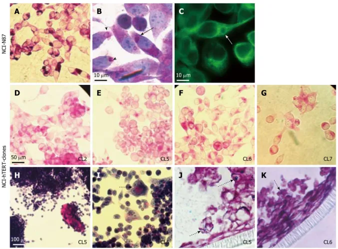
PAS-staining revealed the expression of neutral mucins by the parental N87 cell line and all the NCI-hTERT clones. The intense vesicular PAS-reactivity exhibited by some cells of the parental cell line (Figure 3A) is supportive of the presence of mucus secreting vesicles that are formed in the endoplasmic reticulum vicinity (arrow head at Figure 3B). Such vesicles, in a close interaction with the microtubule network (white arrow at Figure 3C), migrate towards the cytoplasmic membrane and are exocytosed (black arrows at Figure 3B), following a mucin secretion pathway described by others[42]
. However, such phenotype was only acquired by the NCI-hTERT clones in a more
differentiated state, upon full confluence. Indeed, sub-confluent cultures of NCI-hTERT cell lines, in particular
the NCI-hTERT-CL2, CL5, CL6 and CL7, exhibited a less intense cytoplasmatic PAS reactivity (Figure 3D-G, respectively). A similar pattern was described by Chailler and Ménard[23]
, who have linked it with an enhanced ability for mucin-secretion exhibited by their non-transfected NCI-N87-derived cell lines. This hypothesis was further supported by the continuous
Figure 1 Relative telomerase activity measured for 8 NCI-N87 derived clones (NCI-CL1 to NCI-CL8), a pool of the remaining hTERT-expressing NCI-N87 derived clones (NCI-hTERT-pool cell line) and for the parental cell line (NCI-N87, ATCC CRL-5822). These values were obtained using the TeloTAGGG Telomerase PCR ELISAPLUS (Roche Diagnostics), according to the manufacturer’s instructions. The dashed line indicates the average level of the relative telomerase activity measured for the clones.
800
700
600
500
400
300
200
100
0
CL1
Relative telomerase activity
CL2 CL3 CL4 CL5 CL6 CL7 CL8
NCI-hTERT-pool NCI-N87
observation of extracellular accumulation of PAS-reactive mucins in some NCI-hTERT clones, namely in CL5 and CL6 (Figure 3H). This event was sometimes accompanied by the appearance of more organized cellular structures, suggesting a more differentiated state for the cells (dashed arrow at Figure 3I). Additionally, traces of Alcian Blue-reactive mucins (acid mucins) were detected for both NCI-hTERT-CL5 and CL6 when grown in polarization favouring culture conditions (semi wet interphase culture for 28
d post confluency), indicated the presence of residual
amounts of acidic mucins (dotted arrows at Figure 3J and K). Despite these important data, the tested clones were only able to generate a loose/partial mucus layer, as determined by observation of PAS/
Alcian blue stained fixed slides (data not shown).
By immunodetection (Figure 4), we were able to
confirm the presence of MUC5AC among the secreted
and intracellular PAS-reactive mucins. Indeed, our immunocytochemistry data on paraffin-embedded cell lines cultured on Snapwell membranes pointed to an equivalent number of MUC5AC-positive cells in both NCI-hTERT-CL5 and -CL6 (about 25% and about 20%, respectively) (Table 1). Slot-blot analysis proved the expression of MUC5AC by NCI-hTERT-CL6
(Figure 4A), but showed a much lower abundance of this mucin in protein extracts of both NCI-hTERT-CL5 and the parental cell lines. These discrepancies in the abundance of MUC5AC may be explained by the use of different anti-MUC5AC antibodies in each of these two approaches, the 45M1 mAb (Sigma-Aldrich) more suitable for immunocytochemistry and the 2H7 mAb (Sigma-Aldrich) more suitable for western-blot analysis, both recognizing different epitopes. Despite the traces of Alcian-blue-reactive mucins, we were not able to immunodetect MUC6 with none of the used anti-MUC6 antibodies (H5 mAb[35] for western-blot and LUM6-3 pAb[36] for immunocytochemistry), in any of the tested cell lines, i.e., NCI-hTERT-CL5, CL6 and parental cell line (Table 1). Nevertheless, MUC2 is counting for those traces of Alcian-Blue-reactive mucins, since it was detected in about 2% of the cells of both clones 5 and 6. Regarding the membrane-bound mucins, we observed by immunocytochemistry analysis that about 2% of the cells of both clones were positive for MUC13 expression and that about 90% and about 60% of the cells of NCI-hTERT-clones 5 and 6, respectively, were positive for MUC1 (an acidic mucin counting for the membrane/intracellular PAS-reactive mucins) (Table 1). This is a quite interesting
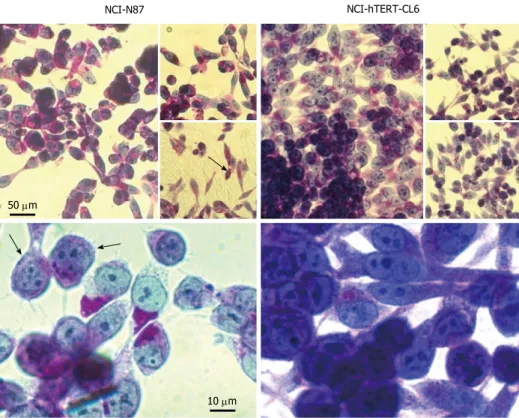
Figure 2 Microscopy analysis of NCI-N87 and NCI-hTERT-clones CL5 and CL6 cell lines. Confluent cultures (A, D, G); square in (A and B), subconfluent cultures (E
and H); E-cadherin immunodetection (C, F and I) (green; 1:1000, E-cadherin mAb plus FITC-conjugated secondary Ab). White bar: 100 μm. NCI-N87 NCI-hTERT-CL5 NCI-hTERT-CL6
100 μm
A
D
G
B
E
H
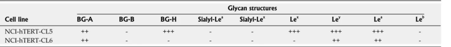
result because of the importance of MUC1 for the interaction with H. pylori[43]. These two cell lines differ in their glycan content (Table 2); the NCI-hTERT-CL5 cells were all positive for blood group H type 2 (BG-H)
and Lewis x (Lex
) antigens that were not detected in CL6 cells. Moreover, Lewis a (Lea
) and Lewis y (Ley ) antigens were found in a smaller percentage of NCI-hTERT-CL6 (21 to 70%) than CL5 (100%) cells. Both cell lines exhibited BG-A (blood group A) antigens. Further analysis on the glycan status of these mucins led us to conclude that, if present at all, both cell lines display undetectable levels of BG-B (blood group B), Leb
, Sialyl-Lea
and Sialyl-Lex
(Table 2). Chailler and Ménard[23]
described the less intense cytoplasmatic PAS-reactivity, similar to that observed in the NCI-hTERT clones (Figure 3), as being typical of the cells with mixed phenotype, i.e., able of simultaneous production of mucins and zymogens. Further corroborating this assumption, the majority of the NCI-hTERT clones exhibited a homogenous positive
PAS/haematoxylin staining (in Figure 5 it is exemplified
with NCI-hTERT-CL6), suggesting the presence of basic molecules, such as zymogens, in the cell cytoplasm, in addition to neutral mucins (PAS-reactive mucins). Such PAS/haematoxylin positive pattern was observed in all subconfluent and postconfluent NCI-hTERT transfected cells, in contrast to the parental
Figure 3 Cell staining analysis for mucins detection on the NCI-N87 parental cell line (A-C) and NCI-hTERT-CL2 (D), CL5 (E, H, I and J), CL6 (F and K) and -CL7 (G). For neutral mucins detection (stained in pink): PAS-staining (A, D-I); and PAS/haematoxylin staining (B). For acidic mucins detection (J and K) PAS/Alcian
blue staining (Alcian positive/PAS negative mucins stained in blue; PAS/Alcian positive mucins stained in purple). α-Tubulin immunodetection (C; green) (1:1000 α-Tubulin mAb plus FITC-conjugated secondary Ab). Black arrow, mucus secreting vesicles that are formed in the endoplasmic reticulum vicinity. Arrow heads, mucus secreting vesicles migrating towards the cytoplasmic membrane and being exocytosed. White arrow, mucus secreting vesicles in close interaction with the microtule network. Dashed arrow suggestive of a more differentiated state for the cells. Dotted arrows, acidic mucins staining.
Figure 4 Evaluation of the expression of epithelial gastric markers by the parental NCI-N87 and NCI-hTERT-clones 5 and 6 cell lines. A: Slot-blot of
100 μg of total protein extracts for detection of mucin 5AC (MUC5AC) with the anti-MUC5AC mAb (1:1000 diluted); B: Western blot of 100 μg of total protein extracts separated in a 12.5% (v/v) SDS-PAGE, with the anti-Pg5 mAb (1:800 diluted) and the anti-HGL pAb (1:800 diluted).
NCI-hTERT
MUC5AC
Pg5 HGL NCI-N87 CL5 CL6
52 kDa 42 kDa
52 kDa 42 kDa NCI-hTERT
NCI-N87 CL5 CL6
NCI-hTERT
NCI-N87 CL5 CL6
A
B
NCI-hTERT-clones NCI-N87
A
B
C
D
E
F
G
H
I
J
K
10 μm 10 μm
50 μm CL2 CL5 CL6 CL7
cell line in which this phenotype was observed only in some cell subpopulations (black arrows in Figure 5). Such observation led us to conclude that the NCI-hTERT-CL5 and CL6 derive from these NCI-N87 cell subpopulations. With this staining procedure, it has also become evident (Figure 5) that the cells of both clones exhibit relative large nuclei, feature shared by progenitor-like gastric cells (reviewed in[3]
). In contrast, no reactivity to eosin was observed either in the parental cell line or in its hTERT-expressing-derived clones (data not shown), clearly demonstrating the absence of parietal-related cells in these cell lines.
By Western blot analysis, we were able to confirm
the expression of both HGL and Pg5 zymogens in the parental cell line and NCI-hTERT-clones 5 and 6 (Figure 4B). Immunocytochemistry analyses (Figure 6) have
further confirmed the progenitor-like phenotype, i.e., simultaneous expression of mucins (MUC5AC) and zymogens (Pg5 and HGL), of NCI-hTERT-CL6. Confocal
images showed ubiquitous apical vesicular-like distribution of both MUC5AC and Pg5 and cytoplasmic granular-like (dotted signal) distribution for HGL.
Usefulness for in vitro infection assays with H. pylori
To better recreate the natural niche of H. pylori
infection it would be important to carry out the co-culture assays under microaerobic conditions to avoid bacterial death[1,21]
. After 48 h of incubation in this conditions, confluent NCI-hTERT-CL6 monolayers showed only a slight decrease (with no statistical
significance) in cell viability (Figure 7A) and maintained
their integrity (data not shown), favouring its use for
in vitroH. pylori infection assays.The measurement of IL-8 and other secreted cytokines has been important in assessing the cellular response of in vitro models to H. pylori virulence factors[40]
. Therefore, to further assess theusefulness of this hTERT-expressinggastric cell line for H. pylori research, co-culture assays were performed using two H. pylori strains differing in virulence, i.e., the clinical strain 417, positive for
homB, which is a paediatric-ulcer associated biomarker, with proinflammatory characteristics, and its homB
double mutant (strain 417DM), with both homB copies inactivated[10]
. By measuring the IL-8 secretion by NCI-hTERT-CL6 cells upon infection (Figure 7B), it was possible to verify that these cells are quite sensitive to H. pylori, presenting a response dependent on the virulence of the strain. Indeed, 24 h of co-culture with the strain 417 caused a about 66 fold increase
Table 1 Mucin expression of NCI-hTERT-clones 5 and 6 cell lines
Percent of positive cells
Cell line MUC1 MUC2 MUC5AC MUC6 MUC13
NCI-hTERT-CL5 +++ (+) ++ - (+)
NCI-hTERT-CL6 ++ (+) + - (+)
-: Negative; (+): Less than 5% stained; +: Less than 21% stained; ++: 21%-70% stained; +++: More than 71% stained; MUC: Mucin.
Figure 5 PAS/haematoxylin cell staining analysis for simultaneous detection of mucins (stained in pink) and zymogens (stained in blue) on the NCI-N87 parental cell and the NCI-hTERT-CL6. The duple PAS/haematoxylin reactivity was observed in only some cell subpopulations of the parental cell line (black arrows)
and, in contrast, in all cells of the NCI-hTERT-CL6 cell line.
NCI-N87 NCI-hTERT-CL6
50 μm
in IL-8 secretion by NCI-hTERT-CL6 cells, which was dramatically higher than with the homB double-mutant
strain (417DM), in accordance to the pro-inflammatory
characteristics of this virulence factor.
DISCUSSION
Corroborating the findings of others, here we have demonstrated that the NCI-N87 cell line exhibits some of the features of a true gastric epithelium[1,44]
. Deriving from a well differentiated human gastric carcinoma, we
observed that these cells preserve the colony-forming growth pattern, the expression of E-cadherin in cell to cell contact and, only in some cell sub-populations, the expression of PAS-reactive mucins and haematoxylin-reactive zymogens. Although advantageous in closer-resembling the heterogeneity of the native tissue, the presence of several types of cells with marked phenotypic differences among them, may hamper the reproducibility of in vitro studies using this cellular model. Indeed, lab maintenance of heterogenic cell lines may lead to accidental loss of some cell sub-populations, always favouring the presence of those with better adhesion and/or mitotic rates properties, which may not necessarily be the ones that better
Table 2 Glycan structures of NCI-hTERT-clones 5 and 6 cell lines
Glycan structures
Cell line BG-A BG-B BG-H Sialyl-Lea
Sialyl-Lex
Lex
Ley
Lea
Leb
NCI-hTERT-CL5 ++ - +++ - - +++ +++ +++
-NCI-hTERT-CL6 ++ - - - ++ ++
--: Negative; +: Less than 21% stained; ++: 21%-70% stained; +++: More than 71% stained. Le: Lewis.
Figure 6 Gastric markers expression by the NCI-hTERT-CL 6 cell line.
Mucin 5AC (MUC5AC), Pg5 and HGL were respectively immunodetected (green) with the anti-MUC5AC mAb (1:25), the anti-Pg5 mAb (1:25) and the anti-HGL pAb (1:25) plus a FITC-conjugated secondary Ab. Nuclei were counterstained with DAPI. Fluorescent signals were recorded by confocal microscopy. Left panels, vertical sections with apical membrane on top of the image. Right panels, horizontal section of same cell sample.
Figure 7 Usefulness of the NCI-hTERT-CL6 cell line for in vitro infection assays with Helicobacter pylori. A: Viability in percentage of the cells when
grown in a microaerobic environment (bacterial growth conditions) for 48 h compared to those grown at standard atmosphere (taken as 100%). Bars and respective error bars are mean ± SD of the values at each condition for twelve observations; B: IL8 secretion by NCI-hTERT-CL6 cells upon infection with the Helicobacter pylori (H. pylori) strain 417 (virulent strain) or the respective
double-mutant for homB gene strain (strain 417DM) (less virulent strain),
compared to uninfected cells. Bars are values obtained from one observation. NCI-hTERT-CL6
HGL Pg5 MUC5AC
10 μm
125
100
75
50
25
0
Standard atmosphere Microaerophily
NCI-hTERT-CL6 viability (%)
40000
32000
24000
16000
8000
0
Uninfected H. pylori strain H. pylori strain 417 417DM
IL8 (pg/mL)
A
represent the functions of that native tissue. This may explain differences between our NCI-N87 cell line and the equivalent cell line used by others[23,41]
, despite their common origin (ATCC CRL-5822). Indeed, in contrast to the previously described intense immunodetection of MUC6 in some cell sub-populations of the NCI-N87 cell line[1,23]
, we were not able to immunodetect this particular mucin, either in protein extracts or in cell slides. In addition, no ZO-1-expressing cells were detected in the NCI-N87 cell line used in the present study, opposing to the observations of Fiorentino et al[40]
and Lemieux et al[41]
that described the expression of ZO-1 in the all NCI-N87 cell contacts and of Chailler and Ménard[23] that reported the expression of this protein in few NCI-N87 cell sub-populations.
As Chailler and Ménard[23]
, we were thus prompted to generate new NCI-N87-derived cell lines exhibiting more homogenous phenotype and, simultaneously, the functional properties of the gastric glandular epithelial cells. Using a completely different approach, we succeed in isolating eight NCI-N87-derived clones of cells by transduction of the hTERT cDNA, which in average showed a 2.2 times increase in the relative telomerase activity over the endogenous level registered for the parental cell line. In normal circumstances, telomerase is active in embryonic cells, germ cells, and some cell sub-populations in tissues with regenerative capacity, but repressed (partially or fully) in somatic cells. Such somatic cells once in culture, by loosing telomeric DNA at each cell division, eventually enter in senescence, a non-replicative state that can be avoided by transfection with some viral oncogenes[45]
. Nevertheless, when telomeres become critically short, these transformed cells with extended replicative life span eventually cease dividing, a second proliferative blockade named crisis. Therefore, telomeric shortening represents the molecular device that tallies replicative doublings and induces senescence and then crisis[27]
. Accordingly, spontaneous telomerase activation warrants the ability of cancer cells to divide beyond the replicative capacity of normal somatic cells, exhibiting unlimited growth in culture. This is fairly known to be a frequent, although not essential, late event in gastric cancer progression[39]
. Naturally, its origin in a human gastric carcinoma justifies the endogenous relative telomerase activity registered in our parental NCI-N87 cell line. In 1998, the ectopic over-expression of
hTERT was shown to be sufficient to induce cellular immortalization by allowing cells to proliferate beyond crisis[27]
. This strategy has been ever since extensively used in the establishment of novel immortalized cell lines without inducing malignant phenotypic effect, but instead preserving their native features[28-33]
.
hTERT over-expression was also shown to bring better features to the CHO-K1 cell line. Indeed, by presenting higher amounts of hTERT than those endogenously observed for the CHO-K1 cell line, the
hTERT-transfected CHO-K1 cells presented an increase in the maximum cell number in batch culture, reduced apoptosis, prolonged culture duration and reduced serum dependency[34]
. Corroborating these findings, we have shown here that the NCI-hTERT-clones presenting higher levels of relative telomerase activity (CL3, 5 and 6) exhibited better adhesion properties, much higher mitotic rates and much prolonged culture maintenance compared to the parental NCI-N87 cell line. These properties were much more pronounced in the NCI-hTERT-CL6, the one with the highest rate of relative telomerase activity, which we were able to maintain in culture for up to 4 wk post-confluency. Additionally, this clone exhibited more pronounced lamellipodia-like structures at low cell density, protrusions observed by Chailler and Ménard[23]
in their NCI-N87-derived clone HGE-17 (human gastric epithelial-17 cell line), but only under the exogenous growth factors treatment. The NCI-hTERT-CL6 cell line also exhibited a more uniform honeycomb-like pattern of expression for E-cadherin. Probably due to the lack of ZO-1 expression, also registered for the other tested clones and for the parental cell line, only low levels of TEER were achieved even for prolonged cultures (21
d post-confluency), never exceeding 70 ohms × cm2 . Better values were obtained by Chailler and Ménard[23] for their NCI-N87-derived clones, reaching similar TEER values for the clone HGE-17 and higher values
(about 300 ohms × cm2
)for the clone HGE-20, at 14 days post-confluency, both cell lines exhibiting ZO-1 expression at all cells’ periphery. Contrasting with us, but also with these authors, Fiorentino et al[40]
and Lemieux et al[41]
presented independently the NCI-N87 cell line itself, as a gastric epithelial barrier model, able to generate per si much higher TEER values (> 1000
and of about 500 ohms × cm2
, respectively)[40,41] . As mentioned above, in this case, and probably justifying these data discrepancy, the used NCI-N87 cell line exhibited generalized expression of ZO-1 at cell contact surfaces. Future experiments will dictate whether higher TEER values can be generated after ectopic induction of ZO-1 in our NCI-hTERT-CL6 cell line.
PAS-staining revealed huge differences between the parental cell line and its hTERT-expressing derived clones, suggestive of a more differentiated secretory phenotype for the latter. Indeed, for NCI-hTERT-CL5 and -CL6, although presenting slightly distinctive features between them, abundant expression of MUC1 and MUC5AC, and traces of MUC2 and MUC13 were detected by immunohistochemistry, corroborating previous results[46]
. In this way, we demonstrated the ability of both NCI-hTERT-CL5 and -CL6 to produce and secrete the gel-forming glycoprotein MUC5AC (supported by the PAS-positive secreted glycoconjugates and the apical accumulation of fluorescent signal specific for MUC5AC in confocal images), a major component of the mucus layer
an important finding, even considering that these cells
are able to generate only a loose mucus layer (data not shown). In addition to close resemblance of the healthy epithelial native tissue, these cells expressed high amounts of MUC1, the most common membrane-tethered mucin of the healthy gastric mucosa. In contrast, these cell lines did not show reactivity with the used anti-MUC6 antibodies, indicating that MUC6, the second most abundant mucin of the mucus layer, mainly found in the deep glands of the healthy gastric mucosa, if present at all, occurs in very low amounts. It is known that the normal pattern of mucins expression changes during gastric neoplasic transformation, resulting in a decrease of MUC5AC in the late stages of disease and of MUC6 in intestinal metaplasia and/or intestinal-type gastric carcinomas. Moreover, such disease process is also accompanied by an increase in the gastric expression of MUC2 (secreted mucin) and MUC13 (transmembrane mucin), two human intestinal mucins usually absent in the normal gastric mucosa[47]
. Thus, the residual expression of these two mucins by the NCI-hTERT-clones 5 and 6 corroborates the origin of the parental cell line in a well differentiated-type human gastric carcinoma. Mucins are naturally decorated with a vast number of glycans which differ between individuals and change during disease[36,48]
. Indicating the isolation of different cell sub-types, the clones NCI-hTERT-CL5 and CL6 differ in their glycan content. Indeed, the BG-H and Lex antigens were only detected in NCI-hTERT-CL5 cell line (in about 100% of the cells), which also presented higher amounts of Ley
and Lea
compared with the NCI-hTERT-CL6. Notwithstanding, both cell lines exhibited equivalent amounts of BG-A and undetectable levels of BG-B and Leb
. Moreover, in agreement with the residual expression of cancer-associated mucins (MUC2 and MUC13), we were not able to detect the presence of sialyl-Lex
nor sialyl-Lea
, glycans commonly found in tumour samples[36,48]
.
Entailing good gastric functions to these new cell lines, both NCI-hTERT-clones 5 and 6 were shown to express HGL and Pg5. Confocal microscopy showed the accumulation of Pg5 in vesicle-like structures in the apical region of the CL6 cells grown in
polarization-favouring conditions, suggesting the efficient secretion
of this zymogen, as described by Chailler and Ménard[23]
for their HGE-20 clone. Furthermore, a
dotted fluorescence spread through the cell cytoplasm
for HGL, similar to that observed for the HGE cell lines[23]
, proved the production and suggests a not so efficient secretion of this zymogen. Although future experiments are needed to measure the enzymatic activity of both Pg5 and HGL (by specific enzymatic assays) on our new cell lines, based on the literature we may speculate good lipase and pepsin activities[1,4,23,44]
. It is well known that gastric epithelial cells and their organization into pit-glands (Figure 8) play a major role in the normal physiology of the human stomach. These include the foveolar cells of
the surface (MUC5AC-secreting cells) and neck (MUC6-secreting cells), the acid-(MUC6-secreting parietal cells of the neck, and the zymogens (HGL and Pg5)-secreting chief cells (oxyntic glands) or the endocrine cells (pyloric glands) of the base of those tubular structures. Leblond et al, in the late 1940s, have shown that the cellular renewal of the gastric glands upon tissue injury is dependent on a group of undifferentiated cells of the isthmus (reviewed in[2,3]
). These cells migrate bidirectionally up to the mucosal surface and down to the gland base, as they differentiate into mature cells of the gastric unit. Nowadays, it is well established that the perpetual renewal of healthy mucosa is dependent on the presence of isthmal stem (with high nucleus/cytoplasm ratios, open chromatin, small and scant organelles, many ribosomes and mini-granules) and progenitor cells (still presenting relative large nuclei). The latter include those exhibiting dual lineage features (i.e.,uncommitted progenitor cells) and those already presenting features of only one lineage of mature cells (i.e., committed progenitor cells) (reviewed in[2,3]
). One of the cell subpopulations isolated by Chailler and Ménard[23]
from the parental NCI-N87 cell line, the HGE20 clone, presented a pre-zymogenic-like phenotype, being able of packaging zymogens (simultaneously HGL and Pg5) into granule-like structures, just as normal gastric chief cells, and co-synthesized MUC6 (Figure 8). Such type of immature cells correspond to mucous cells of the neck that further differentiate into zymogenic cells, giving rise to mature chief cells during their down-migration into the base of the gland. Interestingly, cells from both NCI-hTERT-clones 5 and 6 accumulate the functions of mature surface foveolar cells and chief cells and present large nuclei, representing a different type of uncommitted progenitor-like cells (Figure 8).
The glandular organization of this tissue, also critical to its role as a barrier to a range of environmental noxious and immunogenic molecules[1,4,44]
, is jeopardized by H. pylori[5]
. During an established infection, the vast majority of H. pylori cells (about 70%) are found in the mucus layer of the superficial gastric mucosa, either motile or adhered to the heavily glycosylated secreted mucins. This location favours the gain of nutrients released from the damaged host cells. In a mucin-type dependent manner, this location is also favourable for the bacteria replication. Indeed, as we have recently shown, higher H. pylori proliferation rates were observed with tumour-derived mucins and mucins from the surface mucosa (MUC5AC) compared with gland-derived mucins (MUC6), with the latter having an antimicrobial activity[48,49]
. Therefore, the lack of MUC6 expression by NCI-hTERT-clones 5 and 6 may somehow be advantageous for in vitro infection studies, facilitating the proliferation of H. pylori in co-cultures. Only a smaller fraction of bacteria (about 30%) is found adhered to the epithelial cells’ surface[50]
eliminated from the stomach by mucus turnover and gastric peristalsis, facilitates evasion from the human
immune system and ensures the efficient delivery of the
bacterial toxic proteins. This interaction is highly affected by the presence of MUC1 in the apical surface of the epithelium[43]
, highlighting once again the usefulness of the NCI-hTERT-clones 5 and 6.
H. pylori expresses a multitude of different outer membrane proteins that are used for bacterial adhesion to the highly diverse glyco-epitopes on the secreted
and superficial mucins. To date, the best characterized
are the blood group antigen-binding adhesin (BabA), a ligand of the host Leb
and H type-1 antigens[51] , and the sialic acid binding adhesin (SabA) which mediates
attachment to the inflammation-associated sialylated
(sialyl-Lex
and -Lea
) antigens[52]
. According to our recently presented data, the binding to Leb
may lead to an increase in H. pylori proliferation[48]
. Although we were not able to detect this type of H. pylori-binding antigens in non-infected NCI-hTERT-clones 5 and 6,
our results point to an efficient adherence of different
H. pylori strains (data not shown). Predictably there are other equally important ligands mediating this
bacteria-host interaction.
Conlin et al[26] demonstrated that compared with the AGS cell line, NCI-N87 cells respond to H. pylori infection in a much closer manner to that of primary cultures of gastric epithelial cells. However, as mentioned by them and other authors[26,40]
, by making a literature review we may easily reach to the puzzling conclusion that the former is still the most commonly used cellular model for in vitro studies of the molecular events involved in this host-bacterium interaction, resulting many times in ambiguous and controversial data. The reliability of this cellular model is, indeed, questionable if we consider that, lacking E-cadherin expression, AGS monolayers may eventually lose their integrity after reaching confluency[1,4,22,44]
. This is a significant bias since H. pylori is known to disrupt the cell-cell adhesion of the gastric epithelium, namely interfering with the E-cadherin distribution[26]
. Moreover, the synthesis of zymogens in AGS cell line is confined to cell sub-populations always presenting a diffused cytoplasmic distribution, an obvious disadvantage when compared with their granular-like distribution observed in the NCI-N87 cell line or its
Figure 8 Oxyntic gastric gland unit. These are organized in four distinctive zones: the pit zone, which is lined by mucin (MUC) 5AC-secreting pit cells; the isthmus,
which houses the stem and progenitor cells; the neck zone, which contains MUC6-secreting neck cells and HCl-secreting parietal cells; and the base zone, with the enzyme-secreting chief cells. Gastric epithelial cells turnover is ensured by differentiation bidirectional migration of progenitor cells of the isthmus to the correct place for the emerging adult cell. The new NCI-hTERT-CL5 and CL6 cell lines present dual features of simultaneous production and secretion of MUC5AC (as pit cells) and HGL and Pg5 zymogens (as chief cells), a progenitor-like phenotype.
Goblet cells (MUC5AC)
NCI-hTERT-CL5 and CL6
Progenitor cells Stem cells
Parietal cells (HCl)
Goblet cells (MUC6)
Pre-zymogenic cells Clone HGE20 (Chailler and Menard, 2005)
Chief cells (zymogens) Isthmus
Neck