EXPLORING THE RELATIONSHIP BETWEEN
TOXIN AND SPORE PRODUCTION IN THE
HUMAN ENTERIC PATHOGEN CLOSTRIDIUM
DIFFICILE
SARA DE CASTRO GONÇALVES RAMALHETE
A THESIS SUBMITTED FOR THE DEGREE OF MASTER IN
MEDICAL MICROBIOLOGY
EXPLORING THE RELATIONSHIP BETWEEN
TOXIN AND SPORE PRODUCTION IN THE
HUMAN ENTERIC PATHOGEN CLOSTRIDIUM
DIFFICILE
SARA DE CASTRO GONÇALVES RAMALHETE
A THESIS SUBMITTED FOR THE DEGREE OF MASTER IN
MEDICAL MICROBIOLOGY
Supervisor
: Prof. Dr. Adriano O. Henriques
Co-supervisor
: Dr. Mónica Serrano
Experimental work performed at the Instituto de Tecnologia Química e Biológica António Xavier/ UNL, Av. da República, Estação Agronómica Nacional 2780-157 Oeiras,
Portugal.
Bibliographic elements resulting from this dissertation
Oral papers:
Carolina P. Cassona, Sara Ramalhete, Wilson Antunes, Bruno Dupuy, Mónica Serrano and Adriano O. Henriques. June 2015. The link between toxinogenesis and sporulation during infection by the human intestinal pathogen Clostridium difficile. 17TH European workshop on bacterial protein toxins. Braga, Portugal.
Carolina P. Cassona, Sara Ramalhete, Wilson Antunes, Bruno Dupuy, Mónica Serrano and Adriano O. Henriques. September 2015. The link between toxin production and spore formation in the intestinal pathogen Clostridium difficile. Clostpath 2015 – 9TH International conference on the Molecular Biology and Pathogenesis of the Clostridia. Freiburg, Germany.
Posters:
Carolina P. Cassona, Sara Ramalhete, Wilson Antunes, Bruno Dupuy, Mónica Serrano and Adriano O. Henriques. October 2015. The link between toxinogenesis and sporulation during infection by the human intestinal pathogen Clostridium difficile. 1º Workshop of Genetics. Lisbon, Portugal.
Acknowledgements
First, I would like to thank Prof. Dr. Adriano O. Henriques and Dr. Mónica Serrano for the opportunity to work in the laboratory and for the guidance they provided me every time that I needed. They were always available to discuss ideas and to give me constructive criticisms, which were crucial for me to learn and think outside the box.
I also would like to thank Carolina Cassona, a very hard worker PhD student that was always ready to help and to dispense some of her time to teach me new techniques, and to all of the MDL colleagues, Aristides Mendes, Carolina Feliciano, Hugo Barreto, Inês Portinha, João Bota, Patrícia Amaral and Wilson Antunes, which were very important to turn the team work into funny and pleasant moments. Also, to Teresa Silva for all the help in routinary tasks which were crucial for me to save some time.
I would like to thank the Scientific Committee of the MSc in Medical Microbiology of UNL for the organization of this master’s course and to Instituto de Tecnologia Química e Biológica António Xavier of Universidade Nova de Lisboa for receiving me as
a master student during this year.
Moreover, I would like to thank my family, Rui Ramalhete, Manuela Ramalhete and Rui Miguel Ramalhete, for the constant support they gave me to proceed my dreams and reach success. To David Braz, which was always ready to give me comfort words that were pivotal for me to be the best that I could and for all the good moments that turned this journey even happier.
Finally, to all of my friends which are also family and helped me to relax and create great memories during the last year.
Abstract
Clostridium difficile is currently the major cause of antibiotic-associated
gastrointestinal diseases in adults. This is a Gram-positive bacterium, endospore-forming and an obligate anaerobe that colonizes the gastrointestinal tract.
Recent years have seen a rise in C. difficile associated disease (CDAD) cases, associated with more severe disease symptoms, higher rates of morbidity, mortality and recurrence, which were mostly caused due to the emergence of “hypervirulent” strains but also due to changing patterns of antibiotics use. C. difficile produces two potent toxins, TcdA and TcdB, which are the main virulence factors and the responsible for the disease symptoms. These are codified from a Pathogenicity Locus (PaLoc), composed also by the positive regulator, TcdR, the holin-like protein, TcdE, and a negative regulator, TcdC. Besides the toxins, the oxygen-resistant spores are also essential for transmission of the organism through diarrhea; moreover, spores can accumulate in the environment or in the host, which will cause disease recurrence.
The expression of the PaLoc genes occurs in vegetative cells, at the end of the exponential growth phase, and in sporulating cells. In this work, we constructed two in-frame deletion mutants of tcdR and tcdE. We showed that the positive auto regulation of tcdR is not significant. However, tcdR is always necessary for the expression of the PaLoc
genes.
A previous work showed that, except tcdC, all the PaLoc genes are expressed in the forespore. Here, we detected TcdA at the spore surface. Furthermore, we showed that the in-frame deletion of tcdE does not affect the accumulation of TcdA in the culture medium or in association with cells or spores. This data was important for us to conclude about the infeccious process: it suggests that the spore may be the vehicle for the delivery of TcdA in early stages of infection, that TcdA may be released during spores germination and that this spore may use the same receptor recognized by TcdA to bind to the colonic mucosa.
Resumo
Clostridium difficile é presentemente a principal causa de doença gastrointestinal
associada à utilização de antibióticos em adultos. C. difficile é uma bactéria Gram-positiva, obrigatoriamente anaeróbica, capaz de formar endósporos. Tem-se verificado um aumento dos casos de doença associada a C. difficile com sintomas mais severos, elevadas taxas de morbilidade, mortalidade e recorrência, em parte, devido à emergência de estirpes mais virulentas, mas também devido à má gestão do uso de antibióticos. C. difficile produz duas toxinas, TcdA e TcdB, que são os principais fatores de virulência e
responsáveis pelos sintomas da doença. Estas são codificadas a partir do Locus de Patogenicidade (PaLoc) que codifica ainda para um regulador positivo, TcdR, uma holina, TcdE, e um regulador negativo, TcdC. Os esporos resistentes ao oxigénio são essenciais para a transmissão do organismo e recorrência da doença.
A expressão dos genes do PaLoc ocorre em células vegetativas, no final da fase de crescimento exponencial, e em células em esporulação. Neste trabalho construímos dois mutantes de eliminação em fase dos genes tcdR e tcdE. Mostrámos que a auto-regulação do gene tcdR não é significativa. No entanto, tcdR é sempre necessário para a expressão dos genes presentes no PaLoc.
Trabalho anterior mostrou que, com a exceção de tcdC, os demais genes do PaLoc são expressos no pré-esporo. Mostrámos aqui que TcdA é detectada à superfície do esporo maduro e que a eliminação do tcdE não influencia a acumulação de TcdA no meio de cultura ou em associação às células ou ao esporo. Estas observações têm consequências para o nosso entendimento do processo infecioso: sugerem que o esporo possa ser também um veículo para a entrega da toxina nos estágios iniciais da infecção, que TcdA possa ser libertada durante a germinação do esporo, e que o esporo possa utilizar o mesmo receptor reconhecido por TcdA para a ligação à mucosa do cólon.
Table of contents:
PageBibliographic elements resulting from this dissertation ... i
Acknowledgements ... ii
Abstract ... iii
Resumo ... iv
Table of contents ... v
List of Figures ... vii
List of Tables ... viii
Symbols and Abbreviations ... ix
1. Introduction 1.1. Clostridium difficile ... 1
1.2. Sporulation ... 3
1.3. Pathogenicity Locus (PaLoc) ... 5
1.3.1. The positive regulator (TcdR) and the “negative” regulator (TcdC) ... 6
1.3.2. C. difficile cytotoxins ... 7
1.3.3. The holin-like protein TcdE ... 10
1.4. Other PaLoc regulators ... 12
1.5. Genetic tools and single-cell analysis of the PaLoc expression ... 14
1.6. Objectives of the work ... 17
2. Material and Methods 2.1. Microbiological techniques ... 19
2.1.1. Bacterial strains and growth conditions ... 19
2.1.2. Bacterial growth... 20
2.2. Biochemical techniques ... 21
2.2.1. Sporefractionation... 21
2.2.2. Western blot ... 22
2.2.3. Dot Blot ... 22
2.2.4. Bradford protein assay ... 23
2.3. Genetics and molecular biology techniques ... 23
2.3.1. Molecular cloning ... 23
2.3.2. Gel electrophoresis of nucleic acids ... 24
2.3.3. Preparation of E. coli competent cells and transformation ... 24
2.3.4. Extraction of plasmid DNA ... 25
2.3.5. C. difficile conjugation ... 25
2.3.6. Allele-Coupled Exchange (ACE) mutagenesis ... 26
2.3.7. Genomic DNA extraction ... 28
2.4. Cell Biology ... 28
2.4.1. Fluorescence microscopy and image analysis ... 28
3. Results 3.1. Construction and in trans complementation of an in-frame deletion mutant of tcdR ... 31
3.2. The tcdR mutation does not affect growth or sporulation ... 36
3.3. The role of TcdR in expression of the PaLoc genes ... 38
3.4. TcdA accumulates at the surface of spores ... 40
3.5. Construction and in trans complementation of an in-frame deletion mutant of tcdE ... 42
3.6. The tcdE mutation does not affect growth or sporulation ... 45
3.7. Absence of TcdE does not affect the release of TcdA from vegetative cells .... 47
3.8. TcdEhas no relevant role on the accumulation of TcdA in spores ... 49
4. Discussion and conclusion ... 51
5. References ... 57
6. Appendix ... 71
List of Figures
Page
Figure 1 - Development of C. difficile disease ... 1
Figure 2 - Morphological stages and compartmentalized gene expression of B. subtilis and C. difficile sporulation ... 4
Figure 3 - Schematic image of the Pathogenecity Locus (PaLoc) ... 6
Figure 4 - Model of the uptake of C. difficile toxins ... 9
Figure 5 - The SNAPCd technology extended to C. difficile ... 15
Figure 6 - Fluorescence microscopy using SNAPCd-tag fused to different promoters of the PaLoc components (tcdR, tcdB, tcdE, tcdA and tcdC) ... 16
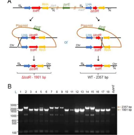
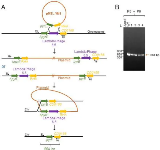
Figure 7 - ACE mutagenesis of the tcdR gene in the 630ΔermΔpyrE strain ... 32
Figure 8 - ACE mutagenesis of the tcdR gene in the 630ΔermΔpyrE strain ... 33
Figure 9 - pyrE reversion using the ACE system ... 34
Figure 10 - tcdR complementation using the ACE system ... 35
Figure 11 - Growth curves of the WT (630Δerm), ΔtcdR, ΔtcdRCand ΔtcdAΔtcdB strains ... 36
Figure 12 - TcdR is not essential for tcdR transcription ... 39
Figure 13 - TcdR protein is necessary for tcdA transcription ... 40
Figure 14 - TcdA accumulates at the spore surface ... 41
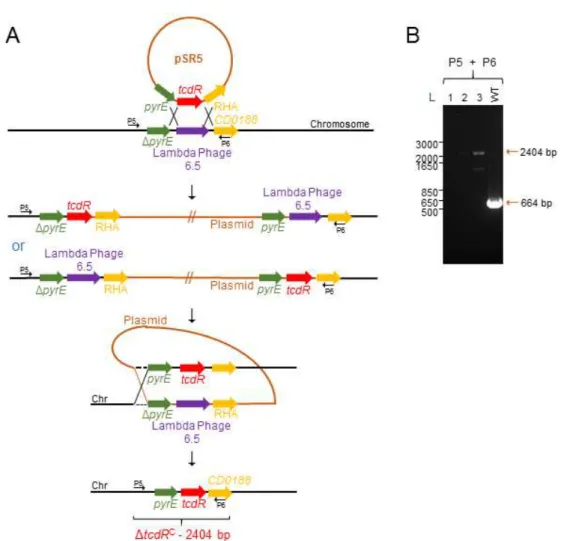
Figure 15 - ACE mutagenesis of the tcdE gene in strain 630ΔermΔpyrE ... 43
Figure 16 - pyrE reversion using the ACE system (A) and in trans complementation of tcdE in strain 630ΔermΔpyrEΔtcdE(C) ... 44
Figure 17 - Growth curves of the WT, ΔtcdE and ΔtcdEC strains ... 46
Figure 18 - TcdE has no major role in TcdA secretion from vegetative cells ... 48
Figure 19 - TcdE has no relevant role on the accumulation of TcdA in spores ... 50
Figure 20 - Regulatory circuits governing tcdR expression ... 53
Figure 21 - Two posssible mechanisms for TcdA accumulation at the spore surface ... 56
List of Tables
Page
Table 1 - Sporulation efficiency of the tcdR mutant and complementation strain, in comparison to the WT, 24, 48 and 72 hours following inoculation into SM ... 37
Table 2 - Sporulation efficiency of tcdE mutant strain, in comparison to the WT, 24, 48 and 72 hours following inoculation into SM ... 47
Symbols and Abbreviations
: : Interruption/junction
°C Degree centigrade
A A
Ampere Absorbance
bp Base pair
cfu Colony-forming unit(s)
DNA Deoxyribonucleic acid
DNase Deoxyribonuclease
DTT Dithiothreitol
EDTA Ethylenediaminetetraacetic acid
g
g
Acceleration of gravity Gram
GTP Guanosine triphosphate
h Hour
kDa Kilodalton
L Liter
M Molar
mg Milligram
min Minutes
ml mM
Mililiter Millimolar
ms Millisecond
PBS PCR
Phosphate-buffered saline Polymerase chain reaction
RNA Ribonucleic acid
RNAse Ribonuclease
SDS Sodium dodecyl sulfate
PAGE PMSF
Polyacrylamide gel electrophoresis Phenylmethylsulfonyl fluoride Tris
UV
Tris(hydroxymethyl)aminomethane Ultraviolet
v Volume
V WT
Volt Wild type
Δ Deletion
nM Nanomolar
μg Microgram
μl Microliter
μm Micrometer
μM Micromolar
σ Sigma
The abbreviations listed are according to the recommendations published by the Journal of Biological Chemistry (JBC). All the other abbreviations are defined in the text.
1.
Introduction
1.1. Clostridium difficile
Clostridium difficile is currently the major cause of antibiotic-associated
gastrointestinal diseases in adults (Rupnik et al., 2009). C. difficile is a Gram-positive bacterium, endospore-forming (hereinafter named spore for simplicity) and an obligate anaerobe that colonizes the gastrointestinal tract. C. difficile infection (CDI) occurs when the gut microbiota is disrupted (Jernberg et al., 2010; Willing et al., 2011;Pérez-Cobas et al., 2013a; Figure 1) and has a range of consequences from asymptomatic carriage to
toxic megacolon, bowel perforation, sepsis, septic shock and death (Rupnik et al., 2009).
Recent years have seen a rise in C. difficile associated disease (CDAD) cases, associated with more severe disease symptoms, higher rates of morbidity, mortality and recurrence, in part because of the emergence of so called hypervirulent strains, mainly, but not exclusively of a specific ribotype, termed 027 (McDonald et al., 2005). Changing patterns of antibiotic use have contributed to the problem (McFarland et al., 2007). While most CDAD cases affect hospitalized patients under antibiotic treatment, in particular clindamycin, aminopenicillins, cephalosporins and fluoroquinolones (Johnson et al., 1999; Gaynes et al., 2004; Loo et al., 2005; Muto et al., 2005; Pépin et al., 2005), cases
Figure 1- Development of C. difficile disease. A: Patients are resistant to CDI if their normal microbiota is not disrupted by antibiotics; B: Once antibiotic treatment starts, infection with a C. difficile strain that is resistant to the antibiotic is more likely; C: When the antibiotic treatment stops, the levels of the antibiotic in the gut diminish rapidly, but the microbiota remains disturbed for a variable period of time (indicated by the break in the graph), depending on the antibiotic given; D: During this time, patients can be infected with either resistant or susceptible C. difficile. Finally, after the microbiota recovers, the resistance to C. difficile is restored (Adapted from Rupnik et al., 2009).
have also been reported without any relation to health care facilities or administration of antibiotics prior to the diagnosis (Rupnik et al., 2009). Therefore, CDI is a growing concern at the community level, as well as in animal husbandry (Rodriguez-Palacios et al., 2006; Songer and Anderson, 2006; Rupnik, 2007).
C. difficile produces two potent toxins, TcdA and TcdB, which are the main virulence
factors and the main causes of the disease symptoms (Rupnik et al., 2009; Burns et al., 2010; Carter et al., 2012; Deakin et al., 2012; Sarker and Paredes-Sabja, 2012). However, the oxygen-resistant spores are essential for transmission of the organism; moreover, spores can accumulate in the environment, and in the host, and are responsible for disease recurrence (Rupnik et al., 2009; Burns et al., 2010; Carter et al., 2012; Deakin et al., 2012; Sarker and Paredes-Sabja, 2012). Infection generally begins with the ingestion of spores; ingested spores will reach the anaerobic colon and germinate. C. difficile responds to unique germinants, such as bile salts (Wilson, 1983). While the bile salt cholate (CA) induces spore germination, another primary bile salt, chenodeoxycholate (CDCA) has been identified as a potent inhibitor of the process (Sorg and Sonenshein, 2008a and b). Upon antibiotic administration, the metabolism of these two compounds is altered and the CA concentration in the gut becomes higher than CDCA, triggering spore germination (Giel et al., 2010). As the organism propagates, it can produce the TcdA and TcdB cytotoxins and more spores (Deneve et al., 2009; Carter et al., 2012). TcdA and TcdB are Rho-glucosylating toxins that cause very typical inflammatory lesions in the colon epithelium, called pseudomembranes (Just et al., 1995; Thelestam and Chaves-Olarte, 2000; Jank et al., 2007; Rupnik et al., 2009). Damage of the colonic mucosa eventually leads to severe diarrhea, which allows shedding of the spores and transmission to new hosts. The treatment recommended by the European Society of Clinical Microbiology and Infectious Diseases (ESCMID) is based on the administration of vancomycin, metronidazole and the recently introduced fidaxomicin; however, these antibiotics may lead to dysbiosis (Debast et al., 2014). Moreover, strains resistant to vancomycin and metronidazole have been isolated (Dworczyński et al., 1991; Pelaez et al., 1994).
The severity of the disease seems linked to the level of dysbiosis (Jernberg et al., 2010; Willing et al., 2011;Pérez-Cobas et al., 2013a). While the microbiota may have an overall protective role, some species-specific interactions may be important to maintain C. difficile in check. A recent study shows that the germination ofC. difficile spores is
inhibited by C. scindens; this occurs because C. scindens is capable of modifying endogenous bile salts which are potent triggers of C. difficile spore germination (Buffie et al., 2015). Therefore, and due to the emerging resistant strains, alternative therapies, as
the faecal microbiota transplantation, are being consider (Matsuoka et al., 2014).
1.2. Sporulation
Spores are central for the pathogenesis of C. difficile. Spores are highly resistant dormant cell types and this resistance is related to their functional architecture. The genome is contained within a central compartment delimited by a lipid bilayer with a layer of peptidoglycan (PG) (germ cell wall) apposed to its external leaflet; this PG layer will serve as the wall of the outgrowing cell that forms when the spore completes germination (Henriques and Moran, 2007; de Hoon et al., 2010; McKenney et al., 2012). The germ cell wall is surrounded by a modified form of PG, called cortex that is essential for the acquisition and maintenance of spore heat resistance (Henriques and Moran, 2007; McKenney et al, 2012). In turn, the cortex is enveloped in a multiprotein coat normally differentiated into two main layers, an inner and an outer coat. In some organisms, including the pathogens B. anthracis, B. cereus and most likely C. difficile, the coat is further enclosed within a structure known as the exosporium. The exosporium is formed by a basal layer from where projections of glycosylated collagen-like proteins emanate (Sylvestre et al., 2002; 2003; Steichen et al., 2003). The coat and exosporium protect the spore cortex from the action of PG-breaking enzymes produced by host organisms or predators, and confer protection to radiation, UV light and small toxic molecules. In addition, the spore surface layers, are required for normal recognition of the molecules that signal spore germination and also mediate spore adhesion to cells and abiotic surfaces (Henriques and Moran, 2007; Panessa-Warren et al., 2007; Oliva et al., 2009; Paredes-Sabja et al., 2012; Paredes-Paredes-Sabja and Sarker, 2012).
The process of spore differentiation has been extensively studied in the model organism B. subtilis but the main morphological stages of sporulation are common to other endospore formers that have been studied in some detail (Henriques and Moran, 2007; de Hoon et al., 2010; McKenney et al., 2012). A hallmark of sporulation is an asymmetric (polar) division that divide the rod-shaped cell into a larger mother cell and a smaller forespore, the future spore. The mother cell then engulfs the forespore and this
process isolates the forespore from the surrounding medium, releasing it as a cell, surrounded by a double membrane, within the mother cell cytoplasm (Hilbert and Piggot, 2004; Higgins and Dworkin, 2012). With the exception of the germ cell wall, which is formed from the forespore, the assembly of the main spore protective structures is mostly a function of the mother cell (Henriques and Moran, 2007; McKenney et al., 2012). At the end of the process, and following a period of spore maturation, the mother cell undergoes autolysis, to release the mature spore (Figure 2).
The developmental regulatory network of sporulation shows a hierarchical organization and functional logic (de Hoon et al., 2010). A master regulatory protein, Spo0A, activated by phosphorylation, governs entry into sporulation, including the switch to asymmetric division (Hilbert and Piggot, 2004; Piggot and Hilbert, 2004). Gene expression in the forespore and mother cell is controlled by 4 cell type-specific sigma factors, which are sequentially activated, alternating between the mother cell and the forespore (Figure 2). When Spo0A-P level reaches a critical threshold in B. subtilis, it
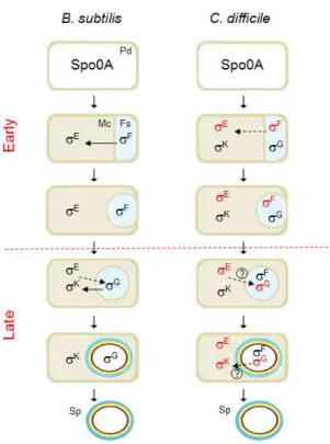
Figure 2 - Morphological stages and compartmentalized gene expression of B. subtilis and C. difficile
sporulation. In a nutrient rich medium, the cell grows and divides by symmetric division (Predivisional cell, Pd). However, upon starvation, the cell enters in sporulation. The process begins with an asymmetric cell division, then, the mother cell (Mc) membrane migrates around the forespore (Fs), engulfing it. At the end of this process, the forespore becomes a free protoplast in the mother cell cytoplasm. Finally, the cortex (brown) and coat (yellow and blue) layers are synthesized and deposited around the developing spore (Sp) and, upon mother cell lysis, a mature spore is released to the surrounding environment, where it remains in a dormant state until good germination conditions. The compartment of activity of the sporulation σF, σE, σG and σK sigma factors is indicated; their main
period of activity in C. difficile cells is indicated in red (Pereira et al., 2013).
activates sporulation genes including spoIIE as well as both the spoIIAA-spoIIAB-sigF and the spoIIGA-sigE operons encoding σF and σE, respectively (Molle et al., 2003). Regarding the B. subtilis model, σF and σE control the early stages of development in the forespore and the mother cell, respectively, and are replaced by σG and σK when engulfment of the forespore is completed (Hilbert and Piggot, 2004; Piggot and Hilbert, 2004; Figure 2). The result is the coordinated deployment of the forespore and mother cell lines of gene expression(Hilbert and Piggot, 2004; Piggot and Hilbert, 2004).
The sporulation pathway of C. difficile and the underlying genetic regulatory network have been recently characterized (Fimlaid et at, 2013; Pereira et al., 2013; Saujet et al., 2013). The main periods of activity of the four cell type-specific sigma factors of C. difficile are conserved in comparison with the B. subtilis model (Pereira et al., 2013).
However, in C. difficile the fact that the activity of σE was partially independent of σF, and that σG or σK did not require σE or σG, respectively, seems to imply a weaker connection between the forespore and mother cell lines of gene expression (Pereira et al., 2013). Relatively to the aerobic Bacilli, the Clostridia represent an older group within the Firmicutes phylum, at the base of which endosporulation has emerged some 2.5 billion years ago, before the initial rise in oxygen level in the earth atmosphere (Stragier, 2002; Paredes et al., 2005; Galperin et al., 2012; Miller et al., 2012; Traag et al., 2012; Abecasis et al., 2013).
1.3. Pathogenicity Locus (PaLoc)
The TcdA and TcdB toxins are encoded by genes located in a Pathogenicity Locus, or PaLoc, which carries the genes for three other proteins, TcdR, TcdE and TcdC (Hammond and Johnson, 1995, Figure 3). TcdR is an RNA polymerase sigma factor that serves as the main positive regulator of expression of the PaLoc (Mani and Dupuy, 2001). TcdE is a holin-like protein thought to be involved in toxin secretion (Govind and Dupuy, 2012). TcdC is a putative TcdR-specific anti-sigma factor that negatively regulates tcdR-dependent transcription, at least in vitro (Matamouros et al., 2007; but see below).
1.3.1.The positive regulator (TcdR) and the “negative” regulator (TcdC)
TcdR is an alternative sigma factor with 22 kDa (Moncrief et al., 1997)that positively regulates toxin production and also activates its own expression (Mani and Dupuy, 2001). This is consistent with the presence of two potential promoters for TcdR-dependent transcription in the region upstream of the tcdR gene (Mani et al., 2002). Recently, El Meouche and co-workers identified a third potential promoter upstream of tcdR and showed that SigD, the main regulatory protein for flagellar biogenesis and motility, positively controls toxin gene expression (El Meouche et al., 2013). Under what conditions are the tcdR promoters utilized is still unclear.
Regarding TcdC, this is an acidic, membrane-associated protein with a predicted molecular weight of 26 kDa (Braun et al., 1996; Govind et al., 2006), which can form dimers (Matamouros et al., 2007). High levels of tcdC transcription may occur during the exponential growth phase of C. difficile , concomitant with low transcription of tcdR and of the tcdA and tcdB genes, whereas during entry into stationary phase transcription of tcdC is low, and transcription of tcdR and the toxin genes is high (Hundsberger et al.,
1997). The reported inverse correlation between the transcription of tcdC and the toxin genes and the expression patterns of the corresponding proteins has led to the prevailing model that TcdC is an important repressor of toxin expression (Hundsberger et al., 1997). This model seems to be supported by the latter finding that the absence of a functional TcdC caused by a frame shift mutation (D117 bp) in the tcdC gene is linked to increased toxin production in certain “hypervirulent” strains (Warny et al., 2005; Curry et al., 2007). Importantly, TcdC can bind to TcdR and inhibit TcdR-directed transcription in vitro, serving as an anti-sigma factor by destabilizing the TcdR-RNA polymerase core
enzyme complex (Matamouros et al., 2007).
Figure 3 - Schematic image of the Pathogenecity Locus (PaLoc). TcdR is a RNA polymerase sigma factor that acts as the positive regulator for PaLoc expression; TcdA and TcdB are the cytotoxins, TcdE is a holin-like protein and TcdC, a possible anti-TcdR factor (Hammond and Johnson, 1995; Mani and Dupuy, 2001; Matamouros et al., 2007; Govind and Dupuy, 2012).
However, recently, some doubts were raised about the importance of TcdC for regulation of toxin expression on the basis of several lines of evidence. First, two studies have found increasing levels of tcdC transcription in time that coincide with increasing transcription of the toxin genes and increasing amounts of toxin production (Merrigan et al., 2010; Vohra and Poxton, 2011). Second, there is a great variability in toxin expression
levels among “hyperirulent” strains, even though these generally carry mutations in tcdC (Curry et al., 2007; Merrigan et al., 2010; Vohra and Poxton, 2011). Third, the prevailing model that TcdC is a negative regulator of toxin expression was supported by the finding that introduction of a functional tcdC gene into an epidemic strain that carries a non-functional tcdC gene (M7404, a PCR ribotype NAP1/027 strain) resulted in decreased toxin production and attenuated virulence in a hamster model (Carter et al., 2011). However, chromosomal complementation in strain R20291, another PCR ribotype NAP1/027 strain with an inactive tcdC gene, resulted in no discernible effect on toxin expression (Cartman et al., 2012). Moreover, other studies showed that disruption of the tcdC gene in the widely used laboratory strain 630Δerm had little if any effect on toxin expression under the conditions tested (Bakker et al., 2012; Cartman et al., 2012). Fourth, recently, van Leeuwen and co-authors showed that TcdC could bind to DNA folded into G-quadruplex structures containing repetitive guanine nucleotides, suggesting that TcdC might also act by destabilizing the open complex formation before transcription initiation; however, no quadruplex-forming motif with multiple G-stretches was found in the PaLoc (van Leeuwen et al., 2013).
The reasons for the conflicting data may relate to experimental variations, including the strain used or the specific growth conditions, either of which might affect the level of TcdC expression or activity (Bouillaut et al., 2015). In conclusion, TcdC might have a modulatory role in regulating toxin expression, but it is probably not a major determinant of the “hypervirulence” of C. difficile (Bakker et al., 2012).
1.3.2.Clostridium difficile cytotoxins
TcdA and TcdB are single-chain proteins with molecular masses of 308 and 270 kDa, respectively, that belong to the group of “Large Clostridial Cytotoxins” (Genth et al., 2008).
Two infection studies in hamsters attempted to clarify the roles of TcdA and TcdB in gastrointestinal disease by using isogenic toxin mutants constructed in the low-virulence clinical isolate 630 (Lyras et al., 2009; Kuehne et al., 2010). The first study found that TcdB alone resulted in disease (Lyras et al., 2009), while the second concluded that both TcdB and TcdA could individually cause severe disease (Kuehne et al., 2010). It is interesting to note that a number of clinical cases of C. difficile infection have been attributed to naturally occurring A−B+ strains (Drudy et al., 2007; 2007), but that there have been no reports of naturally occurring A+B− isolates until now. This would suggest that A+B− strains do not exist, but it may also be an artefact of routine diagnostic testing practices. Either way, if they do not exist in nature already, they may yet evolve.
Thus, both TcdA and TcdB seem to have an enterotoxin activity, however, since TcdB causes several other symptoms outside the gastrointestinal region (Lanis et al., 2013) and has a higher cytopathic potency toward cultured cells (Donta et al., 1982), it is more correct to refer this toxin as cytotoxin.
Both toxins are composed by an N-terminally glucosyltransferase domain followed by a cysteine protease domain, a transmembrane domain and a C-terminally receptor binding domain which harbours repetitive peptide elements called “combined repetitive oligopetides” (CROPs) (Genth et al., 2008). These CROPs exhibit homology to either the carbohydrate-binding regions of glycosyltransferases or to domains for the specific recognition of choline-containing cell-wall components (Just and Gerhard, 2004). In fact, it was shown that TcdA binds to the glycoprotein gp96, a member of the heat shock protein family, and that this binding enhances cellular entry of the toxin (Na et al., 2008). Regarding TcdB, Michelle E. LaFrance et al. (2015) claim that PVRL3 is one of the receptors since they observed a direct binding interaction between PVRL3 and TcdB using purified proteins (LaFrancea et al., 2015). Furthermore, they showed that PVRL3 is independent of the CROPs. This model is compatible with the dual-receptor mechanism, proposed by Schorch et al. (2014) where the CROPs domain allows the toxin to dock onto the cell surface by interacting with oligosaccharides, followed by toxin binding to a high-affinity receptor (Schorch et al., 2014).
Although there are some potential receptors identified for toxin A and B, further studies are required to understand what region(s) of the toxins are binding to what type of receptors.
Upon receptor binding, the toxin is thought to be internalised into the endosome (Florin and Thelestam, 1983). TcdA and TcdB take the “short trip”, which mean they arrange for the glucosyltransferase domain to escape from the endosome, instead of the retrograde transport through the Golgi apparatus (“long trip”) (Genth et al., 2008). In the intermediate part of the delivery domain, a (transmembrane) domain has been postulated to be involved in membrane translocation of the glucosyltransferase domain (Just and Gerhard, 2004). The pH drop in the acidified endosome is thought to induce a structural re-arrangement of the transmembrane domain allowing this domain to form a pore into the endosomal membrane (Giesemann et al., 2006). Once the glucosyltransferase domain has passed the pore and reached the cytosol, it is proteolytically cleaved off from the rest of the protein in the presence of non-proteinaceous cofactors such as inositol phosphates (Egerer et al., 2007; Figure 4). It is free to mono-glucosylate and, thereby, inactivate low molecular mass GTP-binding proteins of the Rho subfamily (avoiding the GDP-GTP exchange) (Herrmann et al., 1998). The Rho subfamilies, which are more probably to be affected by these toxins, are: RhoA, Rac1 and Cdc42. This toxicity will compromise the actin cytoskeleton integrity, resulting rounding cells and cell death (Genth et al., 2008).
Figure 4 - Model of the uptake of C. difficile toxins. Toxins A and B bind to receptors on the surface of target cells and are endocytosed. Acidification of the toxins in endosomes exposes hydrophobic regions of the protein allowing their insertion into the membrane. At this point, the toxin forms a pore, and the N-terminal catalytic domain is translocated from an acidic endosomal compartment into the cytosol. The location of toxin processing is indicated by scissors. In the cytosol, the toxins are capable of glycosylating Rho subfamily proteins. The Figure is according to Rupnik et al., 2009, with minor modifications.
In respect to their role in host–pathogen interactions, Rho proteins essentially participate in epithelial barrier functions and cell–cell contact, in immune cell migration, phagocytosis, cytokine production, wound repair, immune cell signalling, and superoxide anion production (Jank et al., 2007). Rho proteins are regulated by a guanosine triphosphatase (GTPase) cycle and they are inactive in the guanosine diphosphate (GDP)-bound form. Activation occurs after GDP/GTP exchange, which is induced by guanine nucleotide exchange factors (GEFs). In the active form, the Rho proteins interact with numerous effectors and adaptors (Jank et al., 2007).
Toxin-catalysed modification of Rho and Ras proteins have several functional consequences. Firstly, after glucosylation, Rho/Ras proteins are no longer able to interact with their effectors (Herrmann et al., 1998; Sehr et al., 1998). Secondly, glucosylation inhibits the activation of small GTPases by GEFs (Herrmann et al., 1998; Sehr et al., 1998). Finally, glucosylated Rho proteins are associated with the cell membrane and the membrane-cytosol cycle is blocked (Genth et al., 1999). However, the most important structural consequence of glucosylation is probably inhibition of the change into the active conformation of the GTPase.
1.3.3.The holin-like protein TcdE
The tcdE open reading frame encodes a small, hydrophobic protein of 166 amino acids with a short hydrophilic stretch at the N-terminus, a series of charged residues at the C-terminus (Tan et al., 2001) and is predicted to contain three transmembrane domains (Govind and Dupuy, 2012). These structural features and primary sequence similarities strongly suggest that TcdE is a member of the class I holins of which phage λ S protein is a member. Holins are small membrane proteins encoded by double-stranded DNA phages that are required for the lysis of host cells at a programmed time after completion of intracellular phage development (Wang et al., 2000; Young et al., 2000). They form disruptive lesions by oligomerization in the host cell plasma membrane to allow a prophage-encoded endolysin (a muralytic enzyme) to cross the membrane and attack the murein, resulting in cell lysis and release of phage particles (Wang et al., 2000; Young et al., 2000).
Two studies that support this idea are the overexpression of TcdE in Escherichia coli, causing cell death (Tan et al., 2001) and the expression of TcdE from its own promoter
using a multicopy plasmid, which was lethal to C. difficile (Govind and Dupuy, 2012). However, a third study using a controlled expression vector to avoid C. difficile death, has shown that when high concentrations of the inducer were added (>50 ng/ml), the culture supernatant of the complemented tcdE mutant had a higher concentration of toxins than did the parent strain JIR8094 (Govind and Dupuy, 2012). Hence, under normal conditions, C. difficile presumably produces an amount of TcdE sufficient to form pores that allow release of toxin without causing cell lysis (Govind and Dupuy, 2012).
Until now, it is not known how these pores interact with toxins. If the toxins are secreted unfolded, possibly via translationally coupled secretion, only a narrow channel in the cytoplasmic membrane would be needed. Such a channel would not allow cytoplasmic protein leakage (Govind and Dupuy, 2012).On the other hand, if the toxins are secreted as fully folded proteins, a large membrane channel would be needed due to the volumes the large toxin proteins would occupy. Although TcdE has the intrinsic ability to form pores in the membrane that lead to permeability and cell death, as seen in E. coli (Tan et al., 2001), it does not normally do so in C. difficile (Govind and Dupuy, 2012). If TcdE-dependent pores are formed in C. difficile, they should be tightly regulated by a mechanism that could include the toxins themselves. The toxins could, for instance, act as plugs to prevent loss of solutes or proteins from the cells through the TcdE pore. Such a model is consistent with the observation that a tcdA tcdB double mutant lysed more rapidly than the parental and PaLoc negative strains (Govind and Dupuy, 2012).
Finally, TcdE-dependent channels might be formed in association with other proteins that control the opening of the pore or TcdE could form a specific gated channel that only opens in the presence of TcdA/TcdB, without inducing cell lysis (Govind and Dupuy, 2012).
Another study showed that the inactivation of tcdE in the low-virulence strain 630Δerm,did not significantly alter neither the kinetics of release nor the absolute level of secreted TcdA and TcdB (Olling et al., 2012). Thus, the impact of TcdE in toxin secretion is still under debate.
1.4. Other PaLoc regulators
The spectrum of diseases caused by C. difficile is highly variable and depends on the level of toxin produced (Akerlund et al., 2006). This supports the hypothesis that regulation of toxin synthesis is a critical determinant of C. difficile pathogenicity. Toxin synthesis increases as cells enter into stationary phase (Hundsberger et al., 1997), and many environmental factors influence their production. In the presence of phosphotransferase system (PTS) sugars, such as glucose, and of certain aminoacids, like cysteine or proline, toxin production is inhibited (Dupuy and Sonenshein, 1998; Karlsson et al., 2000). Environmental stresses, such as alteration of the redox potential, exposure
to sub-inhibitory concentrations of antibiotics, high temperature, or limitation of biotin, also modulate toxin production (Onderdonk et al., 1979; Yamakawa et al., 1996; Karlsson et al., 2003; Deneve et al., 2009).
Several regulators are now implicated in toxins synthesis: CodY and Spo0A, two regulators that control pre- or post-exponential events, (Dineen et al., 2007; Underwood et al., 2009), CcpA, a glucose-dependent repression mediator (Antunes et al., 2011) and
SigH, a key element in the control of the transition from exponential to the stationary phase and of the initiation of sporulation (Saujet et al., 2011). CodY is the first regulator encoded outside of the PaLoc that has been shown to participate in the regulation of toxin synthesis. Inactivation of the C. difficile codY gene resulted in derepression of all genes of the PaLoc during exponential and stationary growth phases, although this repression was not so pronounced in tcdC expression (Dineen et al., 2007). Moreover, CodY binds to the tcdR promoter region but not to tcdA and tcdB promoters, suggesting that growth phase dependent regulation of C. difficile toxin synthesis is mediated by the effect of CodY on tcdR transcription (Dineen et al., 2007). This binding was also enhanced in the presence of GTP and branched-chain amino acids, thus, regulation by CodY may provide a nutritional link to the pathogenicity of C. difficile (Dineen et al., 2007).
Repression of toxin synthesis in the presence of glucose or other rapidly metabolizable carbon sources suggests that the toxin genes are subject to a form of catabolic repression (Dupuy and Sonenshein, 1998). In low G+C Gram-positive bacteria, carbon catabolite repression (CCR) is mediated by CcpA. To test this correlation, crude extracts of the parental strain were obtained from cells collected during stationary phase (14 h) and the total toxin levels were assayed by Vero cell cytotoxicity assays. In cells grown in the
presence of glucose, the cytotoxic activity was low as compared with that of cells grown without glucose. Moreover, using specific antibodies against TcdA, researchers also showed by Western blot that toxin A accumulation was strongly repressed in the presence of glucose (Antunes et al., 2011). Moreover, the cytotoxic activity and the quantity of TcdA detected in crude extracts of the ccpA mutant grown in the absence or in presence of glucose were the same, which indicates that CcpA mediates glucose repression of toxin synthesis (Antunes et al., 2011). Furthermore, the same study showed that the effect of CcpA is direct since this regulator interacts with the tcdB promoter region and the 5′ end of the tcdA-coding sequence (Antunes et al., 2011).
Taking this into consideration, CodY and CcpA act by monitoring the nutrient sufficiency of the environment, directly repressing the PaLoc genes, and releasing this repression during stationary phase, when nutrient condition become limited (Dineen et al., 2007; Antunes et al., 2011).
Regarding Spo0A, this protein is conserved in all spore-forming bacteria, essential in the initiation of the developmental pathway of spore formation, and was also reported to be a PaLoc repressor (Zhao et al., 2002; Underwood et al., 2009). However, recent studies have failed to elucidate the role of Spo0A in TcdA and TcdB production by C. difficile, with conflicting data published to date (Underwood et al., 2009; Deakin et al., 2012; Rosenbusch et al., 2012; Mackin et al., 2013). In “hypervirulent” strains, as R20291, Spo0A acts as a negative regulator of TcdA and TcdB production (Deakin et al., 2012). In contrast, Spo0A does not appear to regulate toxin production in the low-virulence strain 630Δerm (Rosenbusch et al., 2012). Moreover, in other strains (as JGS6133), Spo0A appears to negatively regulate toxin production during early stationary phase, but has little effect on toxin expression during late stationary phase (Mackin et al., 2013). These data suggest that Spo0A may differentially regulate toxin production in distinct C. difficile strain types. In any case, as no Spo0A boxes are present upstream of the tcdA and tcdB genes, an indirect effect of Spo0A on their transcription may happen via a still uncharacterized regulator (Underwood et al., 2009).
Another study established a correlation between the PaLoc components and sigH expression in the stationary phase. tcdR, tcdB and tcdA showed increased transcription in the sigH mutant than in the 630Δerm strain, while in the sigH complemented strain, the expression was restored to the level observed in 630Δerm strain for all the three genes.
Therefore, the expression of these three genes is negatively controlled by σH (Saujet et al., 2011). However, no σHconsensus sequences are present in the PaLoc, suggesting an indirect effect of σH in the inhibition of the PaLoc transcription. Because the expression of tcdC was similar in the 630Δerm strain and in the sigH mutant, authors suggested that the absence of modulation of tcdC transcription in the sigH mutant indicates that the σH -dependent control of tcdA, tcdB, and tcdR expression is not mediated by the regulation of TcdC synthesis (Saujet et al., 2011). However, they could not exclude the possibility that σHmay influence factors controlling TcdC stability and/or activity (Saujet et al., 2011).
In conclusion, there are four negative regulators encoded outside the PaLoc that participate in the regulation of toxin production. These are some examples of the possible PaLoc regulators, which indicates us that the toxins production is a process that needs to be extremely well controlled, since these are the major virulence factors of C. difficile.
1.5. Genetic tools and single-cell analysis of the PaLoc expression
Although a very important pathogen, only recently a solid platform of genetic and cell biology tools was developed for C. difficile. Studies to understand in more detail C. difficile colonization, virulence and pathogenesis are now possible. First, directed mutants
could only be made using insertional mutagens, reliant either on replication deficient (Liyanage et al., 2001) or defective (O’Connor et al., 2006; Dineen et al., 2007) plasmids, or on the deployment of the ClosTron and group II intron re-targeting (Heap et al., 2007; Heap et al., 2010). Recently, the cytosine deaminase gene (codA) of E. coli was developed as a negative/counter selection marker for C. difficile, which enabled precise manipulation of the C. difficile chromosome for the first time (Cartman et al., 2012). In parallel, a second method (Allele-Coupled Exchange, ACE) has been formulated that allows the rapid insertion of heterologous DNA, of any size or complexity, into the genome (Heap et al., 2012). Because the ACE mutagenesis system allows a specific in-frame deletion, which reduces the polar effects, this was the chosen system for this work. In other hand, the autofluorescence proteins (AFP’s) that have enable noninvasive imaging in living cells of reporter gene expression and therefore have become indispensible tools in cell and development biology, cannot be applied in strict anaerobes like C. difficile. Recently, our laboratory has implemented a system based on a mutant form of the human DNA repair enzyme O6-alkylguanine-DNA alkyltransferase
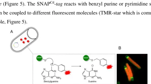
(SNAPCd-tag) (Pereira et al., 2013) to examine gene expression in single cells of C. difficile (Figure 5). The SNAPCd-tag reacts with benzyl purine or pyrimidine substrates that can be coupled to different fluorescent molecules (TMR-star which is commercially available, Figure 5).
Although a wealth of literature has addressed the process of toxinogenesis in C. difficile, expression of the tcdA and tcdB genes, and indeed of the other three PaLoc genes,
was never studied at the single-cell level. This kind of study is very important given the increase evidence that exist cell-to-cell differences at the gene expression level in bacterial populations. Such gene expression heterogeneity determines the fate of individual cell and can also ultimate the fate of the population as a whole.
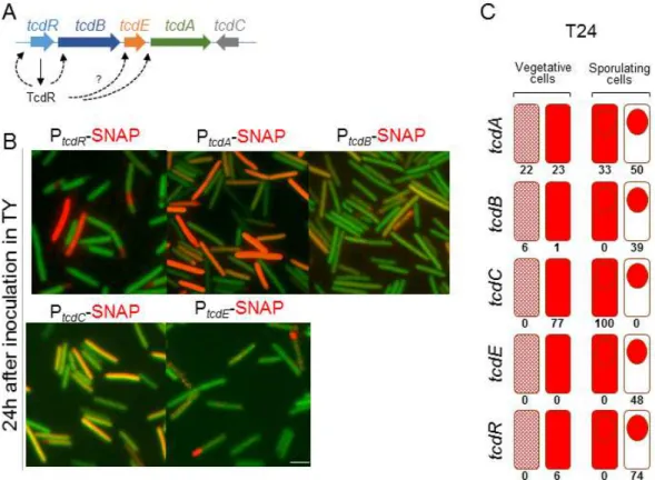
In preliminary work made in the laboratory we have fused the tcdR, tcdB, tcdE, tcdA and tcdC promoters (all the regulatory regions of the PaLoc genes) to the SNAPCd-tag and assessed gene expression in a medium that support toxin production in the standard laboratory strain 630Δerm (Figure 6). Two cellular populations expressing tcdR during stationary phase were observed, a smaller one consisted of cells without signs of asymmetric division; a larger one consisted of sporulating cells in which, strikingly, tcdR is expressed in the forespore (Figure 6B). As expected, fluorescence was not detected during exponential growth (data not shown). These results already show that the PaLoc expression is spatially and temporally regulated. Since we found tcdR to be expressed in the forespore, we therefore anticipated accumulation of the toxin in the developing spore.
Figure 5 - The SNAPCd technology extended to C. difficile. A: Schematic representation of SNAPCd labelling. C.
difficile cells carry a replicative plasmid where the SNAPCd sequence is under the control or fused to a gene of interest. These cells are cultivated under appropriate conditions that allow production of the SNAPCd protein, time at which a cell permeable label (red) should be added. The permeable label enters into the cell, where a covalent modification occurs, allowing labelling and visualization of the SNAPCd protein by fluorescence. B: Microscopy analysis of C.
difficile cells producing SNAPCd visualized by the red color after labelling with the TMR-Star cell-permeable SNAPCd substrate (Pereira et al., 2013).
tcdB and tcdE have similar patterns of expression to tcdR. tcdA is also expressed in two
populations during stationary phase, in cells without signs of asymmetric division and also in sporulating cells, both in the mother cell and in the forespore (Figure 6).
In contrast, tcdC expression was detected during exponential growth (data not shown). During stationary phase, tcdC is also expressed in either cells without signs of asymmetric division or in the mother cell during sporulation. However, expression of tcdC was never detected in the forespore.
Given these results, it is pertinent to go depth in the study of the cell population dynamics with respect to the circuits governing toxinogenesis and sporogenesis.
Figure 6 - Fluorescence microscopy using SNAPCd-tag fused to different promoters of the PaLoc components
(tcdR, tcdB, tcdE, tcdA and tcdC). A: Regulatory interactions in the Pathogenicity Locus. TcdR is thought to be auto-regulatory; TcdR also activates transcription from the indicated PaLoc promoters; B: Cells were incubated 24h in TY medium and observed by fluorescence microscopy. The green color corresponds to C. difficile autofluorescence while the red one corresponds to the reaction of the SNAPCd reporter with the TMR-Star substrate. tcdR expression is mainly detected in the forespore, as is that of tcdB. The expression of tcdA appears mostly in vegetative and mother cells but also in some forespores. tcdC expression seems to be excluded from the forespore. Finally, tcdE was mainly expressed in the forespore. Scale bar: 1µm; C: Percentages of each of the following cell classes: vegetative cells with little expression (light red) and high expression (red) and sporangia with whole cell expression or only in the forespore.
1.6. Objectives of the work
The preliminary results described above show that sporulation and toxinogenesis are interconnected, with expression of the PaLoc genes during sporulation. To increase our knowledge on the relationship between toxinogenesis and sporogenesis, we focused our attention on the regulation of tcdR expression and on the role of tcdE on TcdA secretion and localization. Our specific aims were:
1. To construct a tcdR mutant, and to study the impact of this mutation on tcdA and tcdR expression, on growth and on sporulation. Since tcdR expression was detected
in the forespore, we followed this clue to study the relationship between sporulation and toxin production. Furthermore, this mutant was crucial to understand the relevance of the tcdR positive feedback and to confirm tcdA regulation by TcdR. 2. To analyze the accumulation and localization of TcdA in the mature spore, since
expression of tcdA was detected in both the mother cells and in the forespore. 3. To construct a tcdE mutant, and to study the impact of the mutation on toxin
secretion and on the localization of the toxin in the mature spore.
2.
Material and methods
2.1. Microbiological techniques
2.1.1. Bacterial strains and growth conditions
Escherichia coli strain DH5α was used as a host for molecular cloning and for plasmid propagation (Appendix 1); E. coli strain HB101 was used for conjugation of plasmids into C. difficile due to the presence of the conjugational plasmid RP4. Luria Bertani (LB) medium (1% tryptone, 0.5% yeast extract, 0.5% NaCl, pH 7.0) was routinely used for growth and maintenance of E. coli. When necessary, agar (Agar nº2 bacteriological, from LAB M) was added to a final concentration of 1.6%. When appropriate, ampicillin (100 µg/mL) or chloramphenicol (20 µg/mL) was added to the culture medium.
All the strains of C. difficile used in this work are congenic with 630Δerm (Hussain et al., 2005), the reference strain in the field, and are listed in Appendix 2. Strain 630Δerm
is hereinafter referred to as the wild type (WT) for simplicity and belongs to the ribotype 012, which is not classified as “hypervirulent”. All strains were stored at -80ºC in 20% glycerol. C. difficile strains were routinely grown anaerobically (5% H2, 15% CO2, 80% N2) at 37ºC in brain heart infusion (BHI; from Oxoid) or in BHI supplemented with 0.1% L-cysteine and 5 mg/ml yeast extract (BHIS). Bacto agar (from BD) was normally used at a final concentration of 1.6%. A C. difficile minimal medium (CDMM; Karasawa et al., 1995) solidified with 1.5% agar (from BD) was required for the ACE mutagenesis
(see below; Appendix 3). CDMM was supplemented with 5-fluoroorotic acid (2 mg/ml) and uracil (5 µg/ml) when appropriate. For some experiments, C. difficile was grown in Tryptone Yeast Extract medium (TY: 3% bacto tryptone, 2% bacto yeast extract). When necessary, cefoxitin (25 µg/ml) and thiamphenicol (15 µg/ml) were added to C. difficile cultures. For tcdE complementation anhydrotetracycline (ATc) was used at a concentration of 20 ng/ml.
Sporulation assays were performed in Sporulation Medium (SM): 9% bacto tryptone, 0.5% bacto peptone, 1% (NH4)2SO4 and 0.15% Tris base, pH 7.4 (Wilson et al., 1982). The pre-inoculumn was inoculated in SM medium (1:200 dilution) and, after 24, 48 and 72 hours the sporulation efficiency was analysed. Serial dilutions were performed (up to
10-6) and three spots (20 µl) of each of the 10-6, 10-5, 10-4 and 10-3 dilutions were spotted onto BHI plates containing 0.1% taurocholate acid sodium salt (from Roth) to induce spore germination. The 10-1, 10-2, 10-3 and 10-4 diluted cultures were removed from the chamber and incubated 30 minutes at 60°C to kill vegetative cells. Finally, these were introduced again inside the chamber and three spots of 20 µl were spotted onto a BHI plate containing 0.1% taurocholate. All plates were incubated overnight under anaerobic conditions. The colonies counting was concluded after 24h of the plates inoculation and the sporulation efficiency was calculated according to the following formula:
cfu/ml=number of colonies× 1 Dilution×
1 Volume
Dilution – Dilution factor(e. g., 10-2, 10-3). Volume – Plated volume in ml.
For spore production, C. difficile was grown in BHI. An overnight culture was added to BHI liquid medium in a T-flask with a final dilution of 1:100. Cultures were incubated anaerobically in the horizontal for 7 days at 37ºC. Cells were collected by centrifugation at 4700 x g, resuspended in cold water and stored over 48h at 4ºC, for lysis of mother cells and release of the spores. The suspension was centrifuged again for 10 minutes at 4700 x g, 4ºCand the sediment was suspended in 1 ml of 1x PBS (137 mM NaCl, 2.7 mM KCl, 4.3 mM Na2HPO4, 1.4 mM KH2PO4, pH 7.6) 0.1% tween 20 (PBS-T). This suspension was applied on top of 25 ml of a 42% Gastrografin solution (from Bayer) and centrifuged for 20 minutes at 4700 x g, 4ºC. The supernatant was then aspirated using a vacuum apparatus and the sediment suspended again in 1 ml of PBS-T. Two more washes in PBS-T were performed (to remove traces of Gastrografin) at 4700 x g during 3 minutes at room temperature. Lastly, the sediment was washed two times in water and, finally, suspended in 500 μl of water.
2.1.2. Bacterial growth
Cultures were incubated under anaerobic conditions and growth was followed by measuring the optical density at 600 nm (OD600) at hourly intervals. The growth rate was calculated from the slope of the part of the curve that corresponds to the exponential growth phase, while the generation time was determined according to the following equation: generation time = ln(2)/growth rate.
2.2. Biochemical techniques
2.2.1. Spore fractionation
The volume of a purified spore suspension used in fractionation experiments was determined by measure of spore suspension OD580, previously diluted 1:200 in bi-distiled water (ddH2O), and using the following formula:
Volume of suspension (µl) = (3600/OD580) x 200
The spores were resuspended in 50 µl of 2X decoating buffer (10% glycerol, 4% SDS, 10% β-mercaptoethanol, 1 mM DTT, 250 mM Tris, pH 6.8). Samples were boiled for 5 minutes and centrifuged 2 minutes at 16200 x g. Finally, supernatants were collected to a new tube and 2 µl of 1% bromophenol blue were added. 25 µl of the samples were applied on a 12.5% SDS-PAGE gel (12.5% resolving gel: 41% distilled water, 25.4% 4x lower Tris buffer, 12.6% bis-acrylamide, 0.1% SDS, 0.1% ammonium peroxydisulphate, 0.05% tetramethylethylenediamine, these last two induced polymerization; 4.5% stacking gel: 61.2% distilled water, 25.5% 4x upper Tris buffer, 10.2% bis-acrylamide, 0.1% SDS, 0.1% ammonium peroxydisulphate, 0.1% tetramethylethylenediamine).
The sediments resulting from the spore decoating step were washed twice in PBS-T. Sediments were suspended in 100 µl of PBS-T and divided in two tubes. These were centrifuged 3 minutes at 4700 x g and one of the two sediments was suspended in 25 µl of 50 mM Tris HCl, pH 8.0 with 2 mg/ml lysozyme, while the other was suspended in 25 µl of 50 mM Tris HCl, pH 8.0. Samples were incubated at 37°C for 2 hours and, after this time, 25 µl of loading buffer 2X (0.125 mM Tris-HCl, 5% β-mercaptoethanol, 2% SDS, 0.025% bromophenol blue, 0.5% mM DTT, 5% glycerol, pH 6.8) were added. For each of the samples, 50 µl were resolved by SDS-PAGE gel (12.5% gels). The Precision Plus Protein™ All Blue Ladder (from BioRad) was used in each run.
The resulting gel was incubated 1 hour in the coomassie solution (0.5 g/ml coomassie Brilliant Blue R-250, 80% absolut ethanol, 20% acetic acid) and then, incubated in destaining solution (30% absolut ethanol, 10% acetic acid) overnight with agitation at room temperature. In the next day, the gel was incubated with new destaining solution until the background was clear.
2.2.2. Western blot
Proteins were electrophoretically transferred from SDS-PAGE gel to nitrocellulose membranes (Supported Nitrocellulose, 0.45 µm; from BioRad) at 100 V for 90 minutes using transfer buffer (14.4 g/L glycine, 3.02 g/L Tris base, 10% ETOH). The membrane was incubated in 20 ml of blocking solution (5% milk in PBS-T) for 1 hour with agitation. Next, the blocking solution was removed and the antibody solution was added in 10 ml of PBS-T with 0.5% milk [An anti-TcdA antibody (from Santa Cruz Biotechnology) was used at a dilution of 1:5000; an anti-CotD antibody (Permpoonpattana et al., 2011) was used at a dilution of 1:1000 and an anti-CotA antibody (Permpoonpattana et al., 2011) was used at a dilution of 1:3000]. The membrane was incubated overnight with the antibody solution at 4°C without agitation. The antibody solution was then discarded and the membrane was washed 3 times in PBS-T (10 minutes each wash). The secondary antibody (mouse peroxidase-conjugated secondary antibody from Sigma) was then added in 10 ml of PBS-T with 0.5% milk at a dilution of 1:2000. The membrane was incubated 30 minutes at room temperature with agitation. Finally, the membrane was washed 3 more times in PBS-T (10 minutes each) and the protein was detected using the detection solution (“SuperSignal West Pico Chemiluminescent” from Thermo Scientific) in the dark.
For reprobing, membranes were incubated in 20 ml of a stripping solution (6.25% Tris-HCl pH 6.8, 2% SDS, 0.7% 2-mercaptoethanol) for 30 minutes at 50ºC (with agitation every 10 minutes). Then, the membrane was washed in distilled running water and incubated in blocking solution. The remaining of the protocol is the same as described above.
2.2.3. Dot blot
This technique represents a simplification of the western blot method. In a dot blot, the biomolecules are not separated by electrophoresis as the western blot requires. Instead, a mixture containing the molecule which we want to detect is applied directly on a membrane as a dot. This is then followed by antibody detection (see above 2.2.2).
C. difficile cultures were centrifuged and two fractions were obtained, the sediment
and the supernatant. 10 ml of the supernatant was concentrated up to 1 ml in an Amicon Ultra-4 Centrifugal Filter Unit with Ultracel-50 membrane and 200 µl of the concentrated
solution were analysed by dot blot, except when the total amount of proteins were normalized (see 2.2.4), in this case, different volumes of each sample were used. The sediment resulting from the centrifugation was suspended in 1 ml of French press buffer (10 mM Tris pH 8.0, 10 mM MgCl2, 0.5 mM EDTA, 0.2 mM NaCl, 10% glycerol, 1 mM PMSF) and cells were lysed at 900 psi. 1 µl of DNAse was added to this extract which was then clarified by centrifugation for 10 minutes at 16200 x g, 4ºC. 20 µl of the supernatant were analysed by dot blot, except when the total amount of proteins were normalized (see 2.2.4).
2.2.4. Bradford protein assay
This is a spectroscopic analytical procedure used to measure the protein concentration in a solution. The protein assay-dye (from BioRad) was diluted 5x in ddH2O. For the reference, 200 µl of this diluted solution were added to 800 µl of ddH2O. In order to measure the protein concentration, 10 µl of each sample were added to 200 µl of the diluted Bradford solution and 790 µl of ddH2O. The absorbance (A) of the mixture was measured at 595 nm and the protein concentration was calculated according to the following formula:
(µg/µl)=� A595 0.0656�
10
2.3. Genetics and molecular biology techniques
2.3.1. Molecular cloning
DNA fragments for cloning were generated by the polymerase chain reaction (PCR) using the high fidelity Phusion DNA polymerase (from Thermo Fisher Scientific). All oligonucleotide primers used in this work are listed in Appendix 4. PCR products were purified and concentrated using the DNA Clean and ConcentratorTM – 5 kit (from Zymo research). General cloning methodologies were as previously described (Sambrook and Green, 2012). All DNA restriction and modification enzymes were obtained from Thermo Fisher Scientific and used according to the manufacturer's guidelines. All the plasmids
used and constructed during this work are listed in Appendix 5. The sequence of all newly constructed plasmids was verified by DNA sequencing.
2.3.2. Gel electrophoresis of nucleic acids
In order to verify the presence of specific DNA fragments, samples were subjected to 1% agarose gels in TAE 1X (50X: 242 g/L Tris base, 5.71% glacial acetic acid, 10% 0.5 M EDTA, pH 8.0) buffer. Before application in the gel, orange G loading buffer (2.5% ficoll-400, 11 mM EDTA, 3.3 mM Tris-HCl, 0.017% SDS, 0.15% orange G, pH 8.0) was added to the sample. These gels run in the presence of ethidium bromide [0.001% (v/v)] at 110 Volts. The DNA was visualized using UV light (205 nm). The size of the fragments was measured by comparison with commercial molecular weight marker 1 Kilo base pair (Kb) Plus DNA Ladder (from Invitrogen).
2.3.3. Preparation of E. coli competent cells and transformation
In order to cells have the ability to uptake extracellular DNA from the environment, they need to be competent. However, when it does not occur naturally in some bacteria, as E. coli, it can be induced. To achieve that, fresh LB medium (100 ml) was inoculated with 200 µl of an overnight E. coli culture and incubated at 37°C to an OD550nm ~ 0.3-0.4. This culture was placed on ice for 15 minutes and centrifuged at 900 x g for 15 minutes at 4°C. Then, the supernatant was removed and the sediment was resuspended in ice-cold RF1 buffer, pH 5.8 (12 mg/ml RbCl, 9.9 mg/ml MnCl2, 1.5 mg/ml CaCl2, 11% glycerol, 3% KAc 1M pH 7.46) by pipetting gently up and down (30 ml per 100 ml of culture). After a 15 minutes incubation on ice, the tube was, again, centrifuged at 900 x g for 15 minutes at 4°C and the supernatant was removed. Then, the sediment was resuspended in 8 ml of ice-cold RF2 buffer, pH 6.8. (1.2 mg/ml RbCl, 8.3 mg/ml CaCl2, 10% glycerol, 2% MOPS 0.5 M, pH 6.8).
The transformation process happens when a cell incorporates exogenous genetic material from the surroundings. First, cells need to contact with the plasmid, therefore, 10 µl of the ligation mixture were added to 200 µl of competent E. coli cells followed by a 40 minutes incubation on ice. A thermal shock was performed during 90 seconds at 42°C and then 2 minutes on ice.
After cells recover in 1 ml of LB at 37°C for two hours, the tube was centrifuged 5 minutes at 3500 x g. 1 ml of the supernatant was discarded and the sediment was resuspended in the remaining volume. All of this content was plated in LA with the appropriate antibiotics and incubated overnight at 37°C.
2.3.4. Extraction of plasmid DNA
In order to detect the transformant colony carrying the plasmid of interest, minipreps were performed to extract the plasmid DNA. The verification of the insert was done using restriction enzymes that digest not only the vector but also the insert.
An isolated colony resulting from E. coli DH5α transformation was incubated overnight in 5 ml of LB with the appropriate antibiotic. 2 ml of this culture were centrifuged 5 minutes at 16200 x g and the sediment was resuspended in 394 µl of a solution containing 360 µl of STET buffer (8% sucrose, 0.5% Triton X-100 (v/v), 50 mM EDTA, 10 mM Tris-HCl, pH 8.0), 24 µl of lysozyme (10 mg/ml) and 10 µl of RNAse (10 mg/ml). The tube was incubated at 37ºC for 30 minutes and after that, it was boiled for 1 minute. Then, it was centrifuged at 16200 x g for 5 minutes and the sediment was removed with a loop. Isopropanol was added to the remaining content at a final concentration of 70% (v/v) and the tube was, again, centrifuged at 16200 x g for 45 minutes at 4ºC. Finally, the supernatant was carefully decanted, the sediment was air dried and 20 µl of ddH2O were added to suspend the sediment.
When the goal was to extract the plasmid to transform HB101 cells and to confirm the DNA sequence by sequencing, the “ZR-plasmid Minipreps kit classic” (from Zymo research) was used, since the efficiency of this technique is much higher than the one presented above. This method is based on the alkaline lysis of cells and adsorption of DNA to a silica matrix immobilized in a column, which is then eluted.
2.3.5. C. difficile conjugation
The conjugation process is another DNA transference method that requires direct cell-to-cell contact and a bridge-like connection between two cells. Using E. coli HB101 (pRP4) strain as a donor, the plasmids were transferred by conjugation into C. difficile strains as described previously (Purdy et al., 2002). A single colony of E. coli HB101 with the plasmid of interest was inoculated in 5 ml of LB with the appropriate antibiotics