doi: 10.3389/fpls.2015.00478
Edited by: Silvia Mazzuca, Università della Calabria, Italy
Reviewed by: Martin Hajduch, Slovak Academy of Sciences, Slovakia Letizia Bernardo, Università Cattolica del Sacro Cuore, Italy
*Correspondence: Leonor Guerra-Guimarães, Centro de Investigação das Ferrugens do Cafeeiro/BioTrop, Instituto de Investigação Científica Tropical, Quinta do Marquês, 2784-505 Oeiras, Portugal [email protected]
Specialty section: This article was submitted to Plant Proteomics, a section of the journal Frontiers in Plant Science
Received:16 April 2015 Accepted:15 June 2015 Published:30 June 2015
Citation: Guerra-Guimarães L, Tenente R, Pinheiro C, Chaves I, Silva MC, Cardoso FMH, Planchon S, Barros DR, Renaut J and Ricardo CP (2015) Proteomic analysis of apoplastic fluid of Coffea arabica leaves highlights novel biomarkers for resistance against Hemileia vastatrix. Front. Plant Sci. 6:478. doi: 10.3389/fpls.2015.00478
Proteomic analysis of apoplastic fluid
of
Coffea arabica
leaves highlights
novel biomarkers for resistance
against
Hemileia vastatrix
Leonor Guerra-Guimarães1, 2*, Rita Tenente1, Carla Pinheiro3, 4, Inês Chaves3, 5, Maria do Céu Silva1, 2, Fernando M. H. Cardoso6, Sébastien Planchon7, Danielle R. Barros1, 8, Jenny Renaut7 and Cândido P. Ricardo3
1Centro de Investigação das Ferrugens do Cafeeiro, Instituto de Investigação Científica Tropical, Oeiras, Portugal,2Linking Landscape, Environment, Agriculture and Food, Instituto Superior de Agronomia, Universidade de Lisboa, Lisboa, Portugal, 3Instituto de Tecnologia Química e Biológica, Universidade Nova de Lisboa (UNL), Oeiras, Portugal,4Faculdade de Ciências e Tecnologia, Universidade Nova de Lisboa, Caparica, Portugal,5Instituto de Biologia Experimental e Tecnológica, Oeiras, Portugal,6Global Health and Tropical Medicine, Instituto de Higiene e Medicina Tropical, Universidade Nova de Lisboa, Lisboa, Portugal,7Luxembourg Institute of Science and Technology, Belvaux, Luxembourg,8Department de Fitossanidade, Universidade Federal de Pelotas, Pelotas, Brasil
A proteomic analysis of the apoplastic fluid (APF) of coffee leaves was conducted to investigate the cellular processes associated with incompatible (resistant) and compatible (susceptible)Coffea arabica-Hemileia vastatrixinteractions, during the 24–96 hai period. The APF proteins were extracted by leaf vacuum infiltration and protein profiles were obtained by 2-DE. The comparative analysis of the gels revealed 210 polypeptide spots whose volume changed in abundance between samples (control, resistant and susceptible) during the 24–96 hai period. The proteins identified were involved mainly in protein degradation, cell wall metabolism and stress/defense responses, most of them being hydrolases (around 70%), particularly sugar hydrolases and peptidases/proteases. The changes in the APF proteome along the infection process revealed two distinct phases of defense responses, an initial/basal one (24–48 hai) and a late/specific one (72–96 hai). Compared to susceptibility, resistance was associated with a higher number of proteins, which was more evident in the late/specific phase. Proteins involved in the resistance response were mainly, glycohydrolases of the cell wall, serine proteases and pathogen related-like proteins (PR-proteins), suggesting that some of these proteins could be putative candidates for resistant markers of coffee toH. vastatrix. Antibodies were produced against chitinase, pectin methylesterase, serine carboxypeptidase, reticuline oxidase and subtilase and by an immunodetection assay it was observed an increase of these proteins in the resistant sample. With this methodology we have identified proteins that are candidate markers of resistance and that will be useful in coffee breeding programs to assist in the selection of cultivars with resistance toH. vastatrix.
Introduction
Coffee leaf rust (CLR), caused by the fungusHemileia vastatrix
Berkeley and Broome, is the most important disease of Coffea arabica L. Since the first reported outbreak of CLR in 1867, that caused the eradication of coffee cultivation in Sri-Lanka, the disease has spread to all the coffee growing regions (Bettencourt and Rodrigues, 1988; Várzea and Marques, 2005). The current highly intense epidemic of CLR in Colombia and Central America has considerably affected coffee production with yield losses estimated as several 100 million dollars (Avelino et al., 2015). Although application of fungicides can provide adequate control, the use of coffee resistant varieties has been the most appropriate and sustainable strategy against this disease (Várzea and Marques, 2005).
H. vastatrix, like other rust fungi, is a biotrophic fungus entirely dependent on plant living cells for growth and reproduction. Rust fungi interact intimately with the plant host cells (by means of haustoria, highly specialized intracellular hyphae) modifying plant metabolism to serve the fungus nutrient needs for completion of its life cycle. This mode of interaction involves a prolonged and effective suppression of the host immune system and, at the same time, the induction of specific host genes for establishing biotrophy (Schulze-Lefert and Panstruga, 2003; Voegele and Mendgen, 2003).H. vastatrixstarts to colonize the plant surface and after developing appressoria penetrates the host tissues through stomata, growing initially in the intercellular space before the formation of the first haustoria inside the subsidiary stomatal cells (Silva et al., 1999). The apoplast (the extracellular space that comprises cell walls and the intercellular fluid) is a metabolically very active cellular compartment, since it serves transport, environmental sensing and defense, as well as the construction and maintenance of cell walls. It is in the apoplast where the pathogen and plant first contact, and the primary defenses are activated (Agrawal et al., 2010; Floerl et al., 2012; Delanois et al., 2014; Guerra-Guimarães et al., 2014).
Plants respond to pathogen infection using a multilayer immune system, consisting of both constitutive and inducible mechanisms. The plant’s ability to discriminate between its own molecules and those of the other organisms represents the first essential line of defense of any immune system (Doehlemann and Hemetsberger, 2013). The eliciting pathogen molecules (pathogen-associated molecular patterns - PAMPs) trigger in plants the first level of induced defenses or PAMP-trigger immunity (PTI). Successful pathogens deliver effectors that interfere with PTI, enabling pathogen nutrition and dispersal, and resulting in effector–triggered susceptibility (ETS). As a second defense layer, plants use resistance (R) genes to activate effector-triggered immunity (ETI) upon detection of effectors. ETI is associated with more sustained and robust immune responses including cell death by hypersensitive reaction (HR) (Jones and Dangl, 2006; Doehlemann and Hemetsberger, 2013; Delanois et al., 2014).
Coffee—H. vastatrix rust interactions are governed by the gene-for-gene relationship (Flor, 1942). The resistance of coffee
plant is conditioned by nine major dominant genes (SH1–
SH9) that have the corresponding virulence genes (v1–v9)
in the pathogen (Rodrigues et al., 1975; Bettencourt and Rodrigues, 1988; Várzea and Marques, 2005). There is no evidence of constitutive defenses in coffee againstH. vastatrix, but several resistance mechanisms are induced upon fungus infection (Silva et al., 2006 and references therein). Previous cytological studies have shown that for a number of coffee genotypes, the first signs of incompatibility (resistance) to H. vastatrix correspond to HR (Rijo et al., 1991; Silva et al., 2002, 2008). During the last decade, information on the molecular processes of the coffee-CLR interactions have been gathered using different approaches (e.g., suppression subtractive hybridization method, 454pyrosequencing and qRT-PCR) what allowed the identification of several genes putatively involved in host resistance (Fernandez et al., 2004, 2012; Ganesh et al., 2006; Diniz et al., 2012). It was thus found that more than one-quarter of the predicted proteins of the expressed sequence tags (ESTs) are disease resistance proteins, stress- and defense-proteins and components of signal transduction pathways (e.g., chitinases, beta-1,3-glucanases, PR10, lipoxygenase, AP2-type, WRKY transcription factors). Activity of oxidative enzymes (lipoxygenase, peroxidase, superoxide dismutase, and germin-like protein), phenylalanine ammonia-lyase, chitinases, and glucanases were detected in the resistance reaction. In the susceptible reaction some of these enzymes are also expressed but later (or slower) in the infection process and, therefore, are ineffective to arrest the pathogen (Maxemiuc-Naccache et al., 1992; Rojas et al., 1993; Silva et al., 2002, 2008; Guerra-Guimarães et al., 2009a,b, 2013).
Proteomics is a valuable analysis when aiming for an overview of the biochemical pathways involved in the defense response. In fact, it is an untargeted approach that provides insight into protein localization, protein-protein interactions, enzymatic complexes, or post-translational modifications (PTMs) that are essential for a better understanding of plant-pathogen interactions (Abril et al., 2011; Delanois et al., 2014; Pinheiro et al., 2014; Jorrín-Novo et al., 2015).
Materials and Methods
Biological Material
Five-year-old Coffea arabica S4 Agaro, genotype SH4SH5, that
resulted from clonally propagated stem cuttings, were grown in 50 L pots in a mixture of soil:peat:sand (1:1:1) under greenhouse conditions as previously stated (Guerra-Guimarães et al., 2014). Two races of the fungus Hemileia vastatrix were used in this study, one that establish a compatible interaction characterized by fungus growth and plant disease (susceptible reaction) and another one that establish an incompatible interaction characterized by a resistance response of the plant that leads to fungus death (Várzea and Marques, 2005). So, when race XV (v4,5) was inoculated the plant showed disease symptoms
indicating it was susceptible to this fungal race and it is said that a compatible plant-fungus interaction was established. Inoculation with H. vastatrix race II (v5) showed resistant symptoms to
this fungal race and it is said that an incompatible plant-fungus interaction occurred (Várzea and Marques, 2005). Fresh uredospores ofH. vastatrix(1 mg/pair of leaves) were spread over the lower surface of young coffee leaves, as previously described (Silva et al., 2002). Healthy leaves sprayed with water and kept in the same conditions as inoculated leaves were used as control. For each coffee—rust interaction, inoculations were performed during September/October on at least three separate occasions, using different batches of spores. Leaves were collected 24, 48, 72, and 96 h after inoculation (hai) for experimental purpose.
Light Microscopy
Cross sections of infected leaf fragments made with a freezing microtome (Leica CM1850) were stained and mounted in cotton blue lactophenol to evaluate fungal growth stages (Silva et al., 2002). To detect autofluorescent cells, cross sections of infected leaf fragments were placed in 0.07 M pH 8.9 phosphate solution (K2HPO4) for 5 min, and mounted in the same solution (Silva et al., 2002). Autofluorescence and/or browning of cell contents, under blue light epifluorescence are thought to indicate plant cell death (Heath, 1998). Autofluorescence can also be used as an indicator of fungal death (Heath, 1984). Observations were made with a Leica DM-2500 microscope equipped with a mercury bulb HB 100W, blue light (excitation filter BP 450–490; barrier filter LP 515). Data were recorded from 75 to 100 infection sites/coffee-rust interaction/observation time/experiment. Since no significant differences were observed between different sets of experiments, data for each coffee-rust interaction were pooled. Arcsine-transformed percentages and Studentt-test for statistical analysis were used.
Plant Protein Extraction
The apoplastic fluid (APF) of the leaves was obtained from samples that represent a pool of 8 pairs of leaves (10 ±2.5 g fresh weight) from 3 to 4 different plants. The leaves were vacuum infiltrated as previously described (Guerra-Guimarães et al., 2009b). Briefly, square sections of about 2 cm2 of
leaves were vacuum infiltrated, in 100 mM Tris-HCl buffer (pH 7.6) solution, containing 50 mM L-ascorbic acid, 500 mM KCl and 25 mM 2-mercaptoethanol (at 4◦C). The blotted sections
were centrifuged at 5000 g, during 15 min at 4◦C, and the
collected APF frozen. This fraction was subsequently desalted, concentrated and purified (Guerra-Guimarães et al., 2014). APF protein quantification was made using a modified Bradford assay method (Ramagli, 1999). The purity of the APF was evaluated, prior to protein denaturing, by measuring the relative activity of malate-dehydrogenase, used as a cytosolic marker (Alves et al., 2006; Guerra-Guimarães et al., 2009b).
2D Electrophoresis
As previously described (Guerra-Guimarães et al., 2014) IEF was performed in IPG strips with slight alterations. One hundred microgram of protein was loaded to 13 cm IPG strips (linear pH gradient of 4–7; GE Healthcare). The Ettan IPGphor (GE Healthcare) was used under the following conditions: a total of 33,000Vh at 20◦C; Step-n-hold 100V-2h; Step-n-hold 30V-10h;
Step-n-hold 250V-250Vh; Step-n-hold 500V-750Vh; Step-n-hold 1000V-1500Vh; Step-n-hold 2500V-2500Vh; Gradient 8000V-4h; Step-n-hold 8000V-40000Vh; maximum current setting of 50µA per strip. After IEF, the proteins in the IPG strip were equilibrated for 15 min on a buffer (100 mM Tris–HCl pH 8.8, 6 M urea, 2% SDS, 30% glycerol, and 0.2 mg/mL bromophenol blue) containing 5 mg/mL DTT (to reduce proteins), followed by another 15 min equilibration in the same buffer but containing 25 mg/mL iodoacetamide (to alkylate proteins) instead of DTT.
The second dimension SDS-PAGE was performed at 20◦C
with 12% resolving gels using the Hoefer SE 600 Ruby apparatus (GE Healthcare) at 10 mA per gel, for the first 15 min, and 20 mA per gel for the next 4 h, or until the bromophenol blue dye front had run off the gel. Precision Plus Protein All Blue Standards (Bio-Rad, Hercules, CA) were used for molecular mass determinations.
Gel Staining and Image Analysis
For informatics analysis gels were first stained with Ruthenium II Tris (bathophenantroline disulfonate) (RuBP) according to
Lamanda et al. (2004)and the images acquired in the FLA-5100 Fluorescent Image Analyzer (FujiFilm), with the LPFR filter and at 550 V and 50µm resolution. For spot picking, the same gels were subsequently stained in Colloidal Coomassie Blue (Neuhoff et al., 1985). The image gel analysis was carried out using the Progenesis SameSpots 2D software v. 4.5 (Nonlinear Dynamics Ltd). The spot volumes were normalized using the mean value of the replicates (Grove et al., 2008) (Supplementary Table S1). One-Way ANOVA was performed between the 3 samples analyzed (resistant, susceptible, and control) using ap-value of 0.05. For the proteins with statistically significant changes (and a fold change>1.5) a principal component analysis (PCA) was carried
out and a hierarchical clustering was performed applying a Pearson correlation using the MeV 4.9 (Supplementary Table S2).
MS-based Spot Identification
Polypeptide spots (n = 169) whose abundance changed significantly between samples (p-value of 0.05 and fold change>
sample was washed initially in a 50 mM ammonium bicarbonate solution containing 50% (v/v) methanol and dehydrated using a 75% (v/v) acetonitrile (ACN) solution and dried at 37◦C. Proteins
were then digested in 8µL of trypsin Gold (Promega), 5 ng/µL trypsin in 20 mM ammonium bicarbonate. After extraction with 50% (v/v) ACN containing 0.1% (v/v) trifluoroacetic acid (TFA), the peptides were dried at 50◦C and spotted on MALDI-TOF target plates. A volume of 0.7µL of 7 mg/mLα -cyano-4-hydroxycinnamic acid in 50% (v/v) ACN containing 0.1% (v/v) TFA was added. A MALDI peptide mass spectrum was acquired using the AB Sciex 5800 TOF/TOF (AB Sciex, Foster City, CA, USA), and the 10 most abundant peaks, excluding known contaminants, were selected and fragmented.
The ProteinPilot
™
software 4.0.8085 was used for database searches with an in-house MASCOT platform (version 2.3, Matrix Science, www.matrixscience.com, London, UK). All proteins were identified by search against 2 databases: an EST database of coffee containing 1527276 sequences and downloaded on September 29, 2014; a NCBInr database with the taxonomyViridiplantae(http://www.ncbi.nlm.nih.gov) containing 40910947 sequences and downloaded on October 30, 2014. All searches (combined MS and 10 MS/MS spectra) were carried out using a mass window of 100 ppm for the precursor and 0.5 Da for the fragments. During the different searches the following parameters were defined: two missed cleavages, fixed carbamidomethylation of cysteine, variable oxidation of methionine or tryptophan, and tryptophan to kynurenine or double oxidation to N-formylkynurenine. The proteins identified without clear annotation have been used for BLAST analysis and the protein with the highest homology (when significant) added in Supplementary Table S3.All identifications were manually validated and extra precursors were selected for fragmentation if the obtained data were judged as insufficient. When high quality spectra were not matched to sequences, a sequence was determined manually and in the current data set could be linked to the identified protein by allowing for more missed cleavages, semitryptic peptides, or specific modifications. Only spots considered for discussion were the ones that have an unique and significant protein identification. The spots which contained more than one protein were not considered in the study, since we don’t know which protein increased/decreased.
Further Data Processing
For the polypeptide spots that only gave one identified protein a subsequent bioinformatic analysis was performed. The basic information was obtained using the InterProt, UniProt, and NCBI databases. The conserved domains of each protein as well as the superfamily were determined using the NCBI tools (http://www.ncbi.nlm.nih.gov). The subcellular location assignment of the proteins were performed using TargetP 1.1, SignalP 4.1 and SecretomeP 2.0 servers (http:// www.cbs.dtu.dk/services/), and the LocTree3 (https://rostlab. org/services/loctree2/) (Emanuelsson et al., 2007; Bendtsen et al., 2004; Petersen et al., 2011; Goldberg et al., 2014). The evaluation of the Transmembrane domains was carried out using Transmembrane Hidden Markov Model analysis on TMHMM
server v2.0 (http://www.cbs.dtu.dk/services/TMHMM-2.0/) and the presence of a glycosylphosphatidylinositol (GPI)-anchor was carried out using GPI-(GPI)-anchor Predictor (http:// gpcr.biocomp.unibo.it/predgpi/pred.htm) and big-PI Plant Predictor (http://mendel.imp.ac.at/gpi/plant_server.html) (Krogh et al., 2001; Eisenhaber et al., 2003; Pierleoni et al., 2008). Assignment for functional annotation of the identified proteins was based on MapMan “Bin” ontology (http://mapman. gabipd.org/web/guest/mapman) using Mercator Automated Sequence Annotation Pipeline (http://mapman.gabipd.org/web/ guest/app/mercator) (Lohse et al., 2014) and Gene Ontology Annotation (GO; http://www.geneontology.org) using Blast2GO software (version 2.8.2, http://www.blast2go.de/) (Conesa and Gotz, 2008). The default parameters were used for all the programs.
Immunodetection Assays
Peptide Selection
In order to produce antisera against the coffee apoplastic protein sequences, peptides with minimal homology (to reduce the chance of non-specific antibody binding) were selected after BLASTp search. With the overall aim to identify protein regions that are most likely accessible on its surface, the hydrophobic status was determined by the software BioEdiT. A hydrophilicity plot (calculated using the Kyte-Doolittle or the Hopp-Woods algorithm) indicates which parts of the protein are probably exposed. Structure predictions were done with Chou-Fasman plots. We selected two potential peptide candidates with typical lengths from 12 to 13 amino acids for each protein (Supplementary Table S4). Peptides were purchased from Thermo Fisher Scientific Inc. (NYSE: TMO).
Peptide Conjugation
To increase the immunogenicity of the peptides they were carrier conjugated to ovalbumin (OVA) or bovine serum albumin (BSA). Coupling was performed using one step glutaraldehyde conjugation (Hermanson, 2013), using a 5:1 ratio peptide/protein. The BSA-peptide conjugates were used in the immunization protocol and the OVA-peptide conjugates were used in the ELISA assay.
Animals
CD1 male mice were obtained from the Breeding Laboratory of IHMT/UNL and were housed in cages and fed autoclaved chow and waterad libitum.
Immunization Protocol
Elisa Procedure
Wells of microtiter plates (Greiner) were coated with plant extract samples (10–100µg/ml) in 50µl of extraction buffer (0.1 M Tris-HCl, 0.5 M KCl, 0.1 mM PMSF, and 0.1% sodium sulphite, pH 7.4) or with peptides conjugated with OVA (10µg/ml) for 1 h at 37◦C. The plates were then blocked with 100µl blocking buffer (PBS with 1% PVA, pH 7.4) for 1 h at room temperature (22◦C). Polyclonal antibodies in
gelatin buffer (PBS, pH 7.4, containing 0.1% gelatin) were then added at 1:500 concentration, and plates were incubated for 1 h. Secondary antibodies (anti-mouse IgM or IgG Alkaline phosphate conjugated, Sigma-Aldrich) in washing buffer [PBS, pH 7.4, containing tween 0.05% (v/v)] were added at a dilution of 1:10000 and incubated for 1 h at room temperature (22◦C). The
plates were incubated with chromogen/substrate [nitrophenyl phosphate (4-NPP), in 10 mM ethanolamine buffer, pH 9.6, containing 0.5 mM MgCl2]. The absorbance at 405 nm was
checked with an ELISA microplate reader. The volume was 50µl/well except for the blocking buffer (100µl/well). For each antigen, the cut-off value, which differentiates positive from negative results, was set by defining the cut-off as the mean value of the normal serum group plus three standard deviations.
Ethics Statement
Animal studies were carried out in strict accordance with the Guidelines for Proper Conduct of Animal Experiments by DGAV (Portugal) and approved (ref 0421/000/000/2013). The animal experiments were conducted in strict compliance with animal husbandry and welfare regulations. Regular veterinary care and monitoring, balanced nutrition, and environmental enrichment were provided by the IHMT-UNL.
Results
Fungal Growth and Hypersensitive Host Cell Death
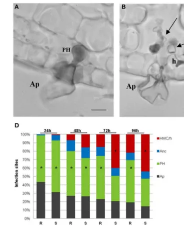
During H. vastatrix growth, after the differentiation of germ tubes and appressoria over stomata, the fungus infected both susceptible and resistant leaf tissues in a similar way, reaching in succession the stages of penetration hypha, anchor, and haustorial mother cell (HMC). The stomatal subsidiary cells were the first plant cells to be invaded by the haustoria. These specialized intracellular hyphae (responsible for fungus nutrients absorption) started to be formed between 24 and 48 hai. In the leaves of the resistant samples, the penetration hypha (Figure 1A) was the fungal growth stage observed with higher frequency during the all time-course of the experiment reaching about 55% at 24 hai and 50% at 96 hai, while HMC with haustorium (HMC/h) only reached 15% of infection sites at 72 hai, and did not exceed 22% at 96 hai (Figure 1D); at this stage the fungus stop growth and died. In the leaves of the susceptible samples, the penetration hypha was also the most representative stage at 24 hai (61%) and 48 hai (41%) but, later on, the HMC/h greatly increased in frequency (40% at 72 hai and 44% at 96 hai), being responsible for the successful fungal growth (Figures 1B,D). The death of the fungus was experimentally assessed by the autofluorescence of the fungal structures that, at 96 hai, reached
100% in the resistant samples and only 45% in the susceptible samples (data not shown).
The first cytological response induced by the fungus in the resistant and susceptible samples is the hypersensitive-like reaction (HR) observed initially in the stomata guard and subsidiary cells and later in mesophyll cells. At 24 hai, HR occurred for both resistant and susceptible samples reaching, respectively, 33 and 20% of infection sites, where the fungus stopped growth (at the stages of appressorium or penetration hypha). HR was always significantly higher in the resistant than in the susceptible samples at all time-points (Figures 1C,E). Only in the resistant samples was the HR observed in subsidiary stomatal cells and mesophyll cells invaded by haustoria, from 72 hai (65%) onwards (71% at 96 h).
APF Protein Expression upon Infection
The APF was obtained from resistant, susceptible and control leaves (mock-inoculated) along the H. vastatrix infection process (24–96 hai). Proteins were separated by 2-DE and statistical analysis of the gel patterns was performed to reveal the polypeptide spots whose volume significantly changed in abundance (p-value ≤ 0.05 and fold change > 1.5) between
FIGURE 1 | Light micrographs ofH. vastatrixinfection sites at 72 hai. (A)Appressorium (Ap) over the stomata and a penetration hypha (PH) in the resistant sample leaves, stained with cotton blue lactophenol.(B)Ap and intercellular hyphae (large arrow) in the susceptible sample leaves, stained with cotton blue lactophenol, being visible an haustorial mother cell (HMC) (small arrow) with a haustorium (h) in the stomatal subsidiary cell.(C)
Autofluorescence, by blue light epifluorescence test, of guard cells (arrows) associated with a PH in the resistant sample leaves. Note that the fungal structures are also autofluorescent. (bars=10µm).(D)Percentage of infection sites with different fungal growth stages (Ap, PH, anchor - Anc, and
HMC/h) in resistant (R) and susceptible (S) sample leaves at 24, 48, 72, and 96 hai. *=mode value. The weighted averages of the different fungal growth stages (Ap, PH, Anc, and HMC/h) were significantly higher in the S than in the R sample leaves at 24 hai (t=4.08;P≤0.001), 48 hai (t=4.47;P≤0.001),
72 hai (t=6.77;P≤0.001) and 96 hai (t=6.91;P≤0.001). (E)Percentage of infection sites with autofluorescent and/or browning cells (HR-like cell death) in R and S leaves, at 24, 48, 72, and 96 hai. The average percentages were significantly higher in the R than in the S leaves, at 24 hai (t=3.71;P≤0.001), 48 hai (t=5.49;P≤0.001), 72 hai (t=12.40;P≤0.001), and 96 hai
(t=8.83;P≤0.001).
(Figure 3B). Analyzing the changes along the infection process it is more evident that the % of proteins involved in proteolysis decreased after an initial increase (53% at 24 hai and 12% at 96 hai) while the % of proteins involved in stress/defense increased along the infection process (9% at 24 hai and 40% at 96 hai). A few proteins are present at all time-points, namely, xylosidases, mannosidases, chitinases, subtilases, and aspartic proteases.
APF Proteins Associated with Resistance and Susceptibility
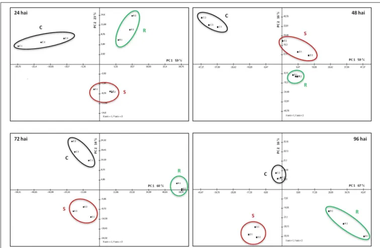
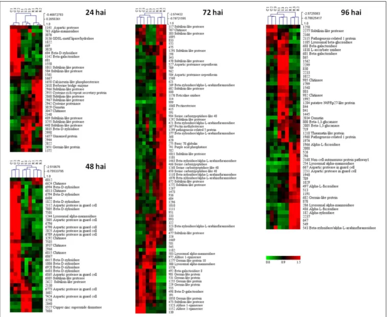
A Principal Component Analysis (PCA) was performed for the spots whose volume significantly changed in abundance during the infection. This analysis revealed a clear separation of the three samples (resistant, susceptible, and control) for each of the four time-points, the two first axes always representing more than 70% of the total variance (Figure 4 and Supplementary Table S2). To visualize the relative accumulation of the spots in the resistant (R) and the susceptible (S) samples, a hierarchical cluster analysis was performed (Figure 5). At 24 hai, the protein
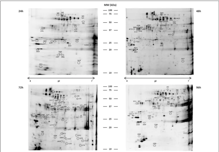
FIGURE 2 | Representative 2DE gels of coffee leaf APF proteins.
Circled spots changed significantly in abundance between samples (control, resistant, and susceptible) at 24, 48, 72, and 96 hai, and the
proteins were successfully identified by MALDI-TOF/TOF-MS (see detailed information inTable 1). Gels were stained with Ruthenium II Tris.
protein, and beta-1,3-glucanase) and beta-galactosidases were observed mostly in the S sample, at 96 hai. There is a noticeable increase in alpha-L-fucosidase proteins in the R sample at 96 hai.
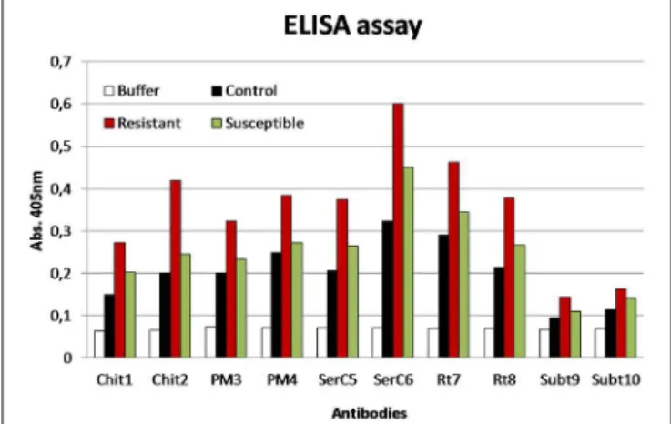
Immunodetection Assay
Some of the identified proteins, referred above, were selected as antigen for the production of antibodies, such as, chitinase, pectin methylesterase, serine carboxypeptidase, reticuline oxidase, and subtilase. Peptides corresponding to these proteins were synthesized and after conjugation with BSA and OVA allowed the production of specific antibodies. The results obtained show a higher level of detection of those proteins in the R than in the S or control samples (Figure 6).
Discussion
We have been studying the APF coffee leaf proteins in response to H. vastatrix infection (Guerra-Guimarães et al., 2009b, 2013), and recently, we have characterized the proteome of APF healthy coffee leaves (Guerra-Guimarães et al., 2014).
With the present study we complement the knowledge on the importance of the proteins present in this sub-cellular compartment, particularly in relation to pathogen defense. In addition to the proteins previously found, a further seven protein superfamilies were now identified in the APF of coffee leaves (control sample), making a total of 29 protein superfamilies. The new identified protein superfamilies are mainly PR proteins, phosphatases and oxi-reductases, highlighting the existence of an important constitutive defense mechanism in C. arabica
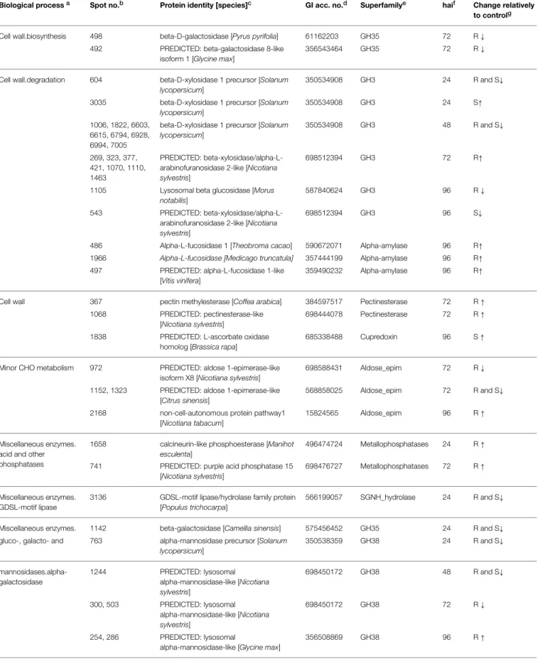
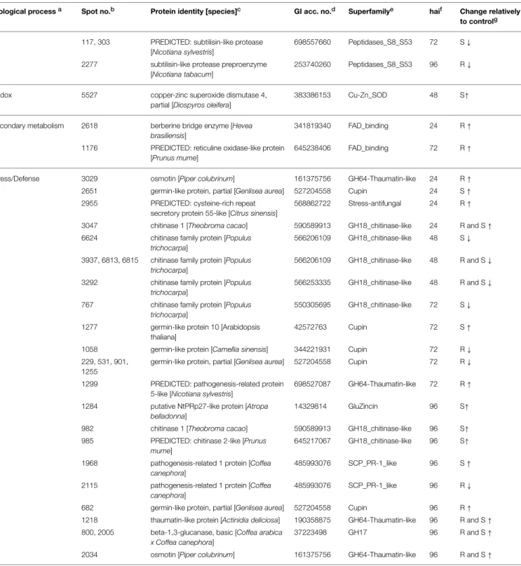
TABLE 1 | Annotation of the coffee leaf apoplastic proteins that changed in abundance along the infection process.
Biological processa Spot no.b Protein identity [species]c GI acc. no.d Superfamilye haif Change relatively
to controlg
Cell wall.biosynthesis 498 beta-D-galactosidase [Pyrus pyrifolia] 61162203 GH35 72 R↓
492 PREDICTED: beta-galactosidase 8-like isoform 1 [Glycine max]
356543464 GH35 72 R↓
Cell wall.degradation 604 beta-D-xylosidase 1 precursor [Solanum lycopersicum]
350534908 GH3 24 R and S↓
3035 beta-D-xylosidase 1 precursor [Solanum lycopersicum]
350534908 GH3 24 S↑
1006, 1822, 6603, 6615, 6794, 6928, 6994, 7005
beta-D-xylosidase 1 precursor [Solanum lycopersicum]
350534908 GH3 48 R and S↓
269, 323, 377, 421, 1070, 1110, 1463
PREDICTED: beta-xylosidase/alpha-L-arabinofuranosidase 2-like [Nicotiana sylvestris]
698512394 GH3 72 R↑
1105 Lysosomal beta glucosidase [Morus notabilis]
587840624 GH3 96 R↓
543 PREDICTED: beta-xylosidase/alpha-L-arabinofuranosidase 2-like [Nicotiana sylvestris]
698512394 GH3 96 S↓
486 Alpha-L-fucosidase 1 [Theobroma cacao] 590672071 Alpha-amylase 96 R↑
1966 Alpha-L-fucosidase [Medicago truncatula] 357444199 Alpha-amylase 96 R↑
497 PREDICTED: alpha-L-fucosidase 1-like [Vitis vinifera]
359490232 Alpha-amylase 96 R↑
Cell wall 367 pectin methylesterase [Coffea arabica] 384597517 Pectinesterase 72 R↑
1068 PREDICTED: pectinesterase-like [Nicotiana sylvestris]
698444078 Pectinesterase 72 R↑
1838 PREDICTED: L-ascorbate oxidase homolog [Brassica rapa]
685338488 Cupredoxin 96 S↑
Minor CHO metabolism 972 PREDICTED: aldose 1-epimerase-like isoform X8 [Nicotiana sylvestris]
698588431 Aldose_epim 72 R↓
1152, 1323 PREDICTED: aldose 1-epimerase-like [Citrus sinensis]
568858025 Aldose_epim 72 R and S↓
2168 non-cell-autonomous protein pathway1 [Nicotiana tabacum]
15824565 Aldose_epim 96 R↑
Miscellaneous enzymes. acid and other phosphatases
1658 calcineurin-like phosphoesterase [Manihot esculenta]
496474724 Metallophosphatases 24 R↑
741 PREDICTED: purple acid phosphatase 15 [Nicotiana sylvestris]
698476727 Metallophosphatases 72 R↑
Miscellaneous enzymes. GDSL-motif lipase
3136 GDSL-motif lipase/hydrolase family protein [Populus trichocarpa]
566199057 SGNH_hydrolase 24 R and S↓
Miscellaneous enzymes. 1142 beta-galactosidase [Camellia sinensis] 575456452 GH35 24 R and S↓
gluco-, galacto- and 763 alpha-mannosidase precursor [Solanum lycopersicum]
350538359 GH38 24 R and S↓
mannosidases.alpha-galactosidase
1244 PREDICTED: lysosomal alpha-mannosidase-like [Nicotiana sylvestris]
698450172 GH38 48 R and S↓
300, 503 PREDICTED: lysosomal alpha-mannosidase-like [Nicotiana sylvestris]
698450172 GH38 72 R↓
254, 286 PREDICTED: lysosomal
alpha-mannosidase-like [Glycine max]
356508869 GH38 96 R↑
TABLE 1 | Continued
Biological processa Spot no.b Protein identity [species]c GI acc. no.d Superfamilye haif Change relatively
to controlg
681, 688 beta-galactosidase [Camellia sinensis] 575456452 GH35 96 S↑
182 Alpha-xylosidase 1 [Theobroma cacao] 590700766 GH31 96 R↑
Protein.degradation. aspartate protease
1141 PREDICTED: protein ASPARTIC PROTEASE IN GUARD CELL 1-like [Solanum tuberosum]
565349288 pepsin_retropepsin 24 R and S↓
1657 unnamed protein product [Coffea canephora]
661898488 pepsin_retropepsin 24 S↑
6773, 7424 PREDICTED: protein ASPARTIC PROTEASE IN GUARD CELL 1-like [Solanum tuberosum]
565349288 pepsin_retropepsin 48 S↑
2005, 2025, 2112, 6789, 6790, 6865
PREDICTED: protein ASPARTIC PROTEASE IN GUARD CELL 1-like [Solanum tuberosum]
565349288 pepsin_retropepsin 48 R and S↓
275 PREDICTED: basic 7S globulin [Vitis vinifera]
225436984 pepsin_retropepsin 72 R↑
504 PREDICTED: aspartic proteinase nepenthesin-1-like [Nelumbo nucifera]
720054046 pepsin_retropepsin 72 R↑
527 PREDICTED: aspartic proteinase nepenthesin-1-like [Solanum tuberosum]
565341835 pepsin_retropepsin 72 R↑
667, 2211 PREDICTED: protein ASPARTIC PROTEASE IN GUARD CELL 1-like [Solanum tuberosum]
565349288 pepsin_retropepsin 96 R↑
Protein.degradation. cysteine protease
2942 cysteine proteinase aleuran type [Nicotiana benthamiana]
71482942 Peptidase_C1 24 R↑
Protein.degradation. serine protease
466 serine carboxypeptidase, putative [Ricinus communis]
255553418 Peptidase_S10 72 R↑
650, 984 PREDICTED: serine carboxypeptidase-like 40-like [Citrus sinensis]
568858842 Peptidase_S10 72 R↑
1268 PREDICTED: serine carboxypeptidase-like 40 isoform X1 [Vitis vinifera]
225449979 Peptidase_S10 72 R↑
Protein.degradation. Subtilases
489 subtilisin-like protease preproenzyme [Nicotiana tabacum]
253740260 Peptidases_S8_S53 24 R and S↑
504 subtilisin-like protease preproenzyme [Nicotiana tabacum]
253740260 Peptidase_S8_S53 24 R and S↓
1011 PREDICTED: subtilisin-like protease [Nicotiana tomentosiformis]
697119321 Peptidases_S8_S53 24 R and S↓
2060, 2966, 2967 PREDICTED: subtilisin-like protease [Nicotiana tomentosiformis]
697119321 Peptidases_S8_S53 24 R↑
3255 PREDICTED: subtilisin-like protease [Solanum lycopersicum]
723696627 Peptidases_S8_S53 24 R and S↑
440 PREDICTED: subtilisin-like protease [Solanum lycopersicum]
723695307 Peptidases_S8_S53 24 R and S↑
2622, 6805 PREDICTED: subtilisin-like protease [Vitis vinifera]
225458653 Peptidases_S8_S53 48 R and S↓
473 PREDICTED: subtilisin-like protease [Nicotiana tomentosiformis]
697119321 Peptidases_S8_S53 72 R↓
477 PREDICTED: subtilisin-like protease [Nicotiana tomentosiformis]
697119321 Peptidases_S8_S53 72 R and S↑
470, 602, 672, 1272, 1291, 1292
PREDICTED: subtilisin-like protease [Nicotiana tomentosiformis]
697119321 Peptidases_S8_S53 72 R↑
1011 PREDICTED: subtilisin-like protease [Nicotiana sylvestris]
698522359 Peptidases_S8_S53 72 R↑
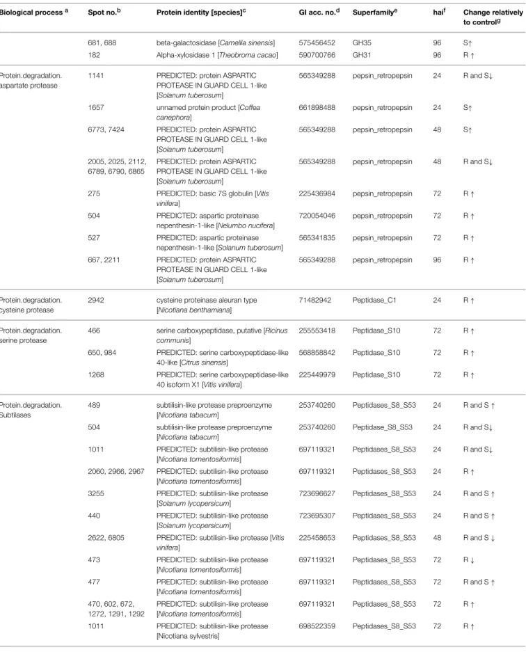
TABLE 1 | Continued
Biological processa Spot no.b Protein identity [species]c GI acc. no.d Superfamilye haif Change relatively
to controlg
117, 303 PREDICTED: subtilisin-like protease [Nicotiana sylvestris]
698557660 Peptidases_S8_S53 72 S↓
2277 subtilisin-like protease preproenzyme [Nicotiana tabacum]
253740260 Peptidases_S8_S53 96 R↓
Redox 5527 copper-zinc superoxide dismutase 4, partial [Diospyros oleifera]
383386153 Cu-Zn_SOD 48 S↑
Secondary metabolism 2618 berberine bridge enzyme [Hevea brasiliensis]
341819340 FAD_binding 24 R↑
1176 PREDICTED: reticuline oxidase-like protein [Prunus mume]
645238406 FAD_binding 72 R↑
Stress/Defense 3029 osmotin [Piper colubrinum] 161375756 GH64-Thaumatin-like 24 R↑
2651 germin-like protein, partial [Genlisea aurea] 527204558 Cupin 24 S↑
2955 PREDICTED: cysteine-rich repeat secretory protein 55-like [Citrus sinensis]
568862722 Stress-antifungal 24 R↑
3047 chitinase 1 [Theobroma cacao] 590589913 GH18_chitinase-like 24 R and S↑
6624 chitinase family protein [Populus trichocarpa]
566206109 GH18_chitinase-like 48 S↓
3937, 6813, 6815 chitinase family protein [Populus trichocarpa]
566206109 GH18_chitinase-like 48 R and S↓
3292 chitinase family protein [Populus trichocarpa]
566253335 GH18_chitinase-like 48 R and S↓
767 chitinase family protein [Populus trichocarpa]
550305695 GH18_chitinase-like 72 S↓
1277 germin-like protein 10 [Arabidopsis thaliana]
42572763 Cupin 72 S↑
1058 germin-like protein [Camellia sinensis] 344221931 Cupin 72 R↓
229, 531, 901, 1255
germin-like protein, partial [Genlisea aurea] 527204558 Cupin 72 R↓
1299 PREDICTED: pathogenesis-related protein 5-like [Nicotiana sylvestris]
698527087 GH64-Thaumatin-like 72 R↑
1284 putative NtPRp27-like protein [Atropa belladonna]
14329814 GluZincin 96 S↑
982 chitinase 1 [Theobroma cacao] 590589913 GH18_chitinase-like 96 S↑
985 PREDICTED: chitinase 2-like [Prunus mume]
645217067 GH18_chitinase-like 96 S↑
1968 pathogenesis-related 1 protein [Coffea canephora]
485993076 SCP_PR-1_like 96 S↑
2115 pathogenesis-related 1 protein [Coffea canephora]
485993076 SCP_PR-1_like 96 R↓
682 germin-like protein, partial [Genlisea aurea] 527204558 Cupin 96 R↑
1218 thaumatin-like protein [Actinidia deliciosa] 190358875 GH64-Thaumatin-like 96 R and S↑
800, 2005 beta-1,3-glucanase, basic [Coffea arabica x Coffea canephora]
37223498 GH17 96 R and S↑
2034 osmotin [Piper colubrinum] 161375756 GH64-Thaumatin-like 96 R and S↑
aFunctional characterization of the proteins based on MapMan “Bin” and GO ontology.
bThe number that identified protein spots on 2-D apoplastic gel.
cThe peptide identification based on homology to proteins characterized in different species by BLASTp. search on NCBI Viridiplantae and ESTcoffee databases.
dThe accession number from GenBank assigned to the polypeptide after MS/MS analysis.
eSuperfamily according to NCBI classification. GH, Glycoside Hydrolase; SGNH_hydrolase, diverse family of lipases and esterases; FAD_binding, flavodoxin binding oxiredutase;
GluZincin, thermolysin-like peptidases including several zinc-dependent metallopeptidases.
fhours after inoculation with H. vastatrix.
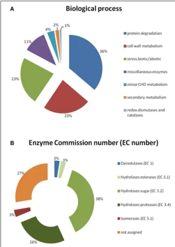
FIGURE 3 | Functional categorization of the identified coffee leaf APF proteins, based on MapMan “Bin” and GO ontology. (A)Biological process;(B)Enzyme Commission number (EC number) of the enzymes.
Initial/Basal Defense Responses
The identification of GDSL-motif lipase/hydrolase (spot #3136) and calcineurin-like phosphoesterase (spot #1658) at 24 hai, suggests the potential involvement of these proteins in pathogen perception and signal transduction cascades. GDSL esterases/lipases are proteins with multifunctional properties, described as having a role in the regulation of plant development, morphogenesis, synthesis of secondary metabolites, and defense response (Chepyshko et al., 2012). In Arabidopsis thaliana a GDSL LIPASE1 protein seems to protect plants fromAlternaria brassicicola attack in two distinct ways: by directly disrupting fungal spore integrity, and by activating defense signaling in the plants (Oh et al., 2005). In our study we have detected a decrease in the accumulation of the protein GDSL-motif lipase/hydrolase at 24 hai, in both infected tissues. According toLee et al. (2009)
such a decrease can be either a negative regulation of proteins to inhibit fungal infection/growth or, in addition, the effect of fungal interacting with the plant cell (by means of effector proteins) by suppressing the host immune system. On the other hand, the increased accumulation of calcineurin-like phosphoesterase (a calcium–dependent phosphatase) can be important in the regulation of various cellular processes with
emphasizes in signal transduction as has already been shown (Kudla et al., 1999; Luan, 2003). It is known that upon perception of microbial signals, kinases and phosphatases target specific proteins, often modifying complex signaling cascades that allow for rapid defense responses (Delanois et al., 2014). The presence of phosphatases in the extracellular proteome of Arabidopsis
infected with Pseudomonas syringae suggests that potential phosphorylation/dephosphorylation reversible regulation could occur in the apoplast (Kaffarnik et al., 2009). Moreover,Ndimba et al. (2003)have shown that chitosan treatment ofArabidopsis
cell-suspentions induced phosphorylation of a receptor-like kinase, and other proteins like chitinases and glucanases (proteins that we have also found to be accumulated at 24 h, particularly in the R samples).
The increased accumulation of PR proteins (chitinases, osmotin and a cysteine-rich repeat secretory protein) in both infected tissues (more markedly in the R samples) also indicates the induction of the basal defense responses, possibly salicylic acid (SA) regulated. Molecular studies on Coffea spp.—H. vastatrix incompatible interaction did show the activation of genes (ex.pr1b and gt) known to be involved in the SA mediating signaling pathway around 21–24 hai (Diniz et al., 2012). Furthermore, SA quantification by HPLC/ESI-MS/MS showed an increase in this signaling compound at 24 hai inCoffea
spp.—H. vastatrixincompatible interaction, suggesting again the involvement of an SA-dependent pathway in coffee resistance to CLR (Sá et al., 2014).
The accumulation of berberine bridge enzyme (a reticuline-like oxidase) in the R sample at 24 hai and a copper-zinc superoxide dismutase (SOD) in S sample at 48 hai, suggests that these “PR-like” proteins may co-regulate basal defenses. Extracellular oxidases have been suggested to catalyze the generation of reactive oxygen species (ROS), such as superoxide anions and hydrogen peroxide during the “oxidative burst” (Martinez et al., 1998; Mika et al., 2004). Indeed, previous cytochemical data in an incompatible C. arabica— H. vastatrix interaction, revealed hydrogen peroxide in the interface between the cuticle and the fungal pre-penetration structures at the infection sites (Silva et al., 2008). Furthermore, the increase in the activity of peroxidases, SOD and oxalate oxidases (germin–like proteins) have already been reported during the resistant response of coffee to CRL (Silva et al., 2006, 2008; Guerra-Guimarães et al., 2009a, 2013). The oxi-reductase activity observed during infection by pathogens indicates that plants were either initiating the production of ROS to fight directly the pathogen or responding to oxidative intermediates produced as a result of cell wall or membrane damage leading to cell death during HR response (Lee et al., 2009).
Late/Specific Defense Responses
FIGURE 4 | Principal Component Analysis (PCA) performed for the spots whose volume significantly changed in abundance (p-value<0.05), for each time-point of the infection (24–96 hai).Distinct groups were obtained per sample: control (C), resistant (R), and susceptible (S).
continued growing with no apparent inhibition, the HR stabilized and protein levels did not change much more than in the control. Most of the proteins that increased in the R sample at 72 hai have hydrolytic activity, being either involved in the cell wall metabolism (beta-xylosidase/alpha-arabinofuranosidases, chitinases and glucanase, pectin methylesterase, purple acid phosphatase, and reticuline oxidase) or in proteolysis (subtilases and serine carboxypeptidases).
It is known that plant glycohydrolases (GH) can play various important functions such as, cell wall expansion, modification during development, defense, and signaling. Since plant cell wall polysaccharides are very heterogeneous and complex polymers, GH activities must be very diverse (Jamet et al., 2008) and with our proteomic approach we identified in the apoplast a total of eight GHs superfamilies (3, 17, 18, 20, 31, 35, 38, 64). According to the carbohydrate-active enzymes database (CAZy; www.cazy.org) (Lombard et al., 2014), the GHs families GH3, GH31 and GH35 comprise enzymes that are mainly involved in the reorganization of cell wall carbohydrates. The other GHs families seem to be involved in glycoprotein post-translational modifications (PTMs), such as alpha-L-arabinofuranosidases (GH3), chitinases (GH18), beta-D-galactosidases (GH35), and alpha-D-mannosidases (GH38) (Jamet et al., 2008).
Alpha-L-arabinofuranosidases are particularly interesting since they accumulate only in the R sample at 72 hai. They are plant enzymes capable of releasing terminal arabinofuranosyl residues from cell wall matrix polymers (Saha, 2000), functioning as a candidate for a role in softening-related depolymerization of the cell wall during the HR response (Cantu et al., 2007). Several other apoplastic proteins identified, are also GHs, and appear to contribute to plant defense. Chitinases (GH18) and beta-1,3-glucanases (GH17) that are PR proteins possess antifungal activity limiting pathogen progression, and their expression is often triggered by pathogen infection (Silva et al., 2006; Guerra-Guimarães et al., 2009b).Leah et al. (1991) and
Mauch et al. (1988)showed that the antifungal proprieties of plant chitinases are enhanced when beta-1,3-glucanases were added in combination with them. In transgenic tobacco plants, susceptibility to fungal attack decreased when chitinase and glucanase genes were both over-expressed (Zhu et al., 1994). Other PR-proteins such as PR-1 and PR-5 also increased in the resistant sample from 72 hai onwards.
FIGURE 5 | Hierarchical cluster analysis of the proteins that significantly changed in abundance (p-value<0.05) between control (C), resistant (R), and susceptible (S) samples, for each
time-point of the infection (24–96 hai).The signals are shown in a red-green color scale, from a gradient of red (higher expression) to green (lower expression).
methyl esterification are critical for the outcome of plant– pathogen infections. The cell walls containing highly methyl esterified pectin are somehow protected against the action of pathogens (Lionetti et al., 2012). Concerning the PAP, it was shown that a PAP5 is required for maintaining basal resistance against Pseudomonas syringaeinArabidopsis, suggesting a role for PAP5 in pathogen triggered immunity (Ravichandran et al., 2013).
Proteolytic enzymes that are thought to be involved in maturation of enzymes, signaling, protein turnover, and defense against pathogens (Jamet et al., 2008) were the proteins that mostly changed in abundance between the R and S samples, at 72 hai. They represent 36% of the total proteins identified and belong to 4 different superfamilies; subtilisin-like protease, serine carboxypeptidase, aspartic protease, and
FIGURE 6 | ELISA assay using the antibodies produced against different proteins: chitinases (Chit), pectin methylesterase (PM), serine carboxypeptidase (SerC), reticuline oxidase (Rt), and subtilases (Subt).
Antigen samples were control, resistant, and susceptible coffee leaf extracts with 72 hai withH. vastatrix(100µg/ml).
In addition to the already referred functions of oxidases in the defense responses, it should be discussed the later increase in reticulin oxidase and germin-like proteins (oxalate oxidase-like) at 96 hai. These proteins can have a role in the oxidative cross-linking of cell wall proteins around the site of infection (Bradley et al., 1992; Silva et al., 2008). Crosslinks between phenolic compounds, the plant cell wall polysaccharides and proteins enhance the protection of the cell wall to digestion by microbial degrading enzymes and, thus, increase the global resistance to fungi (Bily et al., 2003). Deposition of chlorogenic acids and lignin has, indeed, been associated with the resistance of coffee toH. vastatrix(Silva et al., 2002, 2006; Leitão et al., 2011).
Overall, the protein changes occurring in the APF of coffee leaves upon H. vastatrixinfection indicate that cell wall reorganization, accumulation of PR proteins and excretion of hydrolytic enzymes are likely to be important defense mechanisms of coffee. The use of antibodies produced against chitinase, pectin methylesterase, serine carboxypeptidase, reticuline oxidase, and subtilase showed an increased detection of these proteins in the incompatible interaction what strengthens their involvement in the resistant response of coffee againstH. vastatix.
Conclusions
Important constitutive defense proteins were revealed in the APF ofC. arabicaleaves. Upon infection byH. vastatrix, APF
proteins were modulated establishing two distinct phases of defense responses, an initial/basal one (at 24–48 hai) and a late/specific one (at 72–96 hai). The number of proteins detected for the initial/basal phase is essentially half of the number of the proteins for the late/specific phase. When comparing the susceptible and resistant sample it was found that the increase in proteins was always greater in the resistant samples and more markedly in the late/specific phase. The resistant response involves the participation of several important groups of proteins, namely: GH of the cell wall, serine proteases (subtilases and carboxypeptidases) and PR proteins. The GHs confer great plasticity to cell wall polysaccharides, the proteases (together with phosphatases) lead to a complex regulation of cell wall proteins through PTMs and PR proteins are directly involved in antifungal activity. These results suggest that some glycohydrolases, proteases, and PR-proteins are putative candidates for resistant markers of coffee to CLR. The production of antibodies against chitinase, pectin methylesterase, serine carboxypeptidase, reticuline oxidase, and subtilase enabled the validation of the importance of these proteins in the coffee resistance response by immunodetection assay. Reliability of these putative resistant markers will be subsequently tested in several well-known coffee cultivars with commercial value. The genes corresponding to the protein biomarkers can be integrated in marker-assisted breeding programs aiming to assist in the selection of appropriate coffee genotypes with resistance toH. vastatrix.
Acknowledgments
This work was supported by Portuguese Funds through FCT (Fundação para a Ciência e a Tecnologia), under the project PTDC/AGR-GPL/109990/2009 (at CIFC/IICT, ITQB/UNL, and IHMT/UNL) and PEst-OE/EQB/LA0004/2011 (at ITQB/UNL) and GHTM – UID/Multi/04413/2013 (at IHMT/UNL). DRB was on a postdoctoral grant from CNPq (Brazil) and RT received a STSM grant from COST action FA1306. The authors wish to thank Doctor Colin E. McVey (Principal Investigator and Head of Structural Virology Lab, ITQB/UNL) for critically reviewing the manuscript.
Supplementary Material
The Supplementary Material for this article can be found online at: http://journal.frontiersin.org/article/10.3389/fpls.2015. 00478
References
Abril, N., Gion, J.-M., Kerner, R., Mueller-Starck, G., Navarro Cerrillo, R. M., Plomion, C., et al. (2011). Proteomics research on forest trees, the most recalcitrant and orphan plant species.Phytochemistry 72, 1219–1242. doi: 10.1016/j.phytochem.2011.01.005
Agrawal, G. K., Jwa, N.-S., Lebrun, M.-H., Job, D., and Rakwal, R. (2010). Plant secretome: unlocking secrets of the secreted
proteins. Proteomics 10, 799–827. doi: 10.1002/pmic.2009 00514
Alves, M., Francisco, R., Martins, I., and Ricardo, C. P. (2006). Analysis of Lupinus albus leaf apoplastic proteins in response to boron deficiency. Plant Soil 279, 1–11. doi: 10.1007/s11104-005-3154-y
(2008–2013): impacts, plausible causes and proposed solutions.Food Sec. 7, 303–321. doi: 10.1007/s12571-015-0446-9
Bendtsen, J. D., Jensen, L. J., Blom, N., von Heijne, G., and Brunak, S. (2004). Feature-based prediction of non-classical and leaderless protein secretion. Protein Eng. Des. Sel.17, 349–356. doi: 10.1093/protein/gzh037
Bettencourt, A. J., and Rodrigues, C. J. Jr. (1988). “Principles and practice of coffee breeding for resistance to rust and other diseases,” inCoffee Agronomy,Vol. 4, eds R. J. Clarke and R. Macrae (London; New York: Elsevier Applied Science Publishers LTD), 199–234.
Bily, A. C., Reid, L. M., Taylor, J. H., Johnston, D., Malouin, C., Burt, A. J., et al. (2003). Dehydrodimers of ferulic acid in maize grain pericarp and aleurone: resistance factors toFusarium graminearum.Phytopathology93, 712–719. doi: 10.1094/PHYTO.2003.93.6.712
Bradley, D. J., Kjellbom, P., and Lamb, C. J. (1992). Elicitor- and wound-induced oxidative crosslinking of a plant cell wall proline-rich protein: a novel rapid defense response.Cell70, 21–30. doi: 10.1016/0092-8674(92)90530-P Cantu, D., Vicente, A. R., Carl Greve, L., Labavitch, J. M., and Powell, A. L. T.
(2007). Genetic determinants of textural modifications in fruits and role of cell wall polysaccharides and defense proteins in the protection against pathogens. Fresh Produce1, 101–110.
Chepyshko, H., Lai, C.-P., Huang, L.-M., Liu, J.-H., and Shaw, J.-F. (2012). Multifunctionality and diversity of GDSL esterase/lipase gene family in rice (Oryza sativaL.japonica) genome: new insights from bioinformatics analysis. BMC Genomics13:309. doi: 10.1186/1471-2164-13-309
Conesa, A., and Gotz, S. (2008). Blast2GO: a comprehensive suite for functional analysis in plant genomics. Int. J. Plant Genomics 2008:619832. doi: 10.1155/2008/619832
Delanois, B., Jeandet, P., Clément, C., Baillieul, F., Dorey, S., and Cordelier, S. (2014). Uncovering plant-pathogen crosstalk through apoplastic proteomic studies.Front. Plant Sci.5:249. doi: 10.3389/fpls.2014.00249
Diniz, I., Talhinhas, P., Azinheira, H. G., Várzea, V., Medeira, C., Maia, I., et al. (2012). Cellular and molecular analyses of coffee resistance toHemileia vastatrixand nonhost resistance toUromyces vignaein the resistance-donor genotype HDT832/2.Eur. J. Plant Pathol. 133, 141–157. doi: 10.1007/s10658-011-9925-9
Doehlemann, G., and Hemetsberger, C. (2013). Apoplastic immunity and its suppression by filamentous plant pathogens.New Phytol.198, 1001–1016. doi: 10.1111/nph.12277
Eisenhaber, B., Wildpaner, M., Schultz, C. J., Borner, G. H. H., Dupree, P., and Eisenhaber, F. (2003). Glycosylphosphatidylinositol lipid anchoring of plant proteins. Sensitive prediction from sequence- and genome-wide studies for Arabidopsis and rice. Plant Physiol. 133, 1691–1701. doi: 10.1104/pp.103.023580
Emanuelsson, O., Brunak, S., von Heijne, G., and Nielsen, H. (2007). Locating proteins in the cell using TargetP, SignalP and related tools.Nat. Protoc.2, 953–971. doi: 10.1038/nprot.2007.131
Fernandez, D., Santos, P., Agostini, C., Bon, M.-C., Petitot, A.-S., Silva, M. C., et al. (2004). Coffee (Coffea arabicaL.) genes early expressed during infection by the rust fungus (Hemileia vastatrix).Mol. Plant Pathol.5, 527–536. doi: 10.1111/j.1364-3703.2004.00250.x
Fernandez, D., Tisserant, E., Talhinhas, P., Azinheira, H. G., Vieira, A., Loureiro, A., et al. (2012). 454-pyrosequencing of Coffea arabica leaves infected by the rust fungus Hemileia vastatrix revealsin plantaexpressed pathogen secreted proteins and plant functions expressed in a late compatible plant-rust interaction. Mol. Plant Pathol. 13, 17–37. doi: 10.1111/j.1364-3703.2011.00723.x
Figueiredo, A., Monteiro, F., and Sebastiana, M. (2014). Subtilisin-like proteases in plant–pathogen recognition and immune priming: a perspective.Front. Plant Sci. 5:739. doi: 10.3389/fpls.2014.00739
Floerl, S., Majcherczyk, A., Possienke, M., Feussner, K., Tappe, H., Gatz, C., et al. (2012).Verticillium longisporuminfection affects the leaf apoplastic proteome, metabolome, and cell wall properties in Arabidopsis thaliana. PLoS ONE 7:e31435. doi: 10.1371/journal.pone.0031435
Flor, H. H. (1942). Inheritance of pathogenicity inMelampsora lini.Phytopathology 32, 653–669.
Ganesh, D., Petitot, A., Silva, M. C., Alary, R., Lecouls, A. C., and Fernandez, D. (2006). Monitoring of the early molecular resistance responses of coffee (Coffea arabicaL.) to the rust fungus (Hemileia vastatrix) using real-time
quantitative RT-PCR.Plant Sci. 170, 1045–1051. doi: 10.1016/j.plantsci.2005. 12.009
Goldberg, T., Hecht, M., Hamp, T., Karl, T., Yachdav, G., Ahmed, N., et al. (2014). LocTree3 prediction of localization.Nucl. Acids Res.42, W350–W355. doi: 10.1093/nar/gku396
Grove, H., Jorgensen, B. M., Jessen, F., Sondergaard, I., Jacobsen, S., Hollung, K., et al. (2008). Combination of statistical approaches for analysis of 2-DE data gives complementary results.J. Proteome Res. 7, 5119–5124. doi: 10.1021/pr800424c
Guerra-Guimarães, L., Cardoso, S., Martins, I., Loureiro, A., Bernardes, A. S.,Varzea, V., et al. (2009a). “Differential induction of superoxide dismutase inCoffea arabica–Hemileia vastatrixinteractions,” inProceedings of the 22th International Conference on Coffee Science (ASIC2008)(Campinas).
Guerra-Guimarães, L., Silva, M. C., Struck, C., Loureiro, A., Nicole, M., Rodrigues, C. J. Jr., et al. (2009b). Chitinases ofCoffea arabicagenotypes resistant to orange rustHemileia vastatrix.Biol. Plant.53, 702–706. doi: 10.1007/s10535-009-0126-8
Guerra-Guimarães, L., Vieira, A., Chaves, I., Pinheiro, C., Queiroz, V., Renaut, J., et al. (2014). Effect of greenhouse conditions on the leaf apoplastic proteome of Coffea arabica plants. J. Proteomics 104, 128–139. doi: 10.1016/j.jprot.2014.03.024
Guerra-Guimarães, L., Vieira, A., Chaves, I., Queiroz, V., Pinheiro, C., Renaut, J., et al. (2013). “Integrated cytological and proteomic analysis ofCoffea arabica -Hemileia vastatrix interactions,” in Proceedings of the 24th International Conference on Coffee Science (ASIC2012)(San José).
Heath, M. C. (1984). Relationship between heat-induced fungal death and plant necrosis in compatible and incompatible interactions involving the bean and cowpea rust fungi.Phytopathology74, 1370–1376.
Heath, M. C. (1998). Involvement of reactive oxygen species in the response of resistant (hypersensitive) or susceptible cowpeas to the cowpea rust fungus. New Phytol.138, 251–263. doi: 10.1046/j.1469-8137.1998.00897.x
Hermanson, G. T. (2013). “Antibody modification and conjugation, Chapter 20,” inBioconjugate Techniques, 3rd Edn., (Academic Press), 880.
Jamet, E., Albenne, C., Boudart, G., Irshad, M., Canut, H., and Pont-Lezica, R. (2008). Recent advances in plant cell wall proteomics.Proteomics8, 893–908. doi: 10.1002/pmic.200700938
Jones, J. D., and Dangl, J. L. (2006). The plant immune system.Nature444, 323–329. doi: 10.1038/nature05286
Jorrín-Novo, J. V., Pascual, J., Sánchez-Lucas, R., Romero-Rodríguez, M. C., Rodríguez-Ortega, M. J., Lenz, C., et al. (2015). Fourteen years of plant proteomics reflected in proteomics: moving from model species and 2DE-based approaches to orphan species and gel-free platforms.Proteomics15, 1089–1112. doi: 10.1002/pmic.201400349
Kaffarnik, F. A., Jones, A. M., Rathjen, J. P., and Peck, S. C. (2009). Effector proteins of the bacterial pathogenPseudomonas syringaealter the extracellular proteome of the host plant,Arabidopsis thaliana.Mol. Cell. Proteomics8, 145–156. doi: 10.1074/mcp.M800043-MCP200
Krogh, A., Larsson, B., von Heijne, G., and Sonnhammer, E. L. L. (2001). Predicting transmembrane protein topology with a hidden Markov model: application to complete genomes.J. Mol. Biol.305, 567–580. doi: 10.1006/jmbi. 2000.4315
Kudla, J., Xu, Q., Harter, K., Gruissem, W., and Luan, S. (1999). Genes for calcineurin B-like proteins inArabidopsisare differentially regulated by stress signals.Proc. Natl. Acad. Sci. U.S.A.96, 4718–4723.
Lamanda, A., Zahn, A., Roder, D., and Langen, H. (2004). Improved Ruthenium II tris (bathophenantroline disulfonate) staining and distaining protocol for a better signal-to-background ratio and improved baseline resolution. Proteomics 4, 599–608. doi: 10.1002/pmic.200 300587
Leah, R., Tommerup, H., Svendsen, I., and Mundy, J. (1991). Biochemical and molecular characterization of three barley seed proteins with antifungal properties.J. Biol. Chem.266, 1564–1573.
Lee, J., Feng, J., Campbell, K. B., Scheffler, B. E., Garrett, W. M., Thibivilliers, S., et al. (2009). Quantitative proteomic analysis of bean plants infected by a virulent and avirulent obligate rust fungus.Mol. Cell. Proteomics8, 19–31. doi: 10.1074/mcp.M800156-MCP200
coffee leaf rust resistance,” inProceedings of the 24th International Conference on Coffee Science (ASIC2010)(Bali).
Lionetti, V., Cervone, F., and Bellincampi, D. (2012). Methyl esterification of pectin plays a role during plant–pathogen interactions and affects plant resistance to diseases.J. Plant Physiol.169, 1623–1630. doi: 10.1016/j.jplph.2012.05.006 Lohse, M., Nagel, A., Herter, T., May, P., Schroda, M., Zrenner, R., et al.
(2014). Mercator: a fast and simple web server for genome scale functional annotation of plant sequence data.Plant Cell Environ.37, 1250–1258. doi: 10.1111/pce.12231
Lombard, V., Golaconda Ramulu, H., Drula, E., Coutinho, P. M., and Henrissat, B. (2014). The Carbohydrate-active enzymes database (CAZy) in 2013.Nucleic Acids Res.42, D490–D495. doi: 10.1093/nar/gkt1178
Luan, S. (2003). Protein phosphatases in plants.Annu. Rev. Plant Biol. 54, 63–92. doi: 10.1146/annurev.arplant.54.031902.134743
Martinez, C., Montillet, J. L., Bresson, E., Agnel, J. P., Dai, G. H., Daniel, J. F., et al. (1998). Apoplastic peroxidase generates superoxide anions in cells of cotton cotyledons undergoing the hypersensitive reaction toXanthomonas campestris pv.MalvacearumRace 18.Mol. Plant Microbe Interact. 11, 1038–1047. doi: 10.1094/MPMI.1998.11.11.1038
Mauch, F., Mauch-Mani, B., and Boller, T. (1988). Antifungal hydrolases in pea tissue. II. Inhibition of fungal growth by combinations of chitinase and β-1,3-glucanase.Plant Physiol.88, 936–942. doi: 10.1104/pp.88.3.936 Maxemiuc-Naccache, V., Braga, M. R., and Dietrich, S. M. C. (1992). Chitinase
andβ-1,3-glucanase changes in compatible and incompatible combinations between coffee leaf disks and coffee rust (Hemileia vastatrix).Rev. Bras Bot. 15, 145–150.
Mika, A., Minibayeva, F., Beckett, R., and Luthje, S. (2004). Possible functions of extracellular peroxidases in stress-induced generation and detoxification of active oxygen species. Phytochem. Rev. 3, 173–193. doi: 10.1023/B:PHYT.0000047806.21626.49
Monteiro, F., Sebastiana, M., Pais, M. S., and Figueiredo, A. (2013). Reference genes election and validation for the early responses to downy mildew infection in susceptible and resistantVitis viniferacultivars.PLoS ONE8:e72998. doi: 10.1371/journal.pone.0072998
Ndimba, B. K., Chivasa, S., Hamilton, J. M., Simon, W. J., and Slabas, A. R. (2003). Proteomic analysis of changes in the extracellular matrix ofArabidopsiscell suspension cultures induced by fungal elicitors.Proteomics3, 1047–1059. doi: 10.1002/pmic.200300413
Neuhoff, V., Stamm, R., and Eibl, H. (1985). Clear background and highly sensitive protein staining with Coomassie blue dyes in polyacrylamide gels -a system-atic -an-alysis. Electrophoresis 6, 427–448. doi: 10.1002/elps.11500 60905
Oh, I. S., Park, A. R., Bae, M. S., Kwon, S. J., Kim, Y. S., Lee, J. E., et al. (2005). Secretome analysis reveals anArabidopsislipase involved in defence against Alternaria brassicicola.Plant Cell17, 2832–2847. doi: 10.1105/tpc.105.034819 Petersen, T. N., Brunak, S., von Heijne, G., and Nielsen, H. (2011). SignalP 4.0:
discriminating signal peptides from transmembrane regions.Nat. Methods8, 785–786. doi: 10.1038/nmeth.1701
Pierleoni, A., Martelli, P. L., and Casadio, R. (2008). PredGPI: a GPI anchor predictor.BMC Bioinformatics9:392. doi: 10.1186/1471-2105-9-392 Pinheiro, C., Guerra-Guimarães, L., David, T. S., and Vieira, A. (2014). Proteomics:
state of the art to study Mediterranean woody species under stress.Environ. Exp. Bot.103, 117–127. doi: 10.1016/j.envexpbot.2014.01.010
Ramagli, L. S. (1999). Quantifying protein in 2-D PAGE solubilization buffers. 2-D Proteome Analysis Protocols.Methods Mol. Biol.112, 99–103. doi: 10.1385/1-59259-584-7:99
Ramirez, V., Lopez, A., Mauch-Mani, B., Gil, M., and Vera, P. (2013). An extra-cellular subtilase switch for immune priming inArabidopsis.PLoS Pathog. 9:e1003445. doi: 10.1371/journal.ppat.1003445
Ravichandran, S., Stone, S. L., Benkel, B., and Prithiviraj, B. (2013). Purple acid phosphatase5 is required for maintaining basal resistance againstPseudomonas syringaeinArabidopsis.BMC Plant Biol.13:107. doi: 10.1186/1471-2229-13-107
Rijo, L., Rodrigues, C. J. Jr., Silva, M. C., and Vasconcelos, M. I. (1991). Does gene SH5confer to certain coffee-rust associations a reaction near immunity?
A histopathological study.Café Cacao ThéXXXV, 167–176.
Rodrigues, C. Jr., Bettencourt, A., and Rijo, L. (1975). Races of the pathogen and resistance to coffee rust. Annu. Rev. Phytopathol. 13, 49–70. doi: 10.1146/annurev.py.13.090175.000405
Rojas, M. L., Montes de Gómez, V., and Ocampo, C. A. (1993). Stimulation of lipoxygenase activity in cotyledonary leaves of coffee reacting hypersensitively to the coffee leaf rust. Physiol. Mol. Plant Pathol. 43, 209–219. doi: 10.1006/pmpp.1993.1051
Sá, M., Ferreira, J. P., Queiroz, V. T., Vilas Boas, L., Silva, M. C., Almeida, M. H., et al. (2014). A liquid chromatography-electrospray tandem mass spectrometry method for the simultaneous quantification of salicylic, jasmonic, and abscisic acid inCoffea arabicaleaves.J. Sci. Food Agric.94, 529–536. doi: 10.1002/jsfa.6288
Saha, B. C. (2000).α-L-Arabinofuranosidases: biochemistry, molecular biology and application in biotechnology. Biotechnol. Adv. 18, 403–423. doi: 10.1016/S0734-9750(00)00044-6
Schulze-Lefert, P., and Panstruga, R. (2003). Establishment of biotrophy by parasitic fungi and reprogramming of host cells for disease resistance.Annu. Rev. Phytopathol. 41, 641–667. doi: 10.1146/annurev.phyto.41.061002.083300 Silva, M. C., Guerra-Guimarães, L., Loureiro, A., and Nicole, M. R.
(2008). Involvement of peroxidases in the coffee resistance to orange rust (Hemileia vastatrix). Physiol. Mol. Plant Pathol. 72, 29–38. doi: 10.1016/j.pmpp.2008.04.004
Silva, M. C., Nicole, M., Guerra-Guimarães, L., and Rodrigues, C. J. Jr. (2002). Hypersensitive cell death and post-haustorial defence responses arrest the orange rust (Hemileia vastatrix) growth in resistant coffee leaves.Physiol. Mol. Plant Pathol.60, 169–183. doi: 10.1006/pmpp.2002.0389
Silva, M. C., Nicole, M., Rijo, L., Geiger, J. P., and Rodrigues, C. J. Jr. (1999). Cytochemistry of plant-rust fungus interface during the compatible interaction Coffea arabica(cv.Caturra)-Hemileia vastatrix(race III).Int. J. Plant Sci.160, 79–91. doi: 10.1086/314113
Silva, M. C., Várzea, V. M., Guerra-Guimarães, L., Azinheira, H., Fernandez, D., Petitot, A. S., et al. (2006). Coffee resistance to the main diseases: leaf rust and coffee berry disease.Braz. J. Plant Physiol.18, 119–147. doi: 10.1590/S1677-04202006000100010
Vartapetian, A., Tuzhikov, A., Chichkova, N., Taliansky, M., and Wolpert, T. (2011). A plant alternative to animal caspases: subtilisin-like proteases.Cell Death Differ.18, 1289–1297. doi: 10.1038/cdd.2011.49
Várzea, V. M. P., and Marques, D. V. (2005). “Population variability ofHemileia vastatrixvs coffee durable resistance,” inDurable Resistance to Coffee Leaf Rust, eds L. Zambolim, E. Zambolim, and V. M. P. Várzea (Viçosa: Universidade Federal de Viçosa), 53–74.
Voegele, R. T., and Mendgen, K. (2003). Rust haustoria: nutrient uptake and beyond.New Phytol. 159, 93–100. doi: 10.1046/j.1469-8137.2003.00761.x Zhu, Q., Maher, E. A., Masoud, S., Dixon, R. A., and Lamb, C. J. (1994). Enhanced
protection against fungal attack by constitutive co-expression of chitinase and glucanase genes in transgenic tobacco.Nat. Biotechnol.12, 807–812. doi: 10.1038/nbt0894-807
Conflict of Interest Statement: The authors declare that the research was conducted in the absence of any commercial or financial relationships that could be construed as a potential conflict of interest.