Vol.46, n. 3 : pp. 469-477, June 2003
ISSN 1516-8913 Printed in Brazil
%5$=,/,$1$5&+,9(62)
%,2/2*<$1'7(&+12/2*<
$ 1 , 1 7 ( 5 1 $ 7 , 2 1 $ / - 2 8 5 1 $ /
(OLPLQDWLRQ RI 7XUELGLW\ ,QWHUIHUHQFH LQ 6HUXP ,URQ
&RORULPHWULF$VVD\E\(Q]\PDWLF3URWHRO\VLV
/HRQDUGR0&DUGRVR
∗0LOWRQ+*GH$QGUDGH
1'HRFOpFLR$&KLDQFD-XQLRU
10DUFHOR
(XVWiTXLR6LOYD
2DQG0DULD/~FLD3HGURVD
1'HSWRGH&LrQFLDV%LROyJLFDV1~FOHRGH3HVTXLVDHP&LrQFLDV%LROyJLFDV,&(%8QLYHUVLGDGH)HGHUDOGH2XUR
3UHWR2XUR3UHWR0LQDV*HUDLV%UD]LO'HSWRGH$OLPHQWRV(VFRODGH1XWULomR8QLYHUVLGDGH
)HGHUDOGH2XUR3UHWR2XUR3UHWR0LQDV*HUDLV%UD]LO
$%675$&7
:H GHVFULEH D PRGLILFDWLRQ LQ WKH FRPPHUFLDO FRORULPHWULF PHWKRG IRU WKH GHWHUPLQDWLRQ RI VHUXP LURQ E\ XVLQJ )HUUR]LQH 7KH PRGLILFDWLRQ ZDV SURSRVHG EHFDXVH GXULQJ WKH FRQYHQWLRQDO SURFHGXUH WXUELGLW\ REVHUYHG ZKHQ WKHVHUXPRIDQLPDOVVXEPLWWHGWRVXUJHU\ZDVXVHGLQWHUIHUHGZLWKWKHDVVD\:HDGGHGWRWKHRULJLQDOPHWKRGD SUHYLRXVWUHDWPHQWRIWKHVHUXPZLWKSURWHRO\WLFHQ]\PHV7KLVPRGLILFDWLRQZDVDOVRWHVWHGXVLQJSODVPDVDPSOHV DOWKRXJK WKLV ZDV QRW UHFRPPHQGHG ZKHQ WKH RULJLQDO PHWKRG ZDV XVHG 7KH UHVXOWV GHPRQVWUDWHG WKDW D WKH WUHDWPHQWZLWKDPL[WXUHRIWU\SVLQDQGFK\PRWU\SVLQZDVHIIHFWLYHLQRUGHUWRHOLPLQDWHWXUELGLW\EWKHUHZDVQR GLIIHUHQFHEHWZHHQWKHVWDQGDUGFXUYHVREWDLQHGE\WKHFRQYHQWLRQDODQGWKHPRGLILHGPHWKRGIRUFRQWURODVVD\VF WKHDEVRUEHQFLHVRIWKHVDPSOHVRIVHUXPDQGSODVPDVXEPLWWHGWRSURWHRO\VLVHVWLPDWHGE\WKHDGGLWLRQRIGLIIHUHQW FRQFHQWUDWLRQVRILURQZHUHGLUHFWO\SURSRUWLRQDOWRLURQFRQFHQWUDWLRQVGWKHSUHWUHDWPHQWZLWKHQ]\PHVDOORZHG WKHXWLOL]DWLRQRISODVPDHWKHSUHWUHDWPHQWZLWKJXDQLGLQH+&OZDVQRWHIIHFWLYH
.H\ZRUGV: Iron assay, enzymatic proteolysis, interference
∗ Author for correspondence
,1752'8&7,21
The Ferrozine dye-binding method, performed without prior deproteinization is largely used to determine the iron concentration in serum samples. Although extensively used both in clinical and research laboratories, factors such as lyphemia, hemolysis (Valcour et al., 1990), and the presence of anti-coagulant in samples (Bothwell et al., 1979) interfere with the reaction. Turbidity, resulting from precipitation of fibrinogen, has also been described in samples from hemodialysis patients (Ooi et al., 1992). In evaluating the circulating iron levels in laboratory rats submitted to surgical trauma, turbidity was observed in the
reaction medium, making the dosage impossible. The phenomenon was observed in anticoagulant free samples but it was more intense when heparin was given to the animals during the surgical process.
In the present study we used enzymatic proteolysis of plasma and serum samples and compared the results to those obtained with commercially available kits.
0$7(5,$/$1'0(7+2'6
0DWHULDOV Commercial colorimetric kits for
Labtest Diagnostica S.A. and Bioclin Química Básica (Belo Horizonte, MG, Brazil). Both contain buffer 250 mmol/L, pH 4.0 with hydroxylamine 144 mmol/L; iron standard solution 100 µg/dL and Ferrozine 28 mmol/L. Porcine trypsin (activity: 1,870 units Nα-Benzoyl-L-arginine Ethyl Ester/mg) and bovine chymotrypsin (52 units N-Benzoyl-L-tyrosine Ethyl Ester/mg protein), Sigma Co. Tris/HCl buffer 100 mmol/L, pH 8.1, containing Tris (hydroxymethil) aminomethane, Sigma Co. and HCl 100 mmol/L (Merck S.A). Guanidine.HCl was obtained from Merck S.A.
6DPSOHVSerum or plasma samples were obtained from male Fisher rats from three different groups: 1 – Serum from five cannulated animals (CA serum): Under anesthesia with 25 mg/Kg 2,2,2 -tribromoethanol (Aldrich Chemical Company, Inc. Milwaukee, WI USA), a cannula was inserted into the abdominal aorta through the femoral artery for measurement of pulsate arterial pressure. A second cannula was inserted into femoral vein for drug administration (Chianca-Jr and Machado, 1994). Twenty four hours after surgery, the blood was collected through the cannula inserted in the abdominal aorta, in a glass syringe and transferred to polyethylene tubes. After 15 minutes, the blood was centrifuged at 14,000 rpm for 8 min in an Eppendorf 5410 microcentrifuge. The serum was then separated, recentrifuged under the same conditions for the removal of the remaining cells and pooled;
2 – Serum samples of 9 non-cannulated animals (NCA serum): Before blood collection, these animals were anesthetized with 25 mg/Kg 2,2,2 -tribromoethanol (Aldrich Chemical Company, Inc. Milwaukee, WI USA). The aorta artery was exposed and sectioned and the blood was placed in a glass tube. After 15 minutes, the blood was centrifuged at 3,000 rpm in an Excelsa Baby 206 model centrifuge (Fanem), for 10 minutes. The serum was separated, placed in polyethylene tubes and centrifuged at 14,000 rpm for 8 min in an Eppendorf centrifuge and subsequently pooled; 3 – Plasma of 9 cannulated animals (CA plasma): The animals received 20µg of heparin in the systemic circulation 24h after the surgical trauma and 90 minutes before the blood collection. The blood was collected through the cannula inserted in the abdominal aorta in a glass syringe, and
for 8 min in an Eppendorf 5410 microcentrifuge. The plasma was then separated, recentrifuged under the same conditions for the removal of the remaining cells and pooled. All samples were stored at 4oC and assayed within 6 days.
3ULQFLSOH Ferric iron is dissociated from
transferrin in an acid buffer (pH 4.0, 250 mmol/L) and it is reduced to ferrous state by hydroxylamine. The ferrous ion forms a colored complex with Ferrozine [3-(2-pyridyl)-5,6-bis (4-phenyl-sulfonic acid)-1,2,4triazine; Amax 560nm]
with an absorbance that is proportional to the concentration of iron in the sample (Stookey, 1970).
&RQWURO DVVD\ ± DX[LOLDU\ DEVRUEDQFH $ DQG ILQDODEVRUEDQFH$ Pool serum or plasma, 250
µL; water, 40 µL; Tris/HCL buffer 100 mmol/L, pH 8.1, 200 µL were dispensed into tubes, mixed and incubated for 2 h at 37°C. After addition of 1000 µL of kit’s buffer the absorbance of the mixture and standard (250µL of iron standard solution 100 µg/dL) were measured against blank (250 µL of water) at 560 nm. This reading was referred as absorbance A1. Then, 30 µL of
ferrozine were added, mixed and incubated for 10 min at 37oC. The absorbance of sample and standard were read against blank at 560 nm (absorbance A2). Iron concentration was calculated
from the difference between A2 and A1.
*XDQLGLQH DVVD\ Guanidine was added to the kit’s buffer in order to achieve a final concentration of 1.6 mol/L in the reaction mixture. To 250 µL of plasma or serum, 1000 µL of guanidine-containing buffer and 240 µL of water were added. After 20 minutes incubation at 20°C the tubes were centrifuged at 14,000 rpm for 15 min. A second set of tubes was prepared as described, except for the fact that the samples were incubated at 37°C for 20 minutes after the addition of the guanidine buffer. The supernatants were used for the determination of A1 and A2 as
described before. Standard curves similar to the ones described for the proteolytic treatment were prepared for this treatment.
$EVRUEDQFH VSHFWUXP This was performed with
the products from the assays with the non cannulated animal serum samples submitted to proteolysis and control assays. Both were submitted to screenings between 360 and 800 nm, in a Shimadzu UV-1601 spectrophotometer.
&DOFXODWLRQV
I – Iron concentration taken from the assay standard and sample absorbance:
G/ J G/ J $ $ )H
6 100 / /
]
[ ×
µ
=µ
∆ = Where:
[)H] = iron concentration ∆$ A2 − A1
$V= standard absorbance at 560 nm.
II – Iron recovery:
Where:
5 = percentage of iron recovered;
[)H] = iron concentration in the sample with addition of iron standard solution, calculated from the absorbance; [)H] = iron concentration in the sample without addition of iron standard solution, calculated from the absorbance;
[)H]H = expected iron concentration in each sample of serum and plasma.
6WDWLVWLFDO DQDO\VLV Statistical analysis was performed by variance analysis (ANOVA) and results are shown as mean ± standard deviation.
5(68/76
Preliminary observations from our laboratory showed that the utilization of commercially available kits for iron determination with serum from cannulated animals (CA) resulted in the development of turbidity in the reaction tube that interfered with the assay (data not shown). Assay of serum and plasma samples from CA showed turbidity in the reaction medium right after the addition of the iron dissociating buffer. Plasma samples showed more intense turbidity in the final result (A2) than serum samples. This phenomenon
was not observed in the serum from non cannulated animals (NCA)
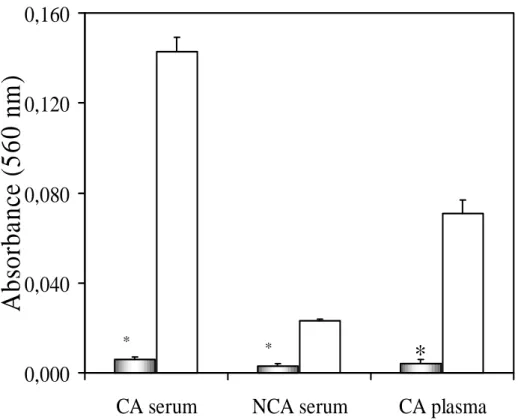
$X[LOLDU\ DEVRUEDQFH $Samples from CA
serum, right after the acid buffer addition, showed significant turbidity in the reaction resulting in high values of auxiliary absorbance (A1). In
contrast, samples of the same serum treated with enzymes showed lower values for A1. The values
were 0.143 ± 0.006 for the control assay and 0.006 ± 0.001 for the enzymes treated one (fig 1). With serum samples from NCA, the control assays showed relatively high values for A1 (fig 1) when
compared to the ones in which the proteolysis was performed: 0.023 ± 0.001 and 0.003 ± 0.001, respectively. Turbidity was noted in plasma samples submitted to the control assay after the addition of the iron dissociating buffer, while no detectable turbidity was noted in treated samples: 0.071 ± 0.006 and 0.004 ± 0.002, respectively) as shown in figure 1. Thus, treatment of serum or plasma samples with proteolytic enzymes significantly decreases the absorbance values for A1.
(
)
100
]
[
]
[
]
[
%
0×
)LJXUH Auxiliary absorbance at 560 nm, without ferrozine, (A1) for serum samples from cannulated
animals (CA serum), serum samples from non cannulated animals (NCA serum) and cannulated animals plasma (CA Plasma); gray columns show the mean ± SD (n = 5 ) of assays with proteolytic treatment and the white ones show the mean ± SD (n = 5) for control assays; significant differences in relation to the control assay: * P<0,05 (ANOVA).
6WDQGDUGFXUYHV Serum samples generated linear curves in the iron concentration range interval used in the experiments. Nevertheless, some differences were noticed in the behavior of these samples in the assay. Fig 2 shows the iron standard curves from CA serum samples. The absorption coefficient in the assays with proteolysis was slightly lower than the control: 1.6×10⁻³ (µg/dL)
-1×
cm-1 and 1.8×10⁻³ (µg/dL)-1× cm-1, respectively. Fig 3 shows the standard curves from NCA serum samples where there was no significant difference between the curves from control and proteolysis assays. Regarding the plasma samples (figure 4), without proteolysis the turbidity is increased after ferrozine addition and incubation for 10 minutes at 37°C. The differences were evident concerning the absorption coefficient, 2.6 times higher in the proteolysis assay compared to the control assay, respectively 3.6×10⁻³ and 1.4×10⁻³ (µg/dL)-1× cm-1. On the other hand, the serum samples treated with
0.089) and non incubated (1.530 ± 0.085) samples when compared with the control assay (0.143 ± 0.006). The centrifugation at 14,000 rpm was not able to remove the turbidity; therefore, the interference problem in CA samples persisted. The standard curve obtained from these assays, although linear, showed that in heated samples the iron concentration was much lower than the expected value, with an absorption coefficient of 5×10⁻⁴ (µg/dL)-1×cm-1. Iron concentration in non heated samples fell above the expected, with an absorption coefficient of 2.2×10⁻³ (µg/dL)-1×cm-1.
$EVRUEDQFH VSHFWUXP Fig 6 shows the
absorbance spectrum after the proteolysis and control assays. This profile showed a coincidence of absorbance peaks in the wavelength of 560 nm. This indicated that the treatment did not promote spectrum deviations within the wavelength interval in which absorbance in this assay is measured.
0,000
0,040
0,080
0,120
0,160
CA serum
NCA serum
CA plasma
A
b
so
rb
a
n
ce
(5
6
0
n
m
)
*
)LJXUH Iron standard curve from CA serum samples were determined by increasing iron concentrations and its respective mean values ± SD (n=2) of corrected absorbance (A1 were subtracted
from the respective A2 values and ∆A values of serum sample without iron standard solution); the lines
were obtained by linear regression of points referring to proteolysis assays, \ [, 5 () and control assay, \ [±, 5 (−−−−).
,URQUHFRYHU\ The percentage of iron recovery is shown in Fig 7. In the control assay performed with plasma samples it reached 370 ± 104%, while in the sample submitted to proteolysis this value was 117 ± 3%, much closer to the expected. With serum samples of cannulated animal, the assays showed a recovery of added iron close to 100%, both for control and modified assay (108 ± 2% and 112 ± 4%, respectively). In the guanidine assays, an iron recovery percentage of 191 ± 19% for non heated and 41 ± 6% for heated ones was observed.
',6&866,21
All serum samples of cannulated animals produced turbidity without previous proteolysis, when colorimetric assay was used. However, for non cannulated animals, serum samples not present turbidity.
0.000
0.050
0.100
0.150
0.200
0
20
40
60
80
100
120
Expected Iron (
µ
g/dL)
A
b
so
rb
a
n
ce
(5
6
0
n
m
)LJXUH Iron standard curve obtained from NCA serum samples were determined by increasing iron concentrations and its respective mean values ± SD (n=2) of corrected absorbance (A1 values were
subtracted from the respective A2 values and ∆A values of serum sample without iron standard solution);
the lines were obtained by linear regression of points referring to proteolysis assay, y = 0.0019x + 0.0005, R2 = 0.9992 () and control assay, y = 0.0019x + 0.0055, R2 = 0.9996 (−−−−).
)LJXUH - Iron standard curve obtained from CA plasma samples were determined by increasing iron concentration and its respective mean values ± SD (n=2) of corrected absorbance (A1 values subtracted
0.000
0.100
0.200
0.300
0.400
0
20
40
60
80
100
120
Expected Iron (
µ
g/dL)
A
b
so
rb
a
n
ce
(
560
n
m
)
0.00 0
0.05 0
0.10 0
0.15 0
0.20 0
0.25 0
0
20
40
6 0
8 0
10 0
120
E xpected Iron (
µ
g /dL)
Ab
so
rb
a
n
ce
(
5
6
0
nm
)LJXUH Iron standard curve from NCA serum samples; after guanidine treatment (1.6 mol/L, final concentration in the mixture), the supernatant was analyzed by iron; lines obtained by linear regression of referring points to assays treated with guanidine and incubated at 37°C, y = 0.0005x + 0.0010, R2 = 0.9979 (ρ), only guanidine treatment, y = 0.0022x − 0.0052, R2 = 0.9954 ( − −) and control assay, y = 0.0018x − 0.0020, R2 = 0.9988 (− −−−). Indicated points were determined by increasing iron concentration and its respective mean values ± SD (n=2) for corrected absorbance (A1 values
subtracted from respective A2 values and ∆A values of serum sample without 100 µg/dL iron standard
solution).
)LJXUH Spectrum scanning performed with samples of proteolytic () and control assays (−−−); assays with NCA serum pool samples. Scanned interval from 360 nm to 800 nm.
0.000
0.050
0.100
0.150
0.200
0.250
0
20
40
60
80
100
12 0
Expected Iron (
µ
g/dL)
Ab
so
rb
anc
e (
5
6
0
n
m
)
0.00 0
0.05 0
0.10 0
0.15 0
0.20 0
0.25 0
360
4 20
4 80
5 40
6 00
6 60
72 0
78 0
W avelen gth (n m )
Ab
so
rb
a
n
)LJXUH - Recovery rate of iron added to serum samples of cannulated animals (AC Serum), non cannulated animals serum (NCA Serum) and cannulated animals plasma (AC Plasma); columns represent means ± SD of four measurements that determined iron standard curve for each assay type. AC represents cannulated animals and NCA represents non cannulated animals; significant difference in relation to control assay: * P< 0.05.
In consequence, the A1 values were higher in
serum CA and plasma samples. Ooi et al (1992) found abnormally increased iron concentrations in serum samples from hemodialysis patients. According to the authors the combination of increased fibrinogen synthesis, commonly seen in renal patients on hemodialysis, and using heparin during the dialysis would lead to the persistence of fibrinogen in serum. On the other hand, the pH of the iron-dissociating buffer is close enough to the pI of fibrinogen to decrease the solubility of the protein, creating turbidity. However, our results with guanidine treatment was not effective. It is possible that other proteins would appear due to the surgical process. In acute aggressions hepatocytes decrease albumin synthesis and increase C reactive protein, ceruloplasmin, α -1-antitrypsin, transferrin, fibrinogen, haptoglobin components of complement (McPherson, 1996). Furthermore, cannulated animals A values
we analyzed the iron recover results (Fig. 7) we observed a tremendous increase in iron recuperation for plasma compared with the serum. At the end of the assay, the plasma sample-buffer mixture was greater than serum sample-buffer mixture. These results indicated that the turbidity of plasma samples were detained. As a matter of fact, Ooi et al (1992) reported that low heparin concentrations retard fibrinogen precipitation in the mixture buffer/sample.
The assay without previous proteolysis offered restrictions in samples of cannulated animals, essential procedure in our experiments. The removal of proteins from samples by TCA precipitation (Williams et al., 1977) was avoided due to the possibility of co-precipitation of the iron. As proposed by Ooi et al. (1992) the iron determination in serum of cannulated animals after previous treatment with guanidine was tried. Our results showed that this procedure did not avoid 0
100 200 300 400 500
CA serum NCA serum CA plasm a
%
of
Iron
R
e
c
o
ve
ry
Control assay Proteolysis assay
Guanidine + Heating Only guanidine
*
*
*
referent of assays did not show coherent absorbance levels for expecting iron concentrations. As a consequence, the iron recovery rates were much higher than expected, due to the medium turbidity, or much lower than the expected due to carrying of iron to the precipitate.
Our results demonstrated that the previous treatment of plasma samples with proteases was effective. In serum samples of cannulated and non cannulated animals, our results also showed good linearity for standard and recovery of external standard curves. The absorbance spectrum showed peak coincidence in the wavelength of interest (560 nm) assured the quality of the result. The significant reduction of A1 values both in plasma
as well as in serum samples permitted the suppression of these auxiliary absorbance readings. Our data also revealed that serum samples of non cannulated animals, without previous enzymatic treatment, showed diminished iron concentration when compared to the treated ones. One possible explanation for these contrast results could be a low auxiliary absorbance values. In conclusion, the enzymatic proteolysis treatment became useful for both plasma and serum of cannulated animals in case of iron determination by colorimetric assays. Besides that our results have shown a decreased of number of steps and lower possibilities of errors due to precipitation interference.
5(6802
Descreveu-se modificação no método colorimétrico comercial para dosagem de ferro sérico que utiliza Ferrozine como reagente de cor. A modificação foi proposta porque durante o procedimento observava-se turvação quando o soro de animais submetidos à cirurgia era utilizado, comprometendo os resultados. Foi acrescentado ao método um tratamento prévio do soro com enzimas proteolíticas. Avaliou-se também a modificação utilizando amostras de plasma, o que não é recomendado na metodologia original. O tratamento das amostras com cloreto de guanidina, descrito anteriormente para amostras de pacientes submetidos à hemodiálise, também foi
avaliado. Os resultados demonstraram que: a) tratamento das amostras com tripsina e quimotripsina eliminou a turvação; b) não houve diferenças entre as curvas padrão obtidas pelo método original ou modificado para amostras de soro provenientes de animais controle; c) as absorbâncias das amostras de soro e plasma submetidas à proteólise foram proporcionais às concentrações de ferro, estimada pela adição de diferentes concentrações do íon; d) o tratamento enzimático permitiu a utilização de plasma; e) o tratamento prévio dos soros, de animais submetidos à cirurgia, com guanidina. HCl, não foi
eficaz.
5()(5(1&(6
Bothwell, T. H.; Charlton, R. W. and Cook, J. D. (1979), Measuremente of the plasma iron concentration, In ,URQ PHWDEROLVP LQ PDQ. Oxford: Blackwell Scientific Publication. pp. 353-362. Chianca-Jr D. A. and Machado B. H. (1994), The
sensitivity of the Bezold-Jarisch reflex is increased in rats with sinoaortic deafferentation. %UD] - 0HG %LRO 5HV., , 775-781.
McPherson, R. A. (1996), Specific Proteins.
In-&OLQLFDO 'LDJQRVLV DQG 0DQDJHPHQW E\ /DERUDWRU\
0HWKRGV ed. 19th Henry, J. B. W.B.. Saunders
Company, Philadelphia. pp. 237-252.
Ooi D. S.; Perkins S. L. and Tokessy, N. E. (1992), Elimination of fibrinogen interference in a dye-binding method for iron. &OLQ&KHP., , 541-544. Stookey, L. L. (1970), Ferrozine – a new
spectrophotometric reagent for iron. $QDO&KHP., , 779-782.
Valcour, A. A.; Krzymowski, G.; Onoroski, M.; Bowers Jr., G. N. and McComb, R. B. (1990), Proposed reference method for iron in serum used to evaluate two automated iron methods. &OLQ &KHP., , 1789-1792.
Williams, H. L.; Johnson, D. J. and Haut, M. J. (1977), Simultaneous spectrophotometry of Fe2+ and Cu2+ in serum denatured with guanidine hydrochloride. &OLQ
&KHP., , 237-240.