Trypanosoma cruzi
ribosomal protein S4: characterization of its
coding locus, analysis of transcripts, and antigenicity of the protein
Mariana Pérez-Escobar, Ana María Cevallos/
+, Bertha Espinoza*, Norma Espinosa,
Ignacio Martínez*, Roberto Hernández
Departamento de Biología Molecular y Biotecnología *Departamento de Inmunologia, Instituto de Investigaciones Biomédicas, Universidad Nacional Autónoma de México, Apartado postal 70-228 CP 04510, México, D. F., México
Two allelic genomic fragments containing ribosomal protein S4 encoding genes (rpS4) from Trypanosoma cruzi (CL-Brener strain) were isolated and characterized. One allele comprises two complete tandem repeats of a sequence encoding an rpS4 gene. In the other, only one rpS4 gene is found. Sequence comparison to the accessed data in the genome project database reveals that our two-copy allele corresponds to a variant haplo-type. However, the deduced aminoacid sequence of all the gene copies is identical. The rpS4 transcripts pro-cessing sites were determined by comparison of genomic sequences with published cDNA data. The obtained sequence data demonstrates that rpS4 genes are expressed in epimastigotes, amastigotes, and trypomastigotes. A recombinant version of rpS4 was found to be an antigenic: it was recognized by 62.5% of the individuals with positive serology for T. cruzi and by 93.3% of patients with proven chronic chagasic disease.
Key words: kinetoplastid - trypanosomatids - Trypanosoma cruzi - ribosome - gene structure
Trypanosoma cruzi is a protozoan parasite that causes Chagas disease, an endemic infection in South and Central America that affects approximately 11 mil-lion people (Guzman-Bracho 2001). T. cruzi species has been classified into two major well defined genomic groups: T. cruzi I and T. cruzi II (Souto et al. 1996). T. cruzi I has been associated with the sylvatic transmis-sion cycle and infection of marsupials whereas T. cruzi
II has been associated with the domestic transmission cycle and infection of placental mammals. The natural history of the disease involves three different phases. The initial acute infection (symptomatic in only 1% of the cases) is characterized by febrile symptoms and infla-mmation at the site of entry of the parasite. This stage is followed by an indeterminate (subacute) stage in which symptoms are not apparent. Finally, after a period of years to decades, the chronic stage of the infection may manifest itself in up to 30% of individuals. A variety of clinical presentations may be seen including digestive tract anomalies (megaesophagus and megacolon) and cardiac enlargement and malfunction (Teixeira 1987). The variability of symptoms has yet to be correlated with specific parasite or host genetic markers, although it is likely that both will affect the outcome of infection.
The ribosomal protein S4 (rpS4) is a basic type pro-tein located at the interface between ribosomal subunits (Uchiumi et al.1986). In agreement with this is the
ob-servation that the rpS4 can be cross-linked to the eu-karyotic initiation factor eIF-3 (Westermann et al.1983). Our early interest in this protein lies with report in which the yeast ribosomal protein S7, homologous to the mam-malian rpS4 was found to be an essential protein (Synetos et al.1992). From the sequence analysis of two epimas-tigote cDNA clones it was deduced that T. cruzirpS4 is a 273 amino acid, conserved basic protein (Hernández et al. 1998). The gene is expressed as a 1 kb transcript and the initial hybridization studies were consistent with the occurrence of two alleles as predicted in a diploid organism. Moreover, Southern blot analysis of pulse field chromosomal gels demonstrated that the genes were present in two homologous chromosomes of different sizes (Hernández et al. 1998).
T. cruzi ribosomes are known to elicit both humoral and cellular immune responses (Teixeira & Santos-Buch 1974, 1975). Furthermore, T. cruzi ribosomesand riboso-mal proteins have been associated with the induction of heart dysfunction in experimental models similar to that seen in patients with chronic disease (Teixeira et al.1975). The acidic ribosomal P proteins have been found to be anti-genic, and the humoral immune responses to these pro-teins have been implicated in the pathogenesis of chagasic chronic heart disease (Elies et al. 1996, Kaplan et al. 1997, Lopez Bergami et al. 2001). However, the role of other ribosomal antigens has not been evaluated.
In addition to a detailed characterization of the rpS4
encoding locus this work evaluates the antigenicity of this basic type ribosomal protein.
MATERIALS AND METHODS
Parasites - T. cruzi II (CL-Brener strain) epimas-tigotes kindly donated in 1995 by Dr Bianca Zingales (Universidade de São Paulo) have been maintained at 27°C in liver-infusion tryptose medium supplemented with 10% new-born calf serum.
+Corresponding author: [email protected] MP-E is CONACyT scholarship
Cloning of the rpS4 genes of T. cruzi - Genomic DNA from our T. cruzi II (CL-Brener strain) was used to construct an EcoR I-digested λZAP II (Stratagene) li-brary (Cevallos et al. 2003). The lili-brary was screened using the EcoR I - Kpn I rpS4 encoding fragment from the T. cruzi I (Tulahuen strain) cDNA clone pS4-2 (Hernandez et al. 1998). Five positive clones were iden-tified which contained two different inserts 8.5 and 7.5 Kb in size. Two clones p811 (with an 8.5 Kb insert) and p812 (with a 7.5 Kb insert) were selected for further study. The regions containing the homologous sequences were subcloned and both strands sequenced by the dye-terminator method on an ABI-PRISM 310 automated se-quencer (Applied Biosystems).
Expression and purification of a recombinant rpS4 and recombinant GST - The entire rpS4 open reading frame (ORF) was amplified from the genomic clone p812 by PCR with primers containing BamH I restriction sites (S4-GST-F 5'-GGGATCCCCATGACCAAGAAGCAC-CTG-3' and S4-GST-R 5'-GGGATCCTATTTTCGTGC-CTTGCG-3'). The amplification product was subcloned in phase into the BamH I site of the bacterial expression vector pGEX-3X (Promega, Madison, Wisconsin), se-quenced and expressed as a GST (glutathione S-trans-ferase)-rpS4 fusion protein in Escherichia coli (BL21 strain). E. coli transformed with the recombinant plas-mid was cultured at 37°C until the optical density reached 0.5 at 600 nm, and the synthesis of recombinant rpS4
was induced with 1 mM isopropylthio-β-galactoside at 37°C for 4 h. The resulting culture was harvested by centrifugation at 12,000 × g for 10 min at 4°C and incu-bated in lysozyme containing buffer [100 µg/ml in 20 mM Tris-HCl, pH 7.5, 1 mM ethylenediaminetetraa-cetic acid (EDTA)] for 30 min at 4°C prior to sonica-tion. A soluble fraction was obtained by centrifugation at 12,000 × g for 10 min at 4°C. Phosphate buffered saline pH 7.2 (PBS, 10 ×) and Triton X-100 were added to the soluble fraction to a final concentration of 1% Triton X and 1 × PBS. The lysate was loaded into a glu-tathione Sepharose 4B column, and the recombinant pro-tein eluted according to the manufacturer’s instructions (Amersham Biosciences, London, UK). The size and purity of the recombinant protein was verified with the use of SDS-PAGE low molecular weight markers (BioRad Laboratories). Sodium dodecyl sulfate-poly-acrylamide gel electrophoresis (SDS-PAGE) were per-formed at room temperature in gels (10 or 12%) con-taining 0.1% SDS, as previously described (Laemmli 1970). Samples were dissolved in sample buffer [1:1 (v/ v); 62.5 mM Tris-HCl pH 6.8, 25% glycerol, 2% SDS, 0.01% bromophenol blue] and the electrophoresis con-duced in running buffer (25 mM Tris pH 8.8, 192 mM glycine, 0.1% SDS).
Since several contaminant proteins remained in the eluted fraction, a second purification was required. The recombinant protein preparations were resolved by SDS-PAGE (10%) and the band of 57 kDa was cut from the gel and electroeluted from it. After dialysis of the eluted protein in 10 mM Tris-HCl pH 8; 100 mM NaCl, 10% glycerol at 4°C, the preparations were concentrated by
ultra filtration in Centricon 30 filter units (Millipore Corporation, Bedforf, US) and stored at –20oC for fur-ther analysis. The GST protein used as a control was ex-pressed directly from the pGEX-3X vector and purified as described above.
Human sera - One hundred human sera obtained with the approval of the Ethical Committee of the Instituto Nacional de Cardiología (México, D. F.), were tested for antibodies against the rpS4 purified recombinant pro-tein. Thirty two sera from infected asymptomatic indi-viduals (indeterminate phase of Chagas disease) previ-ously characterized as seropositive by reactivity to T. cruzi crude antigens in two different serological tests [ELISA (cut off value of positive sera was ≥ 0.5 of OD at 490 nm) and Western blot]; the details of the standard-ization of the assays have been published before (Sánchez et al. 2001). Fifteen sera were obtained from chronic cardiomyopathy chagasic patients with different degrees of electrographic alteration and positive serology to three serological tests (ELISA, Western blot, and indi-rect immunofluorescence). Fifty three samples from serologically negative healthy individuals were used as controls (Rangel-Flores et al. 2001). Sera that were posi-tive to recombinant rpS4 were also tested against puri-fied recombinant GST protein to demonstrate that the antigenic recognition was against the rpS4 portion of the fusion protein and not against the GST portion.
Western blot - Total proteins from T. cruzi, purified recombinant S4 and GST proteins were resolved by SDS-PAGE (12%) and transferred to 0.45 µm nitrocellulose membranes (BioRad Laboratories, California, US) in transfer buffer (25 mM Tris-HCl pH 8.3, 0.19M glycine and 20% methanol), as previously described (Towbin et al. 1979). Western blots were performed as previously described (Sánchez et al. 2001). Briefly, nitrocellulose membranes were blocked with 10% skimmed milk in PBS (16 h at 4ºC). Strips were cut and then individually incubated (2 h at 37ºC) with 1 ml human serum diluted 1:500 in 10% skimmed milk in PBS. Each strip was washed three times with PBS/0.1% Tween 20 and incu-bated with peroxidase-conjugated anti-human IgG (di-luted 1:10 000) for 2 h at room temperature. After wash-ing, the reaction was developed with 0.5 mg/ml of diaminoben-zidine in PBS with 0.02% H2O2. The reac-tion was stopped by the addireac-tion of water. The presence and correct transference of the recombinant protein was verified by probing with an anti-GST antibody (Affinity BioReagents, Inc.). Sera previously identified as reac-tive and non-reacreac-tive to crude T.cruzi antigens were in-cluded in the testing of each membrane. Sera that were positive to recombinant rpS4 were subsequently tested against purified recombinant GST protein to demonstrate that the antigenic recognition was against the rpS4 por-tion of the fusion protein and not against the GST porpor-tion.
RESULTS
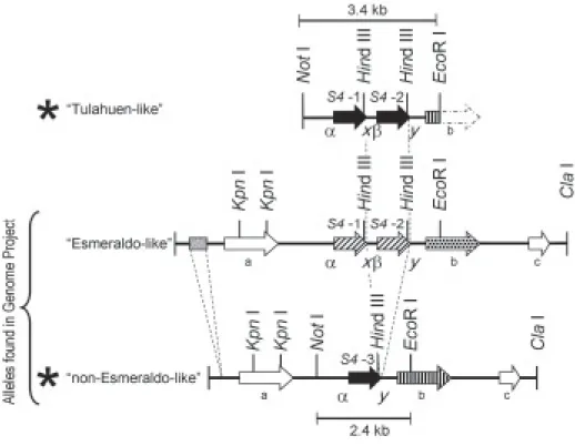
allelic fragments (Hernández et al. 1998). Restriction analysis and Southern hybridizations of the digested clones delimited, in both clones, Not I - EcoR I fragments recognized by the rpS4 cDNA probe. These two regions differed in fragment size: 3.4 kb in one clone and 2.4 kb in the other. Both fragments were sequenced and the nucleotide data was deposited in the GenBank (acces-sion nos. DQ288964 and DQ288965). Sequence analy-ses showed that the difference in size between the two fragments was due to the presence of two rpS4 gene copies in the 3.4 kb fragment (S4-1 and S4-2) and one copy (S4- 3) in the 2.4 kb fragment (see Fig. 1, alleles marked with an asterisk). At the nucleotide level the DNA sequence of both alleles was identical with the excep-tion of a 1072 bp DNA inserexcep-tion in the two-copy allele containing a 250 bp intergenic region and the extra rpS4
gene copy (see the “Tulahuen-like” allele in Fig. 1). Se-quence comparison with our previously reported rpS4
cDNA clones from the T. cruzi I Tulahuen strain
(Gen-Bank accession nos. AF005421 and AF005904), dem-onstrated that the three coding regions of our CL-Brener strain were identical to both of them. In particular, the
rpS4-2 gene copy present in our 3.4 kb cloned genomic fragment is not only identical in its coding region but also in its non-coding untranslated regions to the Tulahuen derived rpS4 cDNA clone. Therefore we have named this allele as “Tulahuen-like”.
The T. cruzi genome project was carried out in the CL-Brener strain which proved to be a genetic hybrid. The alleles in many cases have been named “Esmeraldo-like” and “non-Esmeraldo-“Esmeraldo-like” haplotypes according to their suspected progenitors. When our rpS4 coding re-gion was used to search the T. cruzi Genome Database (http://www.genedb.org/genedb/tcruzi/blast.jsp), two contigs containing rpS4 genes were identified. Our one copy allele corresponds to the “non-Esmeraldo-like” haplotype. On the other hand, our two copy allele did not correspond in sequence to the accessed
“Esmeraldo-Fig. 1: map of the ribosomal protein S4 genomic locus. At the bottom are depicted the two alleles present in the CL-Brener strain accessed in the Genome Project Database. At the top is represented the variant allele “Tulahuen-like” found in our CL-Brener strain that replaces the “Esmeraldo-like” allele reported in the Genome Project Database. Our second allele corresponds to a “non-Esmeraldo-like”. The asterisks mark the two alleles present in our CL-Brener strain and the size of the sequenced fragments are indicated. Arrows represent open reading frames and the shaded box represents a SIRE element. The coding regions for the three copies of the rpS4 gene are labeled as S4-1, S4-2 and S4-3. The rest of the ORFs are labeled with a letter and encode for a Thiamine pyrophosphokinase (a), a nucleobase transporter (b), and for a hypothetical protein (c). There are two insertions in the “Esmeraldo-like” allele not present in the “non-Esmeraldo-like” allele: a SIRE element and an extra copy of the rpS4 gene with the corresponding intergenic region (marked with dashed lines).
Differences in arrow patterns represent differences in the nucleotide sequences of the respective ORF. The rpS4 genes present in the “Tulahuen-like” and in the “non-Esmeraldo-like” alleles (solid arrows) are identical among them but have eight conserved nucleotide differences when compared to the
like” haplotype even though this allele also contains two copies of the rpS4 gene. Differences were found both in the coding and non-coding regions as mentioned in legend to Fig. 1.
A further analysis of the accessed genomic context of the rpS4 genes detected the presence of a short in-terspersed repetitive element (SIRE) as defined by Vazquez et al. (1994) in the “Esmeraldo-like” allele (Fig.1). In addition, three ORFs with the same orienta-tion are found in both accessed alleles (Fig. 1 a, b, c). Gene b (a nucleobase transporter) is different in the “Esmeraldo like” hapotype as detailed in Fig. 1.
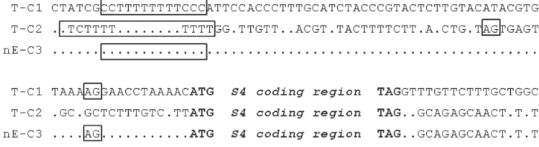
Characterization of rpS4 mRNA processing sites -Productive expression of protein coding genes in trypa-nosomes involve the addition of a trans-spliced capped mini exon (39 nt) next to the 5' UTR, and polyadenylation of the 3' terminus within individual mRNAs. These two reactions occur within the intergenic regions of a pri-mary polycistronic mRNAs transcript and are coupled and directed by a common polypyrimidine tract, which is part of the spliced leader acceptor site. Disruption of this bifunctional signal sequence affects the expression of 5′ and 3′ adjacent flanking genes (Lopez-Estrano et al. 1998, Hummel et al. 2000). Sequence analysis of non coding regions of the rpS4 genes demonstrated that the 5' upstream region of copies 1 and 3 was identical but significantly different from the 5´ upstream sequence of copy 2 (Fig. 1; indicated as α and β). The sequences downstream of the translation stop codon for copies 2 and 3 was similar, and in turn dissimilar to the sequence present downstream of rpS4 gene copy 1 (Fig. 1;
indi-cated as x and y). These differences result in the pres-ence of three types of transcripts that can be readily iden-tified when analyzing cDNA sequence data and allowed us to characterize our previously reported Tulahuen strain derived cDNA clones as copy 2 transcripts.
To determine the sites at which transcript process-ing occurs for the other two gene copies, a search was performed to identify cDNA clones accessed in the NCBI and EMBL databases. A total of 32 rpS4 cDNA clones were identified, 21 derived from T. cruzi epi-mastigotes, 6 from amastigotes and 5 from trypo-mastigotes. The comparison of these sequences with the genomic DNA sequence allowed us to determine the sites where transplicing and polyadenylation occur in each copy (see Fig. 2).
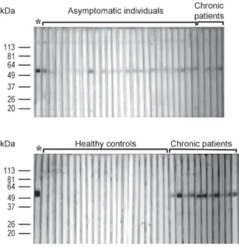
Antigenicity of ribosomal protein S4 - With the aid of a recombinant form of T. cruzi rpS4 we inferred the antibody response to the natural form of this protein in three well characterized groups: patients with chagasic chronic cardiomyopathy, asymptomatic individuals with two positive serological tests and blood bank donors (Fig. 3). Antibodies against rpS4 were common in patients with known exposure to the parasite: 20 out of 32 as-ymptomatic individuals (62.5%) and 14 out of 15 pa-tients with established cardiomyopathy (93.3%). There were no false positives as none of the 53 sera negative for T. cruzi (healthy blood bank donors) was reactive to
rpS4. To analyze the potential recognition of the GST frag-ment of the recombinant protein, all rpS4 positive sera were also tested for antibodies against the GST peptide. None of these sera were GST positive (data not shown).
Fig. 2: ribosomal protein S4 genes, flanking sequences, and mapping of mRNA processing sites. Genomic sequences for the three rpS4 gene copies are aligned (C1- C2, and C3) with their inferred progenitor origin denoted with a T, for a “Tulahuen-like” or nE for “non-Esmeraldo-like”. The rpS4
DISCUSSION
The taxon T. cruzi contains two well defined genomic groups T. cruzi I and T. cruzi II. T. cruzi I has been asso-ciated with the sylvatic transmission cycle and infection of marsupials. T. cruzi II consists of five related sub-groups, termed IIa, IIb, IIc, IId, and IIe, and has been as-sociated with the domestic transmission cycle and in-fection of placental mammals.The T. cruzi strain CL-Brener is a member of subgroup IIe and was selected for the genome sequence project because it was well char-acterized (El Sayed et al. 2005). Although T. cruzi lin-eages have mostly evolved in a clonal fashion, several studies have reported the existence of recombinant geno-types in this species, either in natural populations (Bo-gliolo et al. 1996, Carrasco et al. 1996, Machado & Ayala 2001) or in the laboratory (Gaunt et al. 2003). In vitro, studies have demonstrated that T. cruzi has an extant ca-pacity for genetic exchange. In one report, meticulous quantification analyses demonstrated that biological clones of a single T. cruzi strain had between 30 and 70% more DNA than the parental stock indicating some rapid genetic mechanism for radical change in DNA content (McDaniel & Dvorak 1993). In another report two bio-logical clones were passaged together through the en-tire life cycle and then recovered from the mammalian stage of the life cycle. Comparison of the progeny dem-onstrated fusion of parental genotypes, loss of alleles, homologous recombination, and uniparental inheritance of kinetoplast maxicircle DNA (Gaunt et al. 2003). Not-withstanding these findings, the recombination events
do not seem to be frequent enough as to disrupt the preva-lent clonal pattern of the population (Telleria et al. 2004).
T. cruzi CL-Brener, the strain used in this study, is known to be heterozygous at many loci, with different-sized homologous chromosome pairs. It is believed that its genome is a hybrid from subgroup IIb and subgroup IIc (which itself is also apparently a hybrid derived from
T. cruzi I) (El Sayed et al. 2005). The presence of variant alleles within the same T. cruzi subgroup and the pres-ence of two distinct sequpres-ence classes representative of different subgroups within particular strains has already been reported (Westenberger et al. 2005). In this work we have cloned and sequenced two alleles containing the
rpS4 genes in a laboratory maintained CL-Brener strain. Sequence analyses demonstrated that one of these frag-ments corresponds to a different allele from those de-scribed in the genome project, and that this variant is probably related to a T. cruzi I-Tulahuen strain. The find-ing of T. cruzi I sequences in the CL-Brener strain further supports the proposal of multiple progenitors in the evolu-tion of this T. cruzi hybrid strain (El Sayed et al. 2005).
We have also identified the processing sites of rpS4
primary transcripts by comparing the genomic sequences with published cDNA data. Analyses of the untranslated regions demonstrate that each transcript is different, but can be grouped into two types according to the 3' UTR (copy 1 and copies 2/3). It has been demonstrated that differences in the 3' UTR of β-tubulin mRNAs result in differences in mRNA stability of the specific transcripts in trypomastigotes and amastigotes (Bartholomeu et al. 2002). A working hypothesis would propose that the dis-tinct 3' UTR in the rpS4 transcripts may participate differ-entially in the physiology of expression of this protein.
Finally, it is described here that rpS4 is recognized by two thirds of individuals with positive serologic re-sponse to T. cruzi antigens and more than 90% of pa-tients with proven chagasic disease. There were no false positives as none of the control sera recognized the an-tigen. The prevalence of reactivity to various T. cruzi
proteins in patients with chronic symptomatic chagasic disease by Western blot (Sánchez et al. 2001) has been reported. In this study it was found that the prevalence of reactivity to specific protein bands varied from 23 to 100%. However, only two proteins of 32 and 42 kDa were recognized by 90% of the patients or more.
T. cruzi ribosomes are known to be immunogenic. However the majority of studies have been directed at proteins that elicit the production of autoantibodies. Antibodies against T. cruzi ribosomal P proteins have been shown to cross react with the systemic lupus erythe-matosus ribosomal P protein epitope (Mesri et al. 1990, Levitus et al. 1991, Aznar et al. 1995). Fine epitope map-ping demonstrated that these pathogenic antibodies are directed to the acidic portions of their carboxyl-termi-nal regions. These antibodies also react with the acidic epitope of the second extracellular loop of the β 1-adr-energic receptor stimulating it (Elies et al. 1996, Kaplan et al. 1997, Lopez Bergami et al. 2001), an effect that contributes to the pathogenesis of chagasic cardiomy-opathy. It has also been shown that the T. cruzi riboso-Fig. 3: Western blots against rpS4. Gel purified rpS4 (2.5 µg) was loaded
mal protein L27 has an epitope that cross-reacts with the Sm-epitope present in small nuclear ribonucleopro-teins (Perone et al. 2003). Approximately two thirds of patients with Chagas disease have antibodies against the Sm epitope present in both human and trypanosomal small nuclear ribonucleoproteins (Bach-Elias et al. 1998). The role of these antibodies in the pathogenesis of the disease remains to be determined.
Analysis of expressed sequence tags from T. cruzi
amastigotes, the reproductive stage in humans, has shown that 9% clones of the cDNA library encoded for riboso-mal proteins with more than 30 classes of ribosoriboso-mal pro-teins being identified (Cerqueira et al. 2005). Interest-ingly, an immunosurvey of the same library with sera from patients with chagasic disease revealed that ribo-somal proteins also represent the largest class of anti-gen coding anti-genes expressed in amastigotes (DaRocha et al. 2002). However only a subset of the ribosomal pro-teins expressed appeared antigenic. Besides the previ-ously reported ribosomal P proteins, the ribosomal pro-teins L19 and L7a were also found to be antigenic.
In Leishmania the rpS4 antigen (with a 91% identitity with T. cruzi rpS4) was identified using parasite-specific T cell lines derived from an immune donor and has been proposed as a vaccine candidate (Probst et al. 2001). Further studies should establish the value of rpS4 anti-gen as a diagnostic tool or as a vaccine candidate.
ACKNOWLEDGMENTS
To Dr Imelda López Villaseñor for the critical reading of the manuscript and Juliana Herrera for technical assistance.
REFERENCES
Aznar C, Lopez-Bergami P, Brandariz S, Mariette C, Liegeard P, Alves MD, Barreiro EL, Carrasco R, Lafon S, Kaplan D, Levitus G, Levin MJ 1995. Prevalence of anti-R-13 antibod-ies in human Trypanosoma cruzi infection. FEMS Immunol Med Microbiol12: 231-238.
Bach-Elias M, Bahia D, Teixeira DC, Cicarelli RM 1998. Pres-ence of autoantibodies against small nuclear ribonucleopro-tein epitopes in Chagas’ patients’ sera. Parasitol Res23: 796-799.
Bartholomeu DC, Silva RA, Galvao LM, el-Sayed NM, Donelson JE, Teixeira SM 2002. Trypanosoma cruzi: RNA structure and post-transcriptional control of tubulin gene expression.
Exp Parasitol102: 123-133.
Bogliolo AR, Lauria-Pires L, Gibson WC 1996. Polymorphisms in Trypanosoma cruzi: evidence of genetic recombination.
Acta Trop61: 31-40.
Carrasco HJ, Frame IA, Valente SA, Miles MA 1996. Genetic exchange as a possible source of genomic diversity in syl-vatic populations of Trypanosoma cruzi. AmJ Trop Med Hyg54: 418-424.
Cerqueira GC, DaRocha WD, Campos PC, Zouain CS, Teixeira SM 2005. Analysis of expressed sequence tags from Try-panosoma cruzi amastigotes. Mem Inst Oswaldo Cruz100: 385-389.
Cevallos AM, López-Villaseñor I, Espinosa N, Herrera J, Hernández R 2003. Trypanosomacruzi: Allelic compari-sons of the actin genes and analysis of their transcripts.
Exp Parasitol103: 27-34.
DaRocha WD, Bartholomeu DC, Macedo CD, Horta MF, Cunha-Neto E, Donelson JE, Teixeira SM 2002. Characterization of cDNA clones encoding ribonucleoprotein antigens ex-pressed in Trypanosoma cruzi amastigotes. Parasitol Res 88: 292-300.
Elies R, Ferrari I, Wallukat G, Lebesgue D, Chiale P, Elizari M, Rosenbaum M, Hoebeke J, Levin MJ 1996. Structural and functional analysis of the B cell epitopes recognized by anti-receptor autoantibodies in patients with Chagas’ disease. J Immunol157: 4203-4211.
El-Sayed NM, Myler PJ, Bartholomeu DC, Nilsson D, Aggarwal G, Tran AN, Ghedin E, Worthey EA, Delcher AL, Blandin G, Westenberger SJ, Caler E, Cerqueira GC, Branche C, Haas B, Anupama A, Arner E, Aslund L, Attipoe P, Bontempi E, Bringaud F, Burton P, Cadag E, Campbell DA, Carrington M, Crabtree J, Darban H, da Silveira JF, de Jong P, Edwards K, Englund PT, Fazelina G, Feldblyum T, Ferella M, Frasch AC, Gull K, Horn D, Hou L, Huang Y, Kindlund E, Klingbeil M, Kluge S, Koo H, Lacerda D, Levin MJ, Lorenzi H, Louie T, Machado CR, McCulloch R, McKenna A, Mizuno Y, Mottram JC, Nelson S, Ochaya S, Osoegawa K, Pai G, Par-sons M, Pentony M, Pettersson U, Pop M, Ramirez JL, Rinta J, Robertson L, Salzberg SL, Sanchez DO, Seyler A, Sharma R, Shetty J, Simpson AJ, Sisk E, Tammi MT, Tarleton R, Teixeira S, Van Aken S, Vogt C, Ward PN, Wickstead B, Wortman J, White O, Fraser CM, Stuart KD, Andersson B 2005. The genome sequence of Trypanosoma cruzi, etio-logic agent of Chagas disease. Science309: 409-415. Gaunt MW, Yeo M, Frame IA, Stothard JR, Carrasco HJ, Taylor
MC, Mena SS, Veazey P, Miles GA, Acosta N, de Arias AR, Miles MA 2003. Mechanism of genetic exchange in Ameri-can trypanosomes. Nature421: 936-939.
Guzman-Bracho C 2001. Epidemiology of Chagas disease in Mexico: an update. Trends Parasitol17: 372-376. Hernandez R, Palacios S, Herrera J, Martinez-Calvillo S, Lopez
I 1998. The deduced primary structure of a ribosomal pro-tein S4 from Trypanosoma cruzi. Biochim Biophys Acta 1395: 321-325.
Hummel HS, Gillespie RD, Swindle J 2000. Mutational analysis of 3' splice site selection during trans-splicing. J Biol Chem 275: 35522-35531.
Kaplan D, Ferrari I, Bergami PL, Mahler E, Levitus G, Chiale P, Hoebeke J, Van Regenmortel MH, Levin MJ 1997. Antibod-ies to ribosomal P proteins of Trypanosoma cruzi in Chagas disease possess functional autoreactivity with heart tissue and differ from anti-P autoantibodies in lupus. Proc Natl Acad Sci USA94: 10301-10306.
Laemmli UK 1970. Cleavage of structural proteins during the assembly of the head of bacteriophage T4. Nature 270: 680-685.
Levitus G, Hontebeyrie-Joskowicz M, Van Regenmortel MH, Levin MJ 1991. Humoral autoimmune response to riboso-mal P proteins in chronic Chagas heart disease. Clin Exp Immunol 85: 413-417.
Lopez Bergami P, Scaglione J, Levin MJ 2001. Antibodies against the carboxyl-terminal end of the Trypanosoma cruzi riboso-mal P proteins are pathogenic. FASEB J15: 2602-2612. Lopez-Estrano C, Tschudi C, Ullu E 1998. Exonic sequences in
Machado CA, Ayala FJ 2001. Nucleotide sequences provide evidence of genetic exchange among distantly related lin-eages of Trypanosoma cruzi. Proc Natl Acad Sci USA98: 7396-7401.
McDaniel J P, Dvorak J A 1993. Identification, isolation, and characterization of naturally-occurring Trypanosoma cruzi
variants. Mol Biochem Parasitol57: 213-222.
Mesri EA, Levitus G, Hontebeyrie-Joskowicz M, Dighiero G, Van Regenmortel MH, Levin MJ 1990. Major Trypanosoma cruzi antigenic determinant in Chagas’ heart disease shares homology with the systemic lupus erythematosus ribosomal P protein epitope. J Clin Microbiol28: 1219-1224. Perone D, Santos MA, Peixoto MS, Cicarelli RM 2003.
Trypa-nosoma cruzi: identification and characterization of a novel ribosomal protein L27 (TcrL27) that cross-reacts with an affinity-purified anti-Sm antibody. Parasitology126: 577-583.
Probst P, Stromberg E, Ghalib HW, Mozel M, Badaro R, Reed SG, Webb JR 2001. Identification and characterization of T cell-stimulating antigens from Leishmania by CD4 T cell ex-pression cloning. J Immunol166: 498-505.
Rangel-Flores H, Sánchez B, Mendoza-Duarte J, Barnabé C, Breniére SF, Ramos C, Espinoza B 2001. Serological and parasitological demonstration of Trypanosoma cruzi infec-tions in an urban central area of Mexico: Correlation with electrocardiographic alterations. Am J Trop Med Hyg65: 887-895.
Sánchez B, Monteón V, Reyes P, Espinoza B 2001. Standarization of ELISA and Western blot for detection of Trypanosoma cruzi. Antibodies using extracts of Mexican strains as anti-gens. Concordance between laboratories. Arch Med Res 32: 382-388.
Souto RP, Fernandes O, Macedo AM, Campbell DA, Zingales B 1996. DNA markers define two major phylogenetic lineages of Trypanosoma cruzi. Mol Biochem Parasitol 83:141-152. Synetos D, Dabeva MD, Warner JR 1992. The yeast ribosomal
protein S7 and its genes. J Biol Chem267: 3008-3013.
Teixeira AR, Santos-Buch CA 1974. The immunology of experi-mental Chagas’ disease. I. Preparation of Trypanosoma cruzi antigens and humoral antibody response to their anti-gens. J Immunol113: 859-869
Teixeira AR, Santos-Buch CA 1975. The immunology of experi-mental Chagas’ disease. II. Delayed hypersensitivity to Try-panosoma cruzi antigens. Immunology28: 401-410. Teixeira AR, Teixeira ML, Santos-Buch CA 1975. The
immu-nology of experimental Chagas’ disease. IV. Production of lesions in rabbits similar to those of chronic Chagas’ disease in man. Am J Pathol80: 163-180.
Teixeira ARL 1987. The Stercorarian trypanosomes. In EJL Soulsby, Immune Responses in Parasitic Infections: Im-munology, Immunopathology and Immunoprophylaxis, CRC Press, Boca Raton, FL, p. 25-118.
Telleria J, Barnabe C, Hide M, Banuls AL, Tibayrenc M 2004. Predominant clonal evolution leads to a close parity between gene expression profiles and subspecific phylogeny in Try-panosoma cruzi. Mol Biochem Parasitol137: 133-141. Towbin H, Staehelin T, Gordon J 1979. Electrophoretic transfer
of proteins from polyacrylamide gels to nitrocellulose sheets: procedure and some applications. Proc Natl Acad Sci USA 76: 4350-4354.
Uchiumi T, Kikuchi M, Ogata K 1986. Cross-linking study on protein neighborhoods at the subunit interface of rat liver ri-bosomes with 2-iminothiolane. J Biol Chem261: 9663-9667. Vazquez MP, Schijman AG, Levin MJ 1994. A short interspersed repetitive element provides a new 3' acceptor site for trans-splicing in certain ribosomal P2 beta protein genes of Trypa-nosoma cruzi. Mol Biochem Parasitol64: 327-336. Westenberger SJ, Barnabe C, Campbell DA, Sturm NR 2005.
Two hybridization events define the population structure of Trypanosoma cruzi. Genetics171: 527-543.