CHARACTERIZATION OF THE GENE PRODUCTS
Xiuli Yi 1,2; Yan Shi 1,2; Hui Xu 3; Wei Li 1,2; Jie Xie 1,2; Rongqing Yu 1,2; Jun Zhu 1,2; Yi Cao 1,2; Dairong Qiao 1,2*
1
College of Life Science, Sichuan University, Chengdu 610064, China; 2Microbiology and Metabolic Engineering key Laboratory of Sichuan Province, Chengdu 610064, China; 3Sichuan BioShine Biotechnology Co. Ltd., Chengdu 610064, China.
Submitted: May 11, 2009; Returned to authors for corrections: October 07, 2009; Approved: February 18, 2010.
ABSTRACT
The analysis of individual gene product should enable to clarify the role of a particular enzyme in a
complex xylanase system of A. niger. The two genes encoding precursors of co-produced endo-1,4-
-D-xylanases, xynA1 and xynB, were isolated from Aspergillus niger SCTCC 400264 (SCTCC, China) by
using RT-PCR technique and then successfully expressed in Escherichia coli BL21. The nucleotide
sequences of the xynA1 and xynB genes revealed that they were only 52.5% homology to each other.
Characterization of the recombinant enzymes revealed the different properties: the specific activity of
recombinant XYNA1 was 16.58 U/mg compared to 1201.7 U/mg for recombinant XYNB; The optimum
temperature and pH of the recombinant XYNA1 were 35 ºC and 3.0, respectively, whereas the
corresponding values for the recombinant XYNB were 55 ºC and 5.0, respectively; The recombinant
XYNB showed much more thermostability than recombinant XYNA1; The recombinant XYNB showed
94% of maximal activity after incubating in water for 60 min at 60 ºC compared to no activity for
recombinant XYNA1. Various metal ions had different effects on activity between the two recombinant
xylanases.
Key words:Aspergillus niger; xylanase; prokaryotic expression; enzymatic characterization
INTRODUCTION
Xylan, as the most abundant hemicellulose, accouts for
more than 30% of the dry weight of terrestrial plants and
represents the major renewable carbon resource in nature (6,
11). Xylanolytic enzymes are a group of enzymes that
hydrolyze xylan and arabinoxylan polymers, of which the most
important are the endo-1,4- -xylanases (EC 3.2.1.8) (25).
These enzymes degrade xylan to shortchain
xylo-oligosaccharides varying lengths. As the applications of these
enzymes are valuable in many biotechnological processes, they
gain significant attention, and play important roles in the
animal feed, pulp and paper, textile, and food industries (16).
There are also other potential applications for the xylanases,
such as the conversion of xylan that are from agriculture and
food industries’ wastes to xylose, the production of fuel and the
synthesis of chemical feedstocks (26). Therfore, a number of
xylanolytic enzymes from various sources, especially from
microorganisms, have been studied to understand their
biochemical and physical characteristics (26).
Xylanases have been classified into two families, F/10 and
G/11, based on hydrophobic cluster analysis and sequence
homology (9). Family F/10 are endo- -1,4-xylanses with
higher molecular mass than family G/11 xylanses, and
presenting ( / )8 barrel folds in three-dimensional(3D) structure (7). Family G/11 are xylanases with lower molecular
masses (<30kDa) (12) and are encoded as precursors composed
of signal peptide and a mature xylanase. The 3D structures of
family G/11 xylanases have the overall shape of a “right hand”
as described by Torronen et al. (1994) (29). Fungi have been
recognized as a target for screening and a sourse of new
enzymes with useful and novel characteristics due to their
diversity (24). Filamentous fungus Aspergillus niger produces
a broad spectrum of xylan-degrading enzymes with different
physicochemical properties (8, 20, 15), and most of which
belong to family G/11. However, the inconsistency between the
reported properties of homologous xylanases of family G/11
hampers comprehensive understanding of a xylanase system.
Therefore, it would be of great importance to examine
thoroughly the heterogonous xylanases co-produced by one
strain of A. niger to determine their function. Nevertheless,
detailed analysis of an enzyme was hampered by the presence
of several enzyme activities in the fungal preparation and the
inability to obtain the specific xylanolytic enzyme in a pure
form, the recombinant DNA technology that enables to analyse
the single gene products successfully.
Recently, several genes encoding Aspergillius niger
xylanases have been cloned. Some of these genes were then
either overexpressed in Aspergillus species (13) or
heterologously expressed in the yeast Saccharomyces
cerevisiae and Pichia pastoris (20, 14, 19), but in most cases
the low secretion levels did not allow complete purification and
characterization of the enzymes. Nevertheless, heterologous
expression of A. niger xylanase was of interest for the
production of large quantities of a single xylanolytic enzyme.
The Escherichia coli BL21 due to its clear genetic background,
simple operation, short growth period and high expression
became an attractive expression system of foreign proteins,
including these of eukaryotic origin.
Since many industrial processes need high temperature,
thermostability is one of the most desirable enzyme
characteristics. Moreover, it has long been recognized that
thermophiles represent a source of novel thermostable enzymes
(4). Previous study conducted in our laboratory showed the
crude proteins from a thermoresistant fungus Aspergillus niger
SCTCC 400264 retained high xylanolytic activity after heat
treatment, however, there was little information about the
genes encoded the xylanases from the strain. In this study, we
presented genes retrieval of two highly expressed
endoxylanases of family 11 from A. niger SCTCC 400264 that
were later named: xynA1 and xynB, and contributed greatly to
understand the complexity of the xylanase system of A. niger
through characterization of the two recombinant xylanases.
MATERIALS AND METHODS
Microbial strains and plamids
A. niger SCTCC 400264 from our Institute collection
(SCTCC, China) was grown in a complex medium (22)
containing 0.5% oat spelt xylan as the sole carbon source for
induction of the xylanolytic enzymes. E. coli JM109 was used
as a cloning host and DNA propagation and was grown in LB
medium. E. coli BL21 was used to express recombinant protein
using the expression vector pET32a (Novagen/Merck,
America).
RNA isolation, cDNA synthesis and subcloning of genes encoding xylanases from A. niger SCTCC 400264
Total RNA from A. niger SCTCC 400264 was isolated using
trizol reagent (Invitrogen, America). Copy DNA synthesis was
initiated by cDNA kit [TaKaRa RNA PCR Kit (AMV) ver. 2.1,
Japan] following the instruction. Total RNA concentration was
determined spectrophotometrically. The samples were reverse
transcribed into cDNA immediately or stored at -20 ºC until
used. We designed the primers of xynA1 and xynB encoding
xylanases from A.niger SCTCC 400264 based on the
5’terminal and 3’ terminal acid sequences of xylanases from
AF490982) reported in GenBank. The genes of xylanases were
amplified by the PCR technique (23), using the following
primers for xynA1: 5’- GGAATTCATGAAGGTCACTGCGG
CTT-3’ (forward), 5’- CCGCTCGAGTTAAGAGGAGATCG
TGACACTGG-3’ (reverse) and for xynB: 5' - GGAATTCATG
CTCACCAAGAACCTTCTC -3' (forward), 5'- CCGCTCGAG
TTACTGAACAGTGATGGAGGAAGA-3' (reverse),
containing the EcoRI site and XholI site (shown by
underlining), respectively. DNA fragments were amplified in
50 ul reaction mixtures using Mastercycler (Eppendorf,
Germany). The reaction mixture contained 0.5 l DNA
template, 5 l Ex Taq buffer(10×) (TaKaRa , Japan) , 4 l
MgCl2 (25 mM), 4 l dNTP mixture (each 2.5 mM ), 1 l each primers(20 M) , 0.5 l Ex Taq polymerase(5U/ l ) (TaKaRa ,
Japan) in a total volume of 50 l. The reactions conditions were
optimized until the highly specific products were formed.
Finally, denaturation, annealing, and polymerization were
carried out for 1 min at 94 ºC, 30 s at 50 ºC, and 60 s at 72 ºC,
respectively, for 30 cycles followed by the final extension for
10 min at 72 ºC. The resulting PCR products from two parallel
experiments were analysed by gel electrophoresis in 0.9%
agarose. The amplified segments were ligated into the cloning
vector pMD18-T, sequencing was performed by Invitrogen
Inc., Shanghai, China.
Expression and purification of recombinant xylanases Based on the nucleotide sequenses of xynA1 and xynB , the
entire coding regions of the two genes without their signal
peptides, were amplified by PCR, both the forward primers
incorporated EcoRI endonuclease site (shown by underlining), :
GGAATTCAGTGCCGGTATCAACTACGTG-3’ and
5’-GGAATTCTCGACCCCGAGCTCGACCGGCGAGAA-3’ for
xynA1 and xynB, respectively. The reverse primers were
exactly the same as used for amplification full-length DNA.
The amplified fragments were ligated into pMD18-T vector
and the inserts were subjected to DNA sequencing. The
resulting vectors were digested with EcoRI and XhoI and
ligated at EcoRI -XhoI sites of pET32a vector
(Novagen/Merck, America) previously treated with the same
enzymes. The constructed plasmids were named
pET32a-xynA1 and pET32a-xynB, and the recombinant enzymes named
XYNA1 and XYNB, respectively.
E. coli BL21 strains containing expression vector
pET32a-xynA1 and pET32a-xynB was used to express recombinant
xylanases of A. niger. The two recombinant proteins fused six
histidine residues and S•Tag with xylanases. E. coli BL21grew
in LB media at 37 ºC in the presence of 50 g/ml ampicillin.
Expression was induced at OD600 of 0.6-0.8 by the addition of
0.5 mM isopropylthion- -D-galactoside (IPTG) and culture for
another 4h at 30 ºC for recombinant XYNA1, and for
recombinant XYNB, 0.2 mM IPTG and culture for another 12
h at 20 ºC. Bacteria were pelleted by centrifugation at 5,000
r/min for 10 min and resuspended in 1×Phosphate Buffer (PB).
Cells were ruptured 60 s at 10 s per cycle (150 w, 5 s pulse on,
5 s pulse off) by ultrasonic. The lysate was clarified by
centrifugation at 12,000 r/min for 10 min. The supernatant
containing His-xylanase was purified by Ni(2+)-NTA affinity
chromatography. Recombinant XYNA1 and recombinant
XYNB were both eluted with 100mM imidazole. The solution
was dialyzed by 1×PB.
Enzyme and protein assays, SDS-PAGE analysis
The xylanase activity was assayed by measuring the
reducing groups liberated from 0.8% (w/v) oat spelt xylan
(Sigma, America) by the DNS method using xylose as standard
(21). One unit of enzyme activity was defined as the amount of
enzyme that produced reducing sugars equivalent to 1 mol of
xylose per minute. Kinetic parameters of the enzyme were
determined by measuring the enzymatic activity toward
birchwood xylan at various concentrations (2.5–20 mg ml−1 in
100mM citrate buffer pH 5) (22). The activity determination of
the enzyme was carried out at each optimal temperature of
enzymes for 10 min by the DNS method. The kinetic
parameters were then determined by fitting the initial velocity
data to the Michaelis–Menten equation using the Kaleida
Graph software (Synergy Software, Reading, PA) (5).
Protein was estimated using the method of Bradford (3).
Laemmli (17). Gels were stained for protein with Coomassie
Brilliant Blue R-250.
To investigate the effects of different metal ions on the
recombinant xylanases activities, 10mM CuSO4, 10mM ZnSO4, 10mM CaCl2, 10mM Fe2(SO4)3, 10mM FeSO4, 10mM MgSO4, 10mM MnSO4, 10mM KCl were added separately to the reaction solution, and xylanase activities were measured
under the standard assay.
Sequence analysis and Nucleotide sequences accession numbers
Sequence alignments were performed using a BLAST
program. The xynA1 and xynB sequences were deposited at
GenBank under the accession numbers FJ 785738 and FJ
772090, respectively.
RESULTS AND DISCUSSION
Isolation and analysis of genes encoding xynA1 and xynB The genes encoding two coproduced endo1,4
-xylanases, named: xynA1 and xynB, were isolated from A. niger
SCTCC 400264 by RT-PCR technique. Sequence analysis by
NCBI BLAST (http:// www.ncbi.nlm.nih.gov/blast/) showed
that the sequence has high levels of homology with the other
xylanase sequences available in the GenBank database.
A BLAST search showed that nucleotide sequence of xynB
(AC: FJ772090) coding sequence was 99% identical to the
endoxylanases from A.niger (AC: DQ302412), A.niger (AC:
AM269952), A.niger (AC: D38071), the deduced amino acid
sequence was identical to A.niger (AC: XP 001388522),
A.niger (AC: P55330), A.niger (AC: BAA07265), however,
their properties have not been reported. Besides, xynB was 99%
identical to xylanases produced by other strains of A. niger
(AC:AY126481, AF490982, AY551187, AY 536639) at the
amino acid level. The alignment of xynA1 (AC: FJ785738)
revealed that the gene encodes xylanase that differed from
other known A. niger xylanases at the nucleotide as well as the
protein level. xynA1 was the most homology (99%) to A.niger
(AC: U39784), and in 95.3% homology to A.niger (AC:
P55329), 94% to A.niger (AC: AY536688) and 93% to
A.niger (AC: DQ147775) at the amino acid level.
Most of the detailed informations on enzyme properties,
functions, and structure available were derived from the study
of endoxylanase type A (10, 2, 27). Based on structural
analysis, both endoxylanase A and B belong to family 11.
However, they are classified as type A and B because their
amino acid sequences share only 41% identity (1). The xynA1
gene (AC: FJ785738) showed merely 51.5% homology to the
xynB gene (AC: FJ772090) encoding endoxylanases at the
amino acid level.
The DNA sequences comprising the pre(pro)xylanases
revealed the open reading frames (ORF) coding for 211 amino
acid residues in case of xynA1 and 225 amino acids when it
came to xynB. Analysis of the deduced xynA1 and xynB
proteins sequence using the program SignalP 3.0 Server
(http://www.cbs.dtu.dk/services/SignalP ) predicted a 27 aa
signal sequence for xynA1 and a 37 aa signal sequence for xynB
respectively, which were exactly the same as the reported by
Korona, et al. (14). Analysis using ProtParam Tool
(http://www.expasy.org/tools/protparam.html) revealed that the
calculated molecular weight of the mature protein (without
peptide) containing 184 amino acids was 19.837 kDa and the
deduced isoelectric point was 4.19 for xynA1, compared to 188
amid acids, 20.08 kDa, the deduced isoelectric point 4.57 for
xynB. From the sequence analysis, it would appear the two
recombinant enzymes belonged to the family 11 of glycoside
hydrolases. Moreover, two glutamic acid residues conserved in
all the xylanases can be identified as the active site of xynA1:
E79, E170 (15) and xynB: E121, E212 (13). Taken together,
our studies have confirmed that genes of equivalent xylanases
from different strains of A. niger were diversified.
Construction of expression plasmid and expression in E.coli The entire coding regin of xynA1 and xynB (without
peptide), were amplified by PCR from the cloning plasmids
pMD-xynA1and pMD-xynB respectively, and then cloned into
pET32a vector. They expressed in different condition in E.coli
visible as protein bands of about 41 kDa for recombinant
XYNA1 and recombinant XYNB (Fig.1). They were both in an
excellent agreement with the calculated molecular mass.
Figure 1. SDS-PAGE analysis of the recombinant xylanases
produced by E.coli BL21 .lanes 1 and 2: recombined proteins:
XYNA1 and XYNB, respectively; Lane3: pET32a host; M:
Standard protein molecular weight. The protein gels were
stained with Coomasie Brilliant Blue R250; : interest
proteins
Purification of the recombinant xylanase from E.coli According to SDS-PAGE, there were both only one clear
band of the two recombinant xylanases with molecular weights
41 kDa (Fig. 2). The specific activity of the purified enzymes
showed 13.8-fold (XYNA1) and 14.5-fold (XYNB) increase
compared with the crude proteins in culture supernatant of
E.coli BL21 (DE3). However, the highest specific xylanase
activity for recombinant XYNA1 and XYNB were 16.58 U/mg
protein and 1201.7 U/mg protein respectively. The latter was
also higher than that of the other recombinant xylanases from
A. niger reported (14, 19).
Figure 2. SDS-PAGE analysis of the purified xylanases obtained from E.coli BL21. Lane 1: 100 mM imidazole wash
of the recombined protein XYNB to Ni-nitrilotriacetic acid
resin; Lane2: 100 mM imidazole wash of the recombined
protein XYNA1 to Ni-nitrilotriacetic acid resin; M: Standard
protein molecular weight. The protein gels were stained with
Coomasie Brilliant Blue R250. :interest proteins
Properties of the recombinant xylanase
The kinetic parameters of recombinant XYNA1 and
XYNB were determined. The Km and Vmax values of the
XYNA1 enzyme for oat spelt xylan were 13.8 mg ml−1, and 43.66 U/mg protein, respectively. The XYNB enzyme showed
slightly lower affinity for substrate with Km of 18.7 mg ml−1
and a decrease in Vmax with the value of 1666.7 U/mg protein
compared to XYNA1.
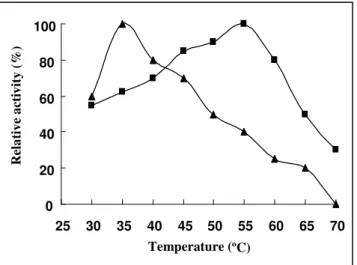
Characterization of the recombinant enzymes revealed the
great difference between the recombinant XYNA1 and XYNB:
The optimum reaction temperature and pH of the recombinant
XYNA1 were 35 ºC and 3.0, respectively, but the recombinant
XYNB were 55 ºC and 5.0(Fig.3, Fig.4). The properties for the
range of that reported for other fungi and bacteria (30).
The XynB from A. niger IBT-90 and XynNB described by
Kinoshita et al. (13) showed the same pH optima of about 5.0.
The pH determined for XYNA1 was exactly the same as
the pH optimum reported for XynI derived from A. niger
(15). As anticipated from the resolved crystal structure of
A. niger XynI (15) and further determined by means of
mutagenesis by Tahir et al. (28), the Asp37 presented in
the active site of acidic -xylanases was the key residue
responsible for low pH optimum (18), so the lower pH
optimum of XYNA1 would be attributed to the presence
of Asp at the mentioned position.
The results of the effects of pH stability on the two
recombinant xylanase revealed that, the recombinant XYNA1
was more stable than recombinant XYNB after being incubated
at pH 2.0-5.0 for 30 min at 37 ºC, however, there had little
difference after being incubated at pH 6.0-10.0 between them ,
and both retained 75%~85% of maximal activity (Fig.5).
Figure 3. Effects of temperature on the activity of
recombinant xylanase. The optimal temperature was
determined by incubating the enzyme in 0.1M citrate
buffer, pH 5, for 10 min at different temperatures as
indicated in the graph. The maximal enzyme activity was
defined as 100% relative activity. Symbols: recombinant
XYNA1 ( ); recombinant XYNB ( ).
Figure 4. Effect of pH on the activity of the recombinant
xylanase. The optimal pH was determined by incubating the
enzyme at 50 ºC for 10 min at different pH. To determine the
optimal pH, pH range from 2 to 10 was used with the
following (100mM) buffers: KCl-HCl (pH 2), sodium citrate
(pH 3–7), Na2HPO4- NaH2PO4 (pH 8.0),glycine -NaOH (pH
9-10). The maximal enzyme activity was defined as 100%
relative activity. Symbols: recombinant XYNA1 ( );
recombinant XYNB ( ).
Figure 5. Acid-alkali stability of recombinant xylanases. To estimate pH stability, the recombinant enzymes were
incubated in the different pH buffers (pH range from 2 to 10)
at 37ºC for 30 min, and then the residual xylanase activity
was determined at optimal pH. The activity determined
under the standard conditions of optimal pH without
incubation was defined as 100% relative activity. Symbols:
recombinant XYNA1 ( ); recombinant XYNB ( ). 0 20 40 60 80 100
25 30 35 40 45 50 55 60 65 70
Temperature (
ºC)
R el a ti v e a ct iv it y ( % ) 0 20 40 60 80 100
1 2 3 4 5 6 7 8 9 10 pH R el a ti v e a ct iv it y ( % ) 0 20 40 60 80 100
1 2 3 4 5 6 7 8 9 10
Moreover, it was worth stressing that the effects of
temperature on xylanases activity and the thermostability
of the recombinant enzymes revealed much more
difference which were presented in Fig.6. The recombinant
XYNB showed much more thermostability than recombinant
XYNA1; the recombinant XYNB showed 94% of maximal
activity after incubating in water for 60 min at 60 ºC compared
to no activity for recombinant XYNA1. In order to futher study
their thermostability, the incubatied temperature was risen
to 85 ºC. The recombinant XYNB remained 94% of maximal
activity after being incubated in water for 10 min at 85 ºC
compared to no activity for recombinant XYNA1. Hence, the
discrepancies between these two low molecular weight
endo-1,4- -D-xylanases co-expressed by A. niger are not
only in the pH optima, but also in temperature optima and
thermostability. It can be implied that the co-existence of
these two similar enzymes may be due to their different
catalytic activity. Moreover, the recombinant XYNB showed
higher thermostability than crude enzyme from A. niger
SCTCC 400264 preparation. Hence, we can deduced that
the xynB coded protein played a key role in the
thermostability of xylanase system of A. niger SCTCC
400264. In addition, comparing to the other xylanases from
A.niger (19, 30), the recombinant XYNB in our research also
showeded higher thermostability.
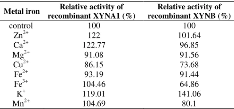
Various metal ions have different effects on activity
between the two recombinant xylanases (Table 1). The
potassium, calcium, zinc, iron had defferent stimulation
effects on recombinant XYNB activity, copper inhibited its
activity. However, manganese, copper and iron had an
inhibitory effect on recombinant XYNA1 activity, only
potassium stimulated its activity by 41%.
In conclusion, this was the first report on isolation of two
genes encoding: xynA1 and xynB from different groups of
glycosyl hydrolase family 11 co-produced by one strain of A.
niger and their subsequent expression in Escherichia coli
BL21. The fact that two xylanases come from the common
origin, the same fungal organism, will make the comparative
studies of both enzymes more accurate. Characterization of the
recombinant enzymes revealed the great differences between
the recombinant XYNA1 and XYNB. The protein encoded by
xynB played a key role to the thermostability of xylanase
system of A. niger SCTCC 400264. Since high temperatures
were used in many industrial processes, and always made the
xylanase activity destroyed, thermostability was one of the
most desirable enzyme characteristics. The excellent
thermostability of recombibant XYNB could establish a
foundation for its industry application.
Figure 6. Thermostability of the recombinant xylanases.The enzyme solutions were incubated at 60ºC and 85ºC without the
presence of substrate for various times, respectively. The residual
xylanases activity were measured by 10 min incubation at optimal
temperature for recombinant XYNA1 and XYNB, respectively.
The activity determined under the standard conditions of optimal
temperature without incubation was defined as 100% relative
activity. Symbols: recombinant XYNB treated by 60ºC ( ),
recombinant XYNB treated by 85ºC (∆), recombinant XYNA1
treated by 60ºC ( ), recombinant XYNA1 treated by 85ºC ( ).
Table 1. The effect of metal ions on recombinant xylanases activity
Metal iron Relative activity of
recombinant XYNA1 (%)
Relative activity of recombinant XYNB (%)
control 100 100
Zn2+ 122 101.64
Ca2+ 122.77 96.85
Mg2+ 91.08 91.56
Cu2+ 86.15 73.68
Fe2+ 93.19 91.44
Fe3+ 104.46 64.86
K+ 119.01 141.06
Mn2+ 104.69 80.1
0 20 40 60 80 100
0 10 20 30 40 50 60
Time (min)
R
el
a
ti
v
e
a
ct
iv
it
y
(
%
ACKNOWLEDGEMENTS
This study was supported by the National Natural Science
Foundation of China (NSFC 30871321, 30740055, 30771312),
and Program for New Century Excellent Talents in University
(NCET-05-0785).
REFERENCES
1. Asano, K.; Sriprang, R.; Gobsuk, J.; Eurwilaichitr, L.; Tanapongpipat, S.; Kirtikara, K. (2005). Endo-1,4- -xylanase B from Aspergillus niger BCC14405 Isolated in Thailand: Purification, Characterization and Gene Isolation. J. Biochem. Mol. Biol., 38, 17-23.
2. Berrin, J.G.; Williamson, G.; Puigserver, A.; Chaix, J.C.; McLauchlan, W.R.; Juge, N. (2000). High-level production of recombinant fungal endo-beta-1,4-xylanase in the methylotrophic yeast Pichia pastoris. Protein Expr. Purif., 19, 179-187.
3. Bradford, M.M. (1976). A rapid and sensitive method for the quantification of microgram quantities of protein utilizing the principle of protein-dye binding. Anal. Biochem., 72, 248–254.
4. Bruins, M.E.; Jasen, A.E.; Boom, R.M. (2001). Thermozymes and their applications: a review of recent literature and patents. Appl. Biochem. Biotechnol., 90, 155-186.
5. Chen, A.P.C.; Chang, S.; Lin, Y.; Sun, Y.; Chen, C.; Wang, A.H.J.; Liang, P. (2005). Substrate and product specificities of cistype undecaprenyl pyrophosphate synthase. Biochem. J., 386, 169–176. 6. Coughlan, M.P.; Hazlewood, G.P. (1993). -1,4-d-Xylan-degrading
enzyme systems:biochemistry, molecular biology and applications. Biotechnol. Appl. Biochem., 17, 259–289.
7. Dominguez, R.; Souchon, H.; Spinelli, S.; Dauter, Z.; Wilson, K.S.; Chauvaux, S.; Beguin, P.; Alzari, P.M. (1995). A common protein fold and similar active site in two distinct families of -glycanases. Nat. Struct. Biol., 2, 569–576.
8. Frederick, M.M,; Kiang, C.; Frederick, J.R.; Reilly, P.J. (1985). Purification and characterization of endo-xylanases from Aspergillus niger. 1. Two isozymes active on xylan backbones near branch points.
Biotechnol. Bioeng., 27, 525–532.
9. Gilkes, N.R.; Henrissat, B.; Kilburn, D.G.; Miller, R.C.; Warren, R.A.J. (1991). Domains in microbial -1,4-glycanases: sequence conservation, function,and enzyme families. Microbiol. Rev., 55, 303–315.
10. Gorbacheva, I.V.; Rodionova, N.A. (1977). Studies on xylan degrading enzymes. I. Purification and characterization of endo-1,4-beta-xylanase from Aspergillus niger str. 14. Biochim.Biophys. Acta., 484, 79-93. 11. Haki, G.D; Rakshit, S.K. (2003). Developments in industrially important
thermostable enzymes: a review. Bioresour. Technol., 89, 17–34 12. Henrissat, B.; Bairoch, A. (1993). New families in the classification of
glycosyl hydrolases based on amino acid sequence similarities. Biochem.
J., 293, 781–788.
13. Kinoshita, K.; Takano, M.; Koseki, T.; Ito, K.; Iwano, K. (1995). Cloning of the xynNB gene encoding xylanase B from Aspergillus niger and its expression in Aspergillus kawachii. J.Ferment. Bioeng., 79, 422–428. 14. Korona, B.; Korona, D.; Bielecki, S. (2006). Efficient expression and
secretion of two co-produced xylanases from Aspergillus niger in Pichia pastoris directed by their native signal peptides and the Saccharomyces
cerevisiae -mating factor. Enzyme Microb. Technol., 39, 683-689. 15. Krengel, U.; Dijkstra, B.W. (1996). Three-dimensional structure of
endo-1, 4-xylanase I from Aspergillus niger: molecular basis for its low pH optimum. J. Mol. Biol., 263, 70–78.
16. Kulkarni, N.; Shendye, A.; Rao, M. (1999). Molecular and biotechnological aspects of xylanases. FEMS. Microbiol. Rev., 23, 411– 456.
17. Laemmli, U.K. (1970). Cleavage of structural protein during the assembly of the head of bacteriophage T4. Nature, 227, 680–685. 18. Liu, L.; Li, X.; Li, X.; Shao, W. (2004). Computational analysis of
responsible dipeptides for optimum pH in G/11 xylanase. Biochem. Biophys. Res. Commun., 321, 391–396.
19. Liu, M.Q.; Weng, X.Y.; Sun, J.Y. (2006). Expression of recombinant Aspergillus niger xylanase A in Pichia pastoris and its action on xylan.
Protein Expr. Purif., 48, 292-299.
20. Luttig, M.; Pretorius, I.S.; VanZyl, W.H. (1997). Cloning of two -xylanase encoding genes from Aspergillus niger and their expression in Saccharomyces cerevisiae. Biotechnol. Lett., 19, 411–415.
21. Miller, G.L. (1959). Use of dinitrosalicylic acid reagent for determination of reducing sugar. Anal. Chem., 31, 426–428.
22. Rogalski, J.; Oleszek, M.; Tokarzewska-Zadora, J. (2001). Purification and characterization of two endo-1,4 beta-xylanase and a 3-xylosidase from Phlebia adiate. Acta. Microbiol. Pol., 50, 117–128.
23. Saiki, R.K.; Gelvand, D.H.; Stoffel, S.; Scharf, S.J.; Higuchi, R.; Horn, G.T.; Mullis, K.B.; Erlich, H.A. (1988). Primer-directed enzymatic amplification of DNA with a thermostable DNA polymerase. Science, 239, 487–491.
24. Singh, S.; Madlala, A.M.; Prior, B.A. (2003). Thermomyces lanuginosus: properties of strains and their hemicellulases. FEMS. Microbiol. Rev., 27, 3-16.
25. Subramaniyan, S.; Prema, P. (2002). Biotechnology of microbial xylanases: enzymology. Crit. Rev. Biotechnol., 22, 33–64
26. Sunna, A.; Antranikian, G. (1997). Xylanolytic enzymes from fungi and bacteria. Crit. Rev. Biotechnol., 17, 39-67.
27. Tahir, T.A.; Berrin, J.G.; Flatman, R.; Roussel, A.; Roepstorff, P.; Williamson, G.; Juge, N. (2002). Specific characterization of substrate and inhibitor binding sites of a glycosyl hydrolase family 11 xylanase from Aspergillus niger. J. Biol. Chem., 277, 44035-44043.
29. Torronen, A.; Harkki, A.; Rouvinen, J. (1994). Three-dimensional structure of endo-1,4- -xylanase II from Trichoderma reesei: two conformational states in the active site. EMBO. J., 13, 2493–2501.