online | memorias.ioc.fiocruz.br
Potential application of rLc36 protein for
diagnosis of canine visceral leishmaniasis
Camila Tita Nogueira1, Mayara Lúcia Del Cistia2, Ana Carolina Urbaczek3, Márcia MG Jusi4,
Angela Maria Arenas Velásquez1, Rosângela Zacarias Machado4, Henrique Ferreira5,
Flávio Henrique-Silva6, Hélio Langoni7, Paulo Inácio da Costa2, Márcia AS Graminha1,2/+
1Universidade Estadual Paulista, Instituto de Química, Campus de Araraquara, Araraquara, SP, Brasil
2Universidade Estadual Paulista, Faculdade de Ciências Farmacêuticas, Campus de Araraquara, Araraquara, SP, Brasil 3Universidade de São Paulo, Instituto de Química, Campus de São Carlos, São Carlos, SP, Brasil
4Universidade Estadual Paulista, Faculdade de Ciências Agrárias e Veterinárias, Campus de Jaboticabal, Jaboticabal, SP, Brasil 5Universidade Estadual Paulista, Instituto de Biociências, Campus de Rio Claro, Rio Claro, SP, Brasil
6Universidade Federal de São Carlos, Departamento de Genética e Evolução, São Carlos, SP, Brasil
7Universidade Estadual Paulista, Faculdade de Medicina Veterinária e de Zootecnia, Campus de Botucatu, Botucatu, SP, Brasil
Visceral leishmaniasis (VL) is fatal if left untreated. Infected dogs are important reservoirs of the disease, and thus specific identification of infected animals is very important. Several diagnostic tests have been developed for canine VL (CVL); however, these tests show varied specificity and sensitivity. The present study describes the recombinant protein rLc36, expressed by Leishmania infantum, as potential antigen for more sensitive and specific diagnosis of CVL
based on an immunoenzymatic assay. The concentration of 1.0 μg/mL of rLc36 enabled differentiation of positive and
negative sera and showed a sensitivity of 85% and specificity of 71% (with 95% confidence), with an accuracy of 76%.
Key words: canine visceral leishmaniasis - immunodiagnosis - recombinant protein
doi: 10.1590/0074-02760170171
Financial support: FAPESP (grant #2010/26732-2), CAPES, PADC. CTN was supported by the FAPESP fellowship (#2011/06995-9) and MLDC by the FAPESP (#2011/15508-4) and CAPES fellowships.
CTN and MLDC contributed equally to the study. + Corresponding author: graminha@fcfar.unesp.br Received 27 April 2017
Accepted 24 October 2017
Kala azar, or visceral leishmaniasis (VL), is a zoo-nosis caused by protozoan parasites of the Leishmania donovani complex. L. donovani and L. infantum are the etiological agents of VL in the Old World and L. infan-tum in the Americas (Ikonomopoulos et al. 2003). In Latin America, VL is transmitted by the bite of infected sand flies Lutzomyia longipalpis (Akhoundi et al. 2016), with domestic dogs (Canis familiaris) as the main reser-voir for leishmaniasis infection. Thus, one of the most important approaches for controlling the incidence of human VL is to identify infected dogs (Lira et al. 2006).
Clinical diagnosis of canine VL (CVL) is difficult to achieve because of the nonspecific symptoms in com-mon with other diseases (Mondal et al. 2010). Para-sitological methods are based on biopsy or aspiration specimens, but these methods are invasive and present variable sensitivity (Silva et al. 2014).
Among the different serological tests available, en-zyme-linked immunosorbent assay (ELISA), indirect im-munofluorescence, direct agglutination test, dot-ELISA, and western blotting are the most widely used (Silva et al. 2014). Several Leishmania antigens have been evaluated for serodiagnosis (Badaró et al. 1996, Jensen et al. 1999, Ak-houndi et al. 2013), with variable sensitivity and specificity.
Here, we characterised the rLc36 protein encoded by the L. infantum gene LinJ.36.4190 as a potential antigen for developing a more sensitive and specific test for CVL based on ELISA.
Lc36 (GenBank: LinJ.36.4190) was identified through bioinformatic analyses using the EupathDB (eupathdb. org) and TriTrypDB (tritrypdb.org) databases. B-Cell epitope prediction was performed using web server-based software (Saha & Raghava 2006), and the B-cell epitope
database Bcipep (Saha et al. 2005; available from: http:// www.imtech.res.in/raghava/bcipep). Hydropathicity and
the presence of transmembrane domains were also anal-ysed (Kyte & Doolittle 1982, Cserzo et al. 2004).
L. infantum Lc36 codes for a protein of 733 residues that does not contain transmembrane domains (Supple-mentary data, Fig. 3). Its product is similar to the pro-teins of L. donovani and Leishmania mexicana; how-ever, when compared to Leishmania braziliensis, Lc36 shows sequence conservation only at the DNA level, indicating that Lc36 is a pseudogene in L. braziliensis (Supplementary data, Fig. 1A-B). Lc36 did not present any sequence similarity when searched against other ge-nome databases including those of Crithidia, Leptomo-nas, Endotrypanum, and Trypanosoma.
Quantitative polymerase chain reaction (PCR) assays were performed to assess the gene expression levels in dif-ferent parasite life-cycle stages. For both quantitative and expression assays, we used L. infantum strain MHOM/
BR/1972/LD, which was donated by the Instituto Oswaldo
For quantitative PCR assays, we used mouse peritoneal macrophages collected from adult male Swiss albino mice, infected with L. infantum as previously described (Ve-lásquez et al. 2017), except that the assay was performed in 25-cm2 cell culture flasks (Nunc™, Roskilde, Denmark). These assays were approved by the Ethics Committee for
Animal Experimentation (Protocol CEUA/FCF/CAr no. 40/2015) in agreement with Sociedade Brasileira de Ciên -cia de Animais de Laboratório and Conselho Nacional de Controle de Experimentação Animal.
For RNA extraction and real-time PCR analysis,
prim-ers RT_36_Forward 5′-GTTCGTCACCGTTGTCTTC-3′ and RT_36_Reverse 5′-GTCGTG CTTCCTGCTATTC-3′
were designed using the software tools GeneRunner
(http://www.generunner.com) and Primer Express ver -sion 3.0. Total promastigotes and infected and non-infected macrophage RNA were extracted using TRIzol® Reagent (Life Technologies, Carlsbad, CA, USA) according to the manufacturer’s protocol. Intracellular amastigote RNA was extracted at 18, 48, and 72 h post-infection. For cDNA
synthesis, we used 1 µg of total RNA, the 3′Race System
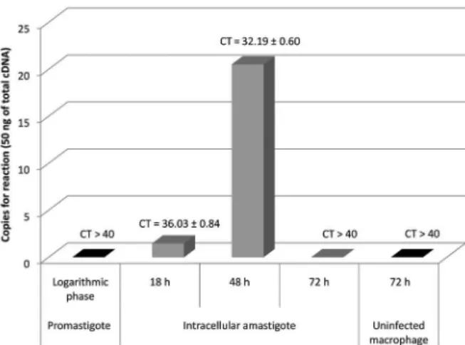
for Rapid Amplification of cDNA ends kit (Life Technolo-gies), and random hexamer primers. Real-time PCR as-says were carried out in a StepOnePlus™ Real-Time PCR System v2.3 (Applied Biosystems, Foster City, CA, USA) with initial denaturation at 95ºC for 20 s followed by 40 denaturation cycles at 95ºC for 15 s, annealing at 60ºC for 1 min, and extension at 95ºC for 15 s. For each reac-tion, 1x Fast Power SYBR® Green Master Mix (Applied Biosystems), 0.1 µM of each primer, and 10 ng of each cDNA sample was added in 10-µL of PCR mixture. A 10-fold dilution series of plasmid DNA (recombinant plasmid pET28a+_Lc36) was used for absolute standard curve construction to determine the efficiency. Assays were carried out in triplicate. Absolute amounts of PCR products were determined based on the threshold cycle (Ct), by interpolating each Ct value from each sample on the corresponding standard curve and by the size of plas-mid DNA template (7588 base pairs) using the equation described in the Supplementary data (Equation 1). For graphics design, we used software Origin® and Excel® software. These assays revealed that the gene is likely ex-pressed in amastigote cells, the form responsible for the disease’s clinical manifestations. The absolute standard curve parameters showed an efficiency of 99.23% and linear regression coefficient (R2) of 0.98 (Supplemen-tary data, Table II, Figs 5, 6). Fig. 1 shows that the cDNA from intracellular amastigotes of 18 h and 48 h post-infection macrophages had CT values of 36.0 ± 0.8 and 32.2 ± 0.6, respectively, while CT values higher than 40 were observed for cDNA from promastigotes and intracellular amastigotes at 72 h post-infection. These data were converted to cDNA copy numbers (Supplementary data, Table II, Figs 5-7) and Fig. 1 illustrates that the cDNA copy number was higher in intracellular amastigotes at 48 h post-infection (20 copies per reaction) compared to in parasites at 18 h post-infection (1 copy per reaction). Thus, the results suggest that this gene has a stage-specific expression pattern and indicate that L. infantum Lc36 is amastigote-stage specific and is expressed at 48 h post-infection.
Leishmania has an intracellular life stage and is likely to encounter reactive oxygen species produced by the macrophage, which can induce DNA damage. Thus, stage-specific amastigote genes may be involved in parasite survival in macrophages (Genois et al. 2014). Interestingly, Lc36 codes for a protein of unknown func-tion containing a nucleotidyl transferase domain which is possibly involved in nucleic acid metabolism, including DNA repair, which may play an important role in protect-ing Leishmania amastigote genome integrity during its exposure to reactive oxygen species produced by
macro-phages (Vaňáčová et al. 2005). For application and diag -nosis, some amastigote stage-specific proteins present antigenic properties recognised by the host, such as the A2 protein, which has been evaluated for VL serodiag-nosis and vaccine application (Coelho et al. 2012, Mar-tins et al. 2015). As these data indicated the importance of rLc36 for the development of VL diagnosis assays, we tested the performance in ELISA. We mapped linear B cell epitopes within the Lc36 coding region. The protein region from residues 1 to 255 was selected and evaluated for serodiagnosis of CVL (Supplementary data, Table I, Fig. 2). The chosen fragment containing 765 base pairs was amplified and cloned into pET28a+ for heterolo-gous expression in Escherichia coli. For expression and purification of rLc36, a recombinant plasmid named as pET28a_Lc36 was constructed (cloning strategy, in Supplementary data). Protein expression was carried out in a 250-mL bacterial culture in LB medium containing
presenting the expected molecular weight, was success-fully purified (Supplementary data, Fig. 4).
rLc36 was subjected to 12% SDS-PAGE and trans-ferred to nitrocellulose membranes. Membranes were blocked with 5% non-fat dry milk in 1x PBS, pH 7.3, and incubated overnight at 4ºC. The membrane was washed with 0.05% Tween 20 in PBS and incubated for 2 h in the presence of monoclonal anti-polyhistidine 1:1500 (Sig-ma, St. Louis, MO, USA). After washing, the membrane was treated for 1 h with peroxidase-conjugated anti-mouse secondary antibody (1:40,000), and subsequently developed successively with luminol [50 mL Tris-HCl 1 M pH 8.5, in H2O, 5 mL of solution A (0.22 g luminol in 5 mL DMSO), 2.2 mL of solution B (0.033 g of p-coumaric acid, 2.2 mL of DMSO)], and 0.3% H2O2. The membranes were placed in contact with a photographic film for 5 min and bands were visualised by the addition
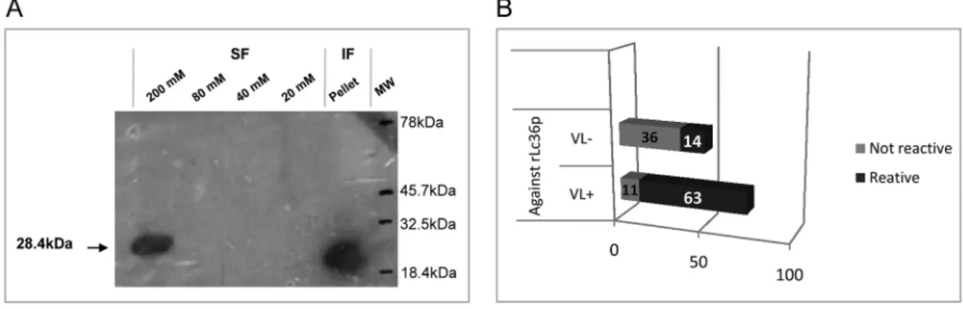
of Step™ NBT/BCIP (Pierce, Rockford, IL, USA). West -ern blotting analyses revealed the presence of the puri-fied His6-tagged protein in the 200 mM imidazole-eluate soluble fraction and in the inclusion bodies (Fig. 2A).
For ELISA, 96-well plates were coated with the re-combinant protein rLc36 in 100 µL of coating buffer for
18 h at 4ºC. For this assay, 1 µg/mL of rLc36 was used,
based on a titration curve developed to determine the optimal recombinant protein concentration. VL-positive sera used in this study were obtained from the area of Campo Grande, Minas Gerais, Brazil, in which CVL is endemic, whereas negative sera were obtained from Jaboticabal, São Paulo, Brazil, a non-endemic area. The sera were selected for CVL based on Leishmania chagasi-specific total IgG and L. chagasi-specific IgG subclass ELISAs with a confidence index of 95%. Both sera groups were confirmed by direct parasitological methods and indirect immunofluorescence. In the ELI-SA for rLc36, the plates were blocked with 2% non-fat dry milk solution for 1 h at 37ºC. After three washes with PBS Tween 80 at 0.05%, the plates were incubated with 100 µL of canine sera for 1 h at 37ºC. Sera samples
were diluted 1:200 in PBS with 2% normal rabbit serum. Plates were then washed three times with PBS Tween 80 and incubated with 1:4000 of alkaline phosphatase con-jugated to anti-dog IgG (Sigma) for 1 h at 37ºC. Plates were washed and 100 µL diethanolamine solution (pH 9.8) containing a substrate for phosphatase (4-nitrophe-nyl phosphate disodium salt hexahydrate, Sigma) was added to each well. The optical density was measured at 405 nm in an ELISA microplate spectrophotometer (Ro-bonik® - Readwell Touch Automatic ELISA Plate Anal-yser). Sensitivity and specificity values were determined by discriminating the absorbance data of sera true-pos-itives and true-negatives, as well as false-postrue-pos-itives and false-negatives, using cut-off method analysis described by Frey et al. (1998). The accuracy predictive value was also determined (Kawamura 2002). For a complete de-scription, see Supplementary data (Equations 2, 3, 4). The His6-tagged protein, confirmed as rLc36, was anal-ysed for antigenic potential against canine sera; 74 sera were previously demonstrated to be positive for CVL while another 50 were negative for this disease (Fig. 2B). Sensitivity and specificity of the rLc36 against the CVL sera were 85% and 72%, respectively, with a cut-off with 95% confidence. Another group of 39 sera samples was separated in symptomatic (n = 33) and asymptomatic (n = 6) subgroups and tested with a cut-off with 99% con-fidence (Fig. 3). All symptomatic sera were confirmed to be positive for CVL, while for asymptomatic sera, the ELISA based on rLC36 detected four of six asymptom-atic samples (Supplementary data, Table V).
Several recombinant proteins have shown variable effectiveness for leishmaniasis serodiagnosis (Badaró et al. 1996, Bhatia et al. 1999, Jensen et al. 1999, Raj et al. 1999) and others present high amino acid sequence
ho-mologies to human and/or canine counterparts, interfer -ing with the applicability of the tests (Maalej et al. 2003, Coelho et al. 2012, Celeste et al. 2014). Thus, based on BLAST analyses revealing a lack of homology to other parasitic sequences, rLc36 may be useful for developing a new and specific diagnostic test for CVL.
In conclusion, the rLc36 protein, which is likely ex-pressed in amastigote forms of L. infantum, was capable of differentiating positive from negative CVL sera and showed a sensitivity and specificity of 85% and 71%, respectively, with a confidence of 95% and accuracy of 76%, in a randomly chosen population. The protein was also able to identify asymptomatic animals. These results indicate that rLc36 is promising for CVL serodiagnosis.
ACKNOWLEDGEMENTS
To professor Paul A Michels for critically reviewing the man-uscript, and Wanderson HC Oliveira for the English revision.
AUTHORS’ CONTRIBUTION
CTN and MLDC provided the experimental data, wrote and reviewed the full manuscript; MASG and PIC designed and re-viewed the full manuscript; ACU, MMGJ, AMAV, RZM, HF, FHS and HL provided assistance with experimental data.
REFERENCES
Akhoundi B, Mohebali, M, Shojaee S, Jalali M, Kazemi B, Ban-dehpour M, et al. Rapid detection of human and canine visceral leishmaniasis: assessment of a latex agglutination test based on the A2 antigen from amastigote forms of Leishmania infantum. Exp Parasitol. 2013; 133(3): 307-13.
Akhoundi M, Kuhls K, Cannet A, Votýpka J, Marty P, Delaunay P, et al. A historical overview of the classification, evolution, and dispersion of leishmania parasites and sandflies. PLoS Negl Trop Dis. 2016; 10(3): e0004349.
Badaró R, Benson D, Eulálio MC, Freire M, Cunha S, Netto EM, et al. rK39: a cloned antigen of Leishmania chagasi that predicts active visceral leishmaniasis. J Infect Dis. 1996; 173(3): 758-61.
Fig. 3: reactivity of 39 positive sera for canine visceral leishmaniasis (CVL) versus rLc36. The dotted line refers to a cut-off of 0.181, calculated based on a confidence of 99%, using 20 negative sera for VL. Source: authors.
Bhatia A, Daifalla NS, Jen S, Badaro R, Reed SG, Skeiky YAW. Clon-ing, characterization and serological evaluation of K9 and K26: two related hydrophilic antigens of Leishmania chagasi. Mol Biochem Parasitol. 1999; 102(2): 249-61.
Celeste BJ, Sánchez MC, Ramos-Sánchez EM, Castro LG, Costa FA, Goto H. Recombinant Leishmania infantum heat shock protein 83 for the serodiagnosis of cutaneous, mucosal, and visceral leish-maniases. Am J Trop Med Hyg. 2014; 90(5): 860-5.
Coelho VTS, Oliveira JS, Valadares DG, Chávez-Fumagalli MA, Du-arte MC, Lage PS, et al. Identification of proteins in promastigote and amastigote-like Leishmania using an immunoproteomic ap-proach. PLoS Negl Trop Dis. 2012; 6(1): e1430.
Cserzo M, Eisenhaber F, Eisenhaber B, Simon I. TM or not TM: transmembrane protein prediction with low false positive rate us-ing DAS-TMfilter. Bioinformatics. 2004; 20(1): 136-7.
Frey A, Di Canzio J, Zurakowski D. A statistically defined endpoint titer determination method for immunoassays. J Immunol Meth-ods. 1998; 221(1-2): 35-41.
Genois MM, Paquet ER, Laffitte MC, Maity R, Rodrigue A, Ouellette M, et al. DNA repair pathways in trypanosomatids: from DNA re-pair to drug resistance. Microbiol Mol Biol Rev. 2014; 78(1): 40-73.
Ikonomopoulos J, Kokotas S, Gazouli M, Zavras A, Stoitsiou M, Gor-goulis VG. Molecular diagnosis of leishmaniosis in dogs: compara-tive application of traditional diagnostic methods and the proposed assay on clinical samples. Vet Parasitol. 2003; 113(2): 99-113.
Jensen ATR, Gasim S, Moller T, Ismail A, Gaafar A, Kemp M, et al. Serodiagnosis of Leishmania donovani infections: assessment of enzyme-linked immunosorbent assays using recombinant L. donovani gene B protein (GBP) and a peptide sequence of L. don-ovani GBP. Trans R Soc Trop Med Hyg. 1999; 93(2): 157-60.
Kawamura T. Interpretação de um teste sob a visão epidemiológica.
Kyte J, Doolittle RF. A simple method for displaying the hydropathic character of a protein. J Mol Biol. 1982; 157(1): 105-32.
Larsen JEP, Lund O, Nielsen M. Improved method for predicting lin-ear B-cell epitopes. Immunome Res. 2006; 2: 2.
Lira RA, Cavalcanti MP, Nakazawa M, Ferreira AGP, Silva ED, Abath FGC, et al. Canine visceral leishmaniosis: a comparative analysis of the EIE-leishmaniose-visceral-canina-Bio-Manguinhos and the IFI-leishmaniose-visceral-canina-Bio-Manguinhos kits. Vet Parasitol. 2006; 137(1-2): 11-6.
Maalej IA, Chenik M, Louzir H, Ben Salah A, Bahloul C, Amri F, et al. Comparative evaluation of ELISAs based on ten recombinant or pu-rified Leishmania antigens for the serodiagnosis of Mediterranean visceral leishmaniasis. Am J Trop Med Hyg. 2003; 68(3): 312-20.
Martins VT, Chávez-Fumagalli MA, Lage DP, Duarte MC, Garde E, Costa LE, et al. Antigenicity, immunogenicity and protective ef-ficacy of three proteins expressed in the promastigote and amas-tigote stages of Leishmania infantum against visceral leishmani-asis. PLoS ONE. 2015; 10(10): e0141496.
Mondal S, Bhattacharya P, Ali N. Current diagnosis and treatment of vis-ceral leishmaniasis. Expert Rev Anti Infect Ther. 2010: 8(8): 919-44.
Raj VS, Ghosh A, Dole VS, Madhubala R, Myler PJ, Stuart KD. Sero-diagnosis of leishmaniasis with recombinant ORFF antigen. Am J Trop Med Hyg. 1999; 61(3): 482-7.
Saha S, Bhasin M, Raghava GPS. Bcipep: a database of B-cell epit-opes. BMC Genomics. 2005; 6: 79.
Saha S, Raghava GP. Prediticion of continuous B-cell epitopes in an an-tigen using recurrent neural network. Proteins. 2006; 65(1): 40-8.
Silva DT, Starke-Buzetti WA, Alves-Martin MF, Paixão MS, Tenório MS, Lopes ML. Comparative evaluation of several methods for canine visceral leishmaniasis diagnosis. Rev Bras Parasitol Vet. 2014; 23(2): 179-86.
Vaňáčová Š, Wolf J, Martin G, Blank D, Dettwiler S, Friedlein A, et al.
A new yeast poly(A) polymerase complex involved in RNA quality control. PLoS Biol. 2005; 3(6): 986-97.