online | memorias.ioc.fiocruz.br
In vitro
evaluation of the anti-leishmanial activity and toxicity of PK11195
Carlos Eduardo Sampaio Guedes1, Beatriz Rocha Simões Dias1, Antonio Luis de Oliveira Almeida
Petersen1, Kercia Pinheiro Cruz1, Niara de Jesus Almeida1, Daniela Rodrigues Andrade2, Juliana
Perrone Bezerra de Menezes1, Valéria de Matos Borges2, Patricia Sampaio Tavares Veras1/+
1Fundação Oswaldo Cruz-Fiocruz, Centro de Pesquisas Gonçalo Moniz, Laboratório de Patologia e Biointervenção, Salvador, BA, Brasil 2Fundação Oswaldo Cruz-Fiocruz, Centro de Pesquisas Gonçalo Moniz, Laboratório Integrado de Microbiologia e Imunoregulação, Salvador, BA, Brasil
BACKGROUND Leishmaniasis, one of the most neglected diseases, is a serious public health problem in many countries, including Brazil. Currently available treatments require long-term use and have serious side effects, necessitating the development of new therapeutic interventions. Because translocator protein (TSPO) levels are reduced in Leishmania amazonensis-infected cells and because this protein participates in apoptosis and immunomodulation, TSPO represents a potential target for Leishmania chemotherapy. The present study evaluated PK11195, a ligand of this protein, as an anti-leishmanial agent.
OBJECTIVE To evaluate the leishmanicidal activity of PK11195 against L. amazonensis in infected CBA mouse macrophages in vitro.
METHODS The viability of axenic L. amazonensis, Leishmania major, and Leishmania braziliensis promastigotes was assessed after 48 h treatment with PK11195 (0.2-400 µM). Additionally, intracellular parasite viability was evaluated to determine IC50 values and the number of viable parasites in infected macrophages treated with PK11195 (50-100 µM). Infected macrophages were then treated with PK11195 (25-100 µM) to determine the percentage of L. amazonensis-infected cells and the number of parasites per infected cell. Electron microscopy was used to investigate morphological changes caused by PK11195. The production of free oxygen radicals, nitric oxide, and pro-inflammatory cytokines was also evaluated in infected macrophages treated with PK11195 and primed or not primed with IFN-γ.
FINDINGS Median IC50 values for PK11195 were 14.2 µM for L. amazonensis, 8.2 µM for L. major, and 3.5 µM for L. braziliensis. The selective index value for L. amazonensis was 13.7, indicating the safety of PK11195 for future testing in mammals. Time- and dose-dependent reductions in the percentage of infected macrophages, the number of parasites per infected macrophage, and the number of viable intracellular parasites were observed. Electron microscopy revealed some morphological alterations suggestive of autophagy. Interestingly, MCP-1 and superoxide levels were reduced in L. amazonensis-infected macrophages treated with PK11195.
MAIN CONCLUSIONS PK11195 causes the killing of amastigotes in vitro by mechanisms independent of inflammatory mediators and causes morphological alterations within Leishmania parasites, suggestive of autophagy, at doses that are non-toxic to macrophages. Thus, this molecule has demonstrated potential as an anti-leishmanial agent.
Key words: PK11195 - Leishmania amazonensis - macrophage - chemotherapy
doi: 10.1590/0074-02760170345 Financial support: FAPESB, CNPq.
PSTV holds a grant from CNPq for productivity in research (307832/2015-5). CESG and BRSD contributed equally to this work.
+ Corresponding author: [email protected] Received 24 August 2017
Accepted 30 November 2017
Leishmaniasis, one of the most challenging neglected diseases faced by developing countries, has an estimated incidence of approximately two million new cases per year and afflicts 98 countries and territories, placing ~310 mil-lion people at risk of infection (Alvar et al. 2012). Taking this into consideration, together with the increasing number of reported HIV-Leishmania co-infections, it is unsurpris-ing that the World Health Organization has labelled this disease a ‘serious public health problem’ (WHO 2010).
Leishmaniasis treatment necessitates the use of high-ly toxic drugs with extended courses of administration, which sometimes leads patients to abandon treatment, increasing the chance of treatment failure. Moreover, co-infection with HIV has been shown to undermine the ef-fectiveness of available treatments (Croft & Olliaro 2011),
making the search for new pharmaceutical compounds an imperative in the ongoing effort to control this disease.
We have previously demonstrated that CBA mouse macrophages can control Leishmania major infection, yet are susceptible to Leishmania amazonensis, and pres-ent differpres-ent immune-inflammatory profiles in response to L. amazonensis and L. major infection, indicating the importance of macrophages in controlling infections aris-ing from these parasites (de Souza et al. 2000, Gomes et al. 2003). We have previously used proteomic analysis to identify proteins that are differentially abundant in CBA mouse macrophages infected with either of these Leish-mania spp., and we hypothesised that some of these pro-teins could serve as novel targets for chemotherapeutic treatment of leishmaniasis (Menezes et al. 2013).
(IBP), which has been renamed TSPO (Papadopoulos et al. 2006), the 32 kDa voltage-dependent anion channel (VDAC), and the 30 kDa adenine nucleotide transport-er (ANT) (McEntransport-ery et al. 1992). TSPO is involved in a range of cellular processes, including apoptosis, transport of cholesterol to the mitochondrial matrix, steroidogen-esis, cell proliferation, chemotaxis, cellular respiration, and immune response (Veenman et al. 2007). The fact that TSPO is a fundamentally important protein for cell function and maintenance of cellular homeostasis pro-vides compelling evidence for its potential as a chemo-therapeutic target. Several specific ligands that modulate TSPO have been described, including benzodiazepines, 1-(2-chlorophenyl)-N-methyl-N -(1-methylpropyl)-3-iso-quinolinecarboxamide (PK11195), and N,N -dihexyl-2-(4-fluorophenyl) indole-3-acetamide (FGIN-1-27) (Veen-man et al. 2007). Studies have demonstrated that TSPO ligands, including PK11195, may act as agonists or antag-onists depending on the ligand concentration and cell type (Totis et al. 1989). Although the underlying mechanisms of action of these TSPO ligands require further clarifi-cation, PK11195 and other ligands are currently being used as markers of neuroinflammation in PET imaging (Folkersma et al. 2011), and they also exhibit anticancer (Shoukrun et al. 2008) and immunomodulatory activities (Domingues-Junior et al. 2000). PK11195 has also been shown to increase free radical production in neuronal cells in a TSPO-dependent manner by promoting opening of the mitochondrial permeability transition pore (Jaya-kumar et al. 2002), and to reduce the proliferation rate of Plasmodium falciparum in infected cells (Dzierszinski et al. 2002, Bouyer et al. 2011). However, TSPO ligands have not been tested against trypanosomatids.
The present study aimed to assess the potential anti-leishmanial effects of a TSPO ligand, PK11195, on CBA mouse macrophages infected with L. amazonensis using an in vitro model. Considering that infected CBA mac-rophages are susceptible to L. amazonensis and express lower levels of TSPO than those infected with L. major (Menezes et al. 2013), we hypothesised that treatment of L. amazonensis-infected macrophages with PK11195, in association with host immune response modulation, would induce killing of the intracellular parasites.
MATERIALS AND METHODS
Ethics statement - CBA mice were obtained from the animal care facility at the Gonçalo Moniz Institute (IGM) - FIOCRUZ, housed in pathogen-free condi-tions, and fed a commercially available diet with water provided ad libitum. All mice were raised under condi-tions in accordance with the International Guiding Prin-ciples for Biomedical Research Involving Animals; all experimental protocols complied with these guidelines, as well as the resolutions established by the Brazilian National Council for the Control of Animal Experimen-tation (CONCEA). The present study was approved by the Institutional Animal Experimentation Review Board (CEUA) under protocol number 18/2010.
Anti-leishmanial drug preparation - The TSPO ligand PK11195 was acquired from Sigma-Aldrich (St Louis, MO,
USA), and a 50 mM stock solution was prepared in 100% ethanol (Sigma, St Louis, MO, USA), then aliquoted and stored at -20ºC until use. For experimental purposes, this stock solution was diluted into culture medium at vary-ing concentrations. Amphotericin B sodium deoxycholate (Fungizone, Gibco) was purchased from Life Technologies (Carlsbad, CA, USA) as a ready-to-use solution (271 µM).
Leishmania culture - Promastigotes of L. amazo-nensis (MHOM/Br88/Ba-125), L. braziliensis (MHOM/ BR/94/H3456), and L. major (MHOM/RI/-/WR-173) were axenically cultured in Schneider’s Insect Me-dium (Sigma, St Louis, MO, USA) supplemented with 50 µg/mL gentamycin (Gibco,Grand Island, NY, USA) and 10% or 20% heat-inactivated foetal bovine serum (Gibco, Grand Island, NY, USA) (Schneider’s complete medium). Cultures were maintained in an incubator at 24ºC, for no more than six consecutive passages, until parasites reached the stationary growth phase.
Viability of axenic Leishmania promastigotes - To determine IC50/48 h, axenic promastigotes from station-ary cultures of L. amazonensis, L. braziliensis, and L. major were cultivated at a density of 2 × 10⁶ cells/mL in 200 µL Schneider’s complete medium in 96-well plates at 24ºC. Parasites were treated with 12 two-fold serial dilutions of PK11195 at concentrations of 400, 200, 100, 50, 25, 12.5, 6.25, 3.13, 1.56, 0.78, 0.39, and 0.20 µM, or with the diluent (ethanol), for 48 h. Next, AlamarBlue® (Invitrogen, Carlsbad, CA, USA) cell viability reagent was added to the parasite cultures to a final concentra-tion of 10% v/v, and the cultures were incubated at 24ºC for 4 h. Reagent absorbance at wavelengths of 570 and 600 nm was measured using a spectrophotometer (Spec-traMax 340 PC, Molecular Devices, Sunnyvale, CA, USA). All experiments were performed in triplicate and individually repeated at least four times.
Macrophage cultivation - Peritoneal washing was per-formed to harvest macrophages from CBA mouse cavities injected with 3% sodium thioglycolate (Sigma, St Louis, MO, USA). All cells were cultured according to the pro-tocol described by Gomes et al. (2003). The peritoneal la-vage was first centrifuged at 300 × g, then resuspended in complete Dulbecco’s modified Eagle’s medium (DMEM) (Gibco, Grand Island, NY, USA) supplemented with 20 mM HEPES (Sigma, St Louis, MO, USA), 42 mM sodi-um bicarbonate (Sigma, St Louis, MO, USA), 10% foetal bovine serum (Gibco, Grand Island, NY, USA), 2 mM glutamine (Gibco, Grand Island, NY, USA), and 10 µg/ mL ciprofloxacin (Isofarma, Precabura, CE, BR). Next, cells were plated and incubated at 37ºC in a 5% CO2, 95% humidity atmosphere for 4 to 6 h, then washed to remove any non-adherent cells. Cells were maintained in 1 mL complete DMEM for further experiments.
50, 25, 12.5, 6.25, 3.13, 1.56, 0.78, 0.39, and 0.20 µM, in parallel with cultures incubated with the diluent (etha-nol) as a negative control. Next, AlamarBlue® was added to the macrophage cultures to a final concentration of 10% v/v, and the plates were then incubated at 37ºC for an additional 4 h. Reagent absorbance was measured as described above in Viability of axenic Leishmania pro-mastigotes. All experiments were performed in triplicate and individually repeated at least four times.
Infection and treatment - To evaluate the anti-leish-manial effect of the TSPO ligand PK11195 on intracellu-lar L. amazonensis parasites, CBA mouse macrophages were infected with stationary-phase promastigotes of L. amazonensis at a ratio of 10:1 for 6 h. The cells were then washed with saline to remove any non-internalised pro-mastigotes. To assess the effect of PK11195 on intracel-lular parasites at early stages of infection, macrophages were subsequently treated with PK11195 at concentra-tions of 25, 50, 75, or 100 µM for 6, 24, or 48 h, whereas control cells were incubated in complete DMEM taining ethanol as a diluent. Additionally, a second con-trol group remained untreated. To evaluate the treatment at later stages of infection, cells that had been washed to remove any non-internalised promastigotes were in-cubated in fresh DMEM medium for a further 96 h, a period of time sufficient to ensure that all parasites had completed the transformation to the amastigote form inside peritoneal macrophages, as previously shown by Courret et al. (2001). Infected cells were then treated with 50 or 75 µM PK11195, or 2.1 µM amphotericin B sodium deoxycholate, for an additional 24, 48, or 72 h, whereas control cells were incubated with ethanol.
To determine the percentage of infected cells and the number of parasites per infected macrophage at early stages of infection, all cells were fixed and stained with haematoxylin and eosin (H & E). Cell counts were de-termined by counting no less than 400 cells in random fields under light microscopy at 1000× magnification.
To assess inhibitory concentration 50 (IC50) values for intracellular parasites at early stages of infection, CBA mouse macrophages were infected with stationary-phase promastigotes of L. amazonensis at a ratio of 10:1 for 6 h. Cells were then washed with saline to remove non-in-ternalised parasites and treated with PK11195 at concen-trations of 6.25, 12, 25, 50, 75, 100, 125, 150, and 175 µM for 48 h, whereas a control group remained untreated. All
cells were then fixed and stained with
4ʹ,6-diamidino-2-phenylindole (DAPI). The percentage of infected cells was determined by counting cells under fluorescence micros-copy as described above. All experiments were performed in quintuplicate and individually repeated at least twice.
Next, the effect of the TSPO ligand on intracellular parasite viability was assessed at early and later stages of infection by measuring the number of viable intracel-lular parasites in treated and untreated cells as described below in Intracellular parasite viability.
Intracellular parasite viability - To assess the effect of PK11195 on intracellular parasite viability at early and latest stages of infection, 2 × 10⁵ thioglycolate-elicited
peritoneal macrophages from CBA mice were culti-vated and infected as described above in Infection and Treatment. After treatment, the cells were washed and the medium was replaced with Schneider’s complete medium to release amastigotes, which, if viable, later transformed into promastigotes. Finally, the cells were incubated at 24ºC for five days, and viable promastigotes were counted in a Neubauer chamber.
Reversibility of the effect of PK11195 treatment - To evaluate whether the effect of the TSPO ligand on para-site viability was reversible, thioglycolate-elicited perito-neal macrophages were infected with L. amazonensis for 6 h and treated with 75 µM PK11195 for 6, 12, 24, or 48 h. All cells were subsequently washed and incubated with PK11195-free complete DMEM for an additional 48 h, and then the reversibility of the effect of TSPO ligand treatment was assessed by counting the number of viable parasites as described above in Intracellular parasite viability.
Quantification of superoxide production - The influence of PK11195 on the production of superox-ide ions (O2●-) by NADPH oxidase in the macrophage plasma membrane was assessed using a lucigenin (N,N ʹ-dimethyl-9,9ʹ-bisacridinium nitrate)-enhanced chemi -luminescence assay to monitor O2●- production during phagocytosis. For this assay, O2●- production was evalu-ated in untreevalu-ated inflammatory peritoneal CBA mouse macrophages and cells pre-treated for 24 h at 37ºC with 75 µM PK11195, 500 ng/mL LPS (Sigma, St Louis, MO, USA), or both 75 µM PK11195 and 500 ng/mL LPS.
To quantify O2●- production by NADPH oxidase in the plasma membrane, baseline O2●- release during phagocytosis was initially measured for 2 min in a lu-minometer by real-time counting of the number of pho-tons emitted per second due to the reaction of O2●- with lucigenin (25 µM) (Sigma, St Louis, MO, USA) prior to the addition of parasites. Next, L. amazonensis pro-mastigotes were added, and photon emissions were mea-sured for a further 20 min, after which 2.5 UI/mL of the enzyme superoxide dismutase (SOD, EC 1.15.1.1) was added to convert O2●- to H2O2. The presented results are representative of four independent experiments.
Quantification of cytokine and NO production - To assess cytokine production in PK11195-treated cells, 106 thioglycolate-elicited peritoneal macrophages were primed with 50 UI/mL IFN-γ for 24 h and subsequently
infected as described above in Infection and treatment. Next, infected cells were treated with 50 µM PK11195 for
IC50, CC50, and selectivity index calculations - After determining parasite (axenic promastigote and intracel-lular amastigote) and uninfected macrophage viability as described above, IC50/48 h and CC50/48 h values were calcu-lated using GraphPad Prism software v6.0. Data were nor-malised and then subjected to nonlinear regression analysis (curve fitting) (de Sá et al. 2009). Selectivity index (SI) val-ues were obtained by calculating the ratio of CC50:IC50.
Transmission electron microscopy - Ultrastructural alterations in infected peritoneal macrophages arising from treatment with the TSPO ligand were assessed by electron microscopy using 75 µM PK11195 for 24 or 48 h. After treatment, all infected macrophages were fixed with Karnovsky fixative (2.5% glutaraldehyde grade II, 2% formaldehyde, and 2.5 mM CaCl2 in 0.1 M sodium cacodylate buffer adjusted to pH 7.4) for at least 1 h, and subsequently post-fixed with 1% osmium tetroxide, 0.8% potassium ferrocyanide, and 5 mM calcium chloride in 0.1 M sodium cacodylate buffer, pH 7.4. All cells were subsequently dehydrated using a graded acetone series and embedded in Polybed resin. Ultrathin sections were cut, stained with uranyl acetate and lead citrate, and then examined under a JEM-1230 transmission electron mi-croscope (JEOL USA, Peabody, MA, USA).
Statistical analysis - GraphPad v6.0 software was used to perform all statistical analyses. Data were test-ed for normality using the Shapiro-Wilk normality test to determine whether the obtained results followed a Gaussian distribution. The Student’s t-test and one-way
analysis of variance (ANOVA) were used to evaluate results with normal distributions, whereas the Mann-Whitney and Kruskal-Wallis tests were applied to non-normally distributed data. Results were considered sta-tistically significant when p< 0.05.
RESULTS
PK11195 kills L. promastigotes - The median IC50 value for axenic L. amazonensis promastigotes was 14.22 µM [interquartile range (IQR) 10.18-18.02] for parasites treated for 48 h with concentrations of PK11195 ranging from 0.20 to 400 µM (Fig. 1A, Supplementary data, Fig-ure A). PK11195 was observed to have similar effects on the viability of promastigotes of two other Leishmania species (L. braziliensis and L. major) treated for 48 h under identical conditions. The most marked inhibition was observed for L. braziliensis, with a median IC50/48 h value of 3.51 µM (IQR = 2.34-5.89; Fig. 1A, Supplemen-tary data, Figure B), followed by L. major, with a median IC50/48 h value of 8.23 µM (IQR = 6.17-9.83; Fig. 1A, Supplementary data, Figure C). Altogether, PK11195 was found to reduce the viability of axenic promas-tigotes of three Leishmania species, which are known to cause cutaneous leishmaniasis, in a dose-dependent manner with low IC50/48 h values (Fig. 1A).
The safety of the TSPO ligand was assessed at the same concentrations that were used for treatment of the axenic promastigotes (0.20 to 400 µM), and host cell viability was evaluated using the AlamarBlue® assay. Treatment of macrophages with PK11195 for 48 h led
to a decrease in viability, with a corresponding median CC50/48 h value of 194.4 µM (IQR = 171.5-239.7; Fig. 1A, Supplementary data, Figure D), giving an SI value of 13.7 for L. amazonensis.
PK11195 reduces parasite infection - The TSPO ligand also demonstrated an anti-leishmanial effect against intracellular parasites, causing a significant re-duction in the percentage of infected macrophages at
ear-ly stages of infection. A pronounced reduction of 97.75% (IQR = 98.69-97.58) was observed in the percentage of infected macrophages treated with 100 µM PK11195 for 24 h (Fig. 1B). Moreover, a significant reduction in the percentage of infected cells was also observed after 48 h, with a median value of 1.37% (IQR = 0.68-2.05) for cells treated with 100 µM PK11195, compared with 86.88% (IQR = 81.5-90.94) for the control group (Fig. 1B).
Similar reductions were also seen in the number of parasites per infected macrophage, with median values of 1.41 parasites per macrophage (IQR = 1.28-2.77) and 1.16 parasites per macrophage (IQR = 1.00-2.22) at PK11195 concentrations of 75 and 100 µM, respectively, compared with 6.37 parasites per macrophage (IQR = 5.95-6.65) for the control group (Fig. 1C). The IC50/48 h value pertaining to the effect on intracellular amastigotes of treatment with PK11195 at concentrations ranging from 6.25 to 175 µM was 46.55 ± 11.88 µM (Fig. 2A, Supplementary data, Fig-ure E), almost four times lower than the CC50/48 h value obtained in macrophages (Fig. 1A).
The effect of PK11195 on parasite load was also as-sessed at early and later stages of infection by comparing the viability of intracellular parasites in PK11195-treated macrophages and ethanol-treated macrophages. At early stages of infection, cells treated with 100 µM PK11195 showed a significant reduction (91.08%) in the number of live intracellular parasites detected after treatment for 24 h (Fig. 2B). Moreover, treatment with PK11195 for 48 h resulted in more pronounced reductions in the number of viable intracellular parasites: 99.09% and 100% in cells treated with 75 µM and 100 µM, respectively (Fig. 2B).
Macrophage cultures were then treated with PK11195 for 24 h and 48 h after a long-term incubation period of 96 h to ensure that all L. amazonensis parasites had completed the intracellular transformation into amasti-gotes. We observed a 100% reduction in the number of viable intracellular parasites in cells treated with 75 µM PK11195 for 48 h and in cells treated with either 50 or 75
µM of this TSPO ligand for 72 h (Fig. 2C). An identical reduction in the number of viable intracellular parasites was also observed in macrophages treated with 2.1 µM amphotericin B at 48 and 72 h (Fig. 2C).
Irreversibility of the effect of PK11195 on Leishmania parasites - Next, we assessed the reversibility of the effect of PK11195 treatment (75 µM) on intracellular parasite survival. A pronounced irreversible reduction in parasite viability of 97.15% (p < 0.001) was achieved as early as 24 h after commencing treatment when infected macro-phages were cultured for an additional 48 h following re-moval of the TSPO ligand. The irreversibility of the effect of PK11195 on parasite viability was found to be 100% (p < 0.0001) after treatment for 48 h (Fig. 2D).
PK11195 reduces the oxidative response of Leish-mania-infected macrophages - Pre-treatment of macro-phages with 75 µM PK11195 prior to Leishmania infec-tion resulted in a significant (3.5-4.5-fold) reducinfec-tion in O2●- production by plasma-membrane NADPH-depen-dent oxidase compared with untreated controls. Similar-ly, when macrophages were pre-treated with LPS (500 ng/mL), pre-treatment with 75 µM PK11195 resulted in a 5.0-fold reduction in O2●- production by plasma-mem-brane NADPH-dependent oxidase in comparison with positive-control macrophages treated with LPS alone, resulting in O2●- levels similar to those produced by un-treated control macrophages (Fig. 3A).
PK11195 reduces MCP-1 production by infected macrophages - Neither changes in the production of NO
nor changes in the levels of the inflammatory cytokines
evaluated (IL-6, IL-10, TNF-α, IL-12, IFN-g) were de -tected in infected macrophages treated with PK11195 for 24 or 48 h. However, PK11195 inhibited production of the chemokine MCP-1 at each time point, with sig-nificant reductions in MCP-1 levels observed after 24 h (55.69%, p < 0.0001) and 48 h (39.39%, p < 0.0001) of treatment. This inhibitory effect of PK11195 also
oc-curred in infected macrophages primed with IFN-γ (50
UI/mL), even after treatment for only 24 h (59.69%, p< 0.0001) or 48 h (56.82%, p< 0.0001) (Fig. 3B).
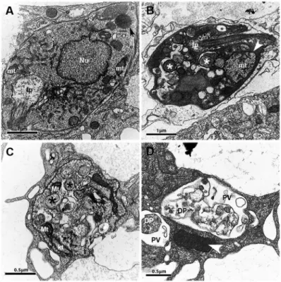
Ultrastructural alterations in L. amazonensis ex-posed to PK11195 - Treatment of infected macrophages with PK11195 caused ultrastructural alterations in in-tracellular parasites suggestive of autophagy induction, including the appearance of double membrane vesicles, compared with parasites within untreated infected con-trol macrophages, Fig. 4A, B, C). In addition, treatment with PK11195 was associated with enhanced mitochon-drial size (Fig. 4B), marked cytosolic disorganisation, and the appearance of multivesicular bodies after 24 h (Fig. 4B) and 48 h (Fig. 4C) of treatment; these features were not observed in intracellular parasites within un-treated macrophages (Fig. 4A). In addition, debris sug-gestive of dead parasites was observed inside parasi-tophorous vacuoles (Fig. 4D), whereas remarkably high electrodensity was observed in the cytosol (Fig. 4B).
Fig. 4: effects of PK11195 on the ultrastructure of Leishmania amazonensis. Macrophages were infected and then not treated (A) or treated (B-D) with 75 µM PK11195 for 24 h (B) or 48 h (C and (B-D). Black and white asterisks indicate examples of vacuoles with double membranes, black arrows indicate lipid droplets in normal cells, and white arrowheads indicate mitochondria with increased volume or high electrodensity. Nu: nucleus; kt: kinetoplast; mt: mitochondria; fp: flagellar pocket; PV: parasitophorous vacuoles; DP: dead parasite.
DISCUSSION
The present study, which endeavoured to assess the anti-leishmanial effect of a specific TSPO ligand in vitro, found that treatment with PK11195 reduced, in a time- and dose-dependent manner, not only the proliferation of axenic promastigotes and the proportion of infected macrophages in CBA mouse macrophages, but also the number of parasites per infected macrophage and the quantity of viable intracellular parasites. The reduction in axenic promastigote proliferation was observed to be generic, as similar results were seen in species causative of tegumentary leishmaniasis belonging to subgenera Leishmania (L. amazonensis and L. major) and Vian-nia (L. braziliensis) (Banuls et al. 2007). With IC50/48 h values of 14.22 µM for L. amazonensis, 3.51 µM for L. braziliensis, and 8.23 µM for L. major, which are lower than the CC50/48 h value of 194.4 µM observed in treat-ed macrophages, PK11195 was found to be capable of inhibiting the growth of all three Leishmania species tested (with SI values of 13.67 for L. amazonensis, 55.38 for L. braziliensis, and 23.62 for L. major), indicating the potential of PK11195 as a chemotherapeutic agent for treatment of cutaneous leishmaniasis.
cyto-toxic effects on a variety of cancer cell types (Shoukrun et al. 2008) have been shown to occur only at elevated (micromolar) concentrations. The fact that PK11195 ex-hibited therapeutic effect only at higher concentrations may be explained by the hydrophobicity of PK11195, which is known to bind other proteins, such as alpha-1-acidglycoprotein, which it binds with a high affin-ity (Lockhart et al. 2003), and albumin, which it binds with low affinity (Dougherty et al. 2002, Lockhart et al. 2003). Our current findings and previous report (Lock-hart et al. 2003) support the notion that PK11195 must be employed at relatively high concentrations or be associ-ated with a delivery system (Vaghela et al. 2017) to exert therapeutic effects (Tanimoto et al. 1999).
PK11195 has been used as a marker of cerebral lesions (Folkersma et al. 2011) and as an immunomodulator (Zav-ala & Lenfant 1987), and has been considered, owing to its pro-apoptotic properties, as a potential chemotherapeutic anticancer agent (Shoukrun et al. 2008). Nonetheless, the literature contains scarce reports on the use of PK11195 as an anti-parasitic molecule. Dzierszinski et al. (2002) demonstrated that PK11195 reduces the proliferation of P. falciparum and Toxoplasma gondiiin vitro. A study by Bouyer et al. (2011) found a reduced proliferation rate of P. falciparum parasites in infected erythrocytes treated with PK11195, showing results similar to those described by Dzierszinski et al. (2002). The effects of PK11195 on these parasites from the protozoan phylum Apicomplexa (Dzierszinski et al. 2002, Bouyer et al. 2011) and on try-panosomatids, as demonstrated in the present study, indi-cate that PK11195 could potentially have an anti-parasitic effect on a wide range of protozoan species.
The direct effect of PK11195 on axenically cultured promastigotes of Leishmania cannot be attributed to interaction of PK11195 with TSPO or homologues of TSPO, because there is no evidence that TSPO or homo-logues of TSPO are present within the Leishmania ge-nome. Recently, Hatty et al. (2014) showed that PK11195 interacts with lipids and is incorporated into lipid bilay-ers, and that incorporation of PK11195 into lipid bilayers alters membrane fluidity. This finding leads us to spec-ulate that the leishmanicidal effect of PK11195 may be in some way associated with alterations in the dynamic properties of the parasite plasma membrane; this pos-sibility deserves further investigation. In addition, it has already been demonstrated that PK11195 has functional effects that are independent of interaction with TSPO (Hatty et al. 2014). However, the exact mechanisms by which this TSPO ligand acts against Leishmania infec-tion require further investigainfec-tion.
In contrast with the observations of Jayakumar et al. (2002) that PK11195 increases free radical productions on neuronal cells, in the present study, PK11195 caused a reduction in the release of O2●- by plasma-membrane NADPH oxidase. Our observations are similar to those of Zavala and Lenfant (1987), who demonstrated that pro-duction of O2●- by P388D1 macrophages was attenuated by treatment with arachidonic acid. In addition, we ob-served that treatment of infected CBA macrophages with PK11195 significantly reduced production of the macro-phage attractant chemokine MCP-1; this finding is
consis-tent with a report published by Bribes et al. (2003), which showed that treatment of MRL/1pr mice with PK11195 reduced the amount of inflammatory infiltrate in a mouse model of pulmonary inflammation. In the future, it will be necessary to use an in vivo model of Leishmania infec-tion to evaluate whether treatment with PK11195 reduces both O2●- and MCP-1 levels and, as a result, dampens the inflammatory response via reduction of O2●- toxicity and inflammatory cell recruitment, respectively. This effect could be particularly beneficial in treating lesions result-ing from L. braziliensis infection, in which ulceration and intense inflammation are observed.
The morphological alterations seen in intracellular L. amazonensis promastigotes following treatment with PK11195 suggest that cell death may have occurred via multiple mechanisms. The observed swelling of mito-chondria and kinetoplasts is suggestive of apoptosis or necrosis, whereas the appearance of double-membrane vacuoles containing degraded material, the presence of multivesicular bodies, and the presence of vesicles in flagellar pockets are alterations suggestive of autophagy. Similar alterations have been described in L. amazonensis (Rodrigues et al. 2005), Trypanosoma cruzi (Braga et al. 2004), and Leishmania infantum (Granthon et al. 2007) treated either with 3-(biphenyl-4-yl)-3-hydroxyquinucli-dine, a potent inhibitor of squalene synthase, which is a key enzyme in the metabolism of ergosterol (Granthon et al. 2007), or with ketoconazole, another drug that affects the availability of protozoan ergosterol (Vannier-Santos et al. 1995). In the present study, ultrastructural analy-sis showed that the intracellular parasites also exhibited double-membrane vesicles, marked cytosolic disorganisa-tion, and the presence of multivesicular bodies, in addi-tion to remarkably high electrodensity in the cytosol and increased amounts of debris suggestive of dead parasites.
Although relatively high doses of PK11195 are re-quired to treat Leishmania infection in vitro, these levels are much lower than those that cause toxicity to mac-rophages. In conclusion, the present in vitro study has demonstrated the potential of PK11195 as an anti-leish-manial candidate. Further studies are required to evalu-ate the activity of this TSPO ligand in vivo.
ACKNOWLEDGEMENTS
To Drs Adriana Lanfredi Rangel, Claudio Pereira Figuei-ra, and Maria Lucia Vieira Moreno, for technical support with electron microscopy imaging and analysis. Finally, we are grateful to Andris K Walter for providing English revision and consulting services.
AUTHORS’ CONTRIBUTION
CESG, BRSD and PSTV - Conceived and designed the ex-periments; CESG, BRSD, ALOAP, KPC, NJA and DRA - per-formed the experiments; CESG, BRSD, ALOAP, JPBM, VMB and PSTV - analysed the data; VMB and PSTV - contributed reagents/materials/analysis tools; CESG, BRSD and PSTV - wrote the paper.
REFERENCES
Banuls AL, Hide M, Prugnolle F. Leishmania and the leishmaniases: a parasite genetic update and advances in taxonomy, epidemiol-ogy and pathogenicity in humans. Adv Parasitol. 2007; 64: 1-109.
Bouyer G, Cueff A, Egee S, Kmiecik J, Maksimova Y, Glogowska E, et al. Erythrocyte peripheral type benzodiazepine receptor/volt-age-dependent anion channels are upregulated by Plasmodium falciparum. Blood. 2011; 118(8): 2305-12.
Braga MV, Urbina JA, de Souza W. Effects of squalene synthase in-hibitors on the growth and ultrastructure of Trypanosoma cruzi. Int J Antimicrob Agents. 2004; 24(1): 72-8.
Bribes E, Bourrie B, Casellas P. Ligands of the peripheral benzodiaz-epine receptor have therapeutic effects in pneumopathies in vivo. Immunol Lett. 2003; 88(3): 241-7.
Courret N, Frehel C, Prina E, Lang T, Antoine JC. Kinetics of the intracellular differentiation of Leishmania amazonensis and in-ternalization of host MHC molecules by the intermediate parasite stages. Parasitology. 2001; 122(Pt 3): 263-79.
Croft SL, Olliaro P. Leishmaniasis chemotherapy - challenges and op-portunities. Clin Microbiol Infect. 2011; 17(10): 1478-83.
de Sá MS, Costa JF, Krettli AU, Zalis MG, Maia GL, Sette IM, et al. Antimalarial activity of betulinic acid and derivatives in vitro against Plasmodium falciparum and in vivo in P. berghei -infect-ed mice. Parasitol Res. 2009; 105(1): 275-9.
de Souza VL, Souza JA, Silva TMC, Veras PST, de-Freitas LAR. Dif-ferent Leishmania species determine distinct profiles of immune and histopathological responses in CBA mice. Microbes Infect. 2000; 2(15): 1807-15.
Domingues-Junior M, Pinheiro SR, Guerra JL, Palermo-Neto J. Ef-fects of treatment with amphetamine and diazepam on Mycobac-terium bovis-induced infection in hamsters. Immunopharmacol Immunotoxicol. 2000; 22(3): 555-74.
Dougherty TJ, Sumlin AB, Greco WR, Weishaupt KR, Vaughan LA, Pandey RK. The role of the peripheral benzodiazepine receptor in photodynamic activity of certain pyropheophorbide ether pho-tosensitizers: albumin site II as a surrogate marker for activity. Photochem Photobiol. 2002; 76(1): 91-7.
Dzierszinski F, Coppin A, Mortuaire M, Dewailly E, Slomianny C, Ameisen JC, et al. Ligands of the peripheral benzodiazepine re-ceptor are potent inhibitors of Plasmodium falciparum and Toxo-plasma gondii in vitro. Antimicrob Agents Chemother. 2002; 46(10): 3197-207.
Folkersma H, Boellaard R, Yaqub M, Kloet RW, Windhorst AD, Lammertsma AA, et al. Widespread and prolonged increase in (R)-(11)C-PK11195 binding after traumatic brain injury. J Nucl Med. 2011; 52(8): 1235-9.
Gomes IN, Calabrich AF, Tavares RS, Wietzerbin J, de Freitas LA, Ve-ras PS. Differential properties of CBA/J mononuclear phagocytes recovered from an inflammatory site and probed with two differ-ent species of Leishmania. Microbes Infect. 2003; 5(4): 251-60.
Granthon AC, Braga MV, Rodrigues JC, Cammerer S, Lorente SO, Gilbert IH, et al. Alterations on the growth and ultrastructure of Leishmania chagasi induced by squalene synthase inhibitors. Vet Parasitol. 2007; 146(1-2): 25-34.
Hatty CR, Le Brun AP, Lake V, Clifton LA, Liu GJ, James M, et al. Investigating the interactions of the 18kDa translocator protein
and its ligand PK11195 in planar lipid bilayers. Biochim Biophys Acta. 2014; 1838(3): 1019-30.
Jayakumar AR, Panickar KS, Norenberg MD. Effects on free radical generation by ligands of the peripheral benzodiazepine receptor in cultured neural cells. J Neurochem. 2002; 83(5): 1226-34.
Lockhart A, Davis B, Matthews JC, Rahmoune H, Hong G, Gee A, et al. The peripheral benzodiazepine receptor ligand PK11195 binds with high affinity to the acute phase reactant alpha1-acid glyco-protein: implications for the use of the ligand as a CNS inflam-matory marker. Nucl Med Biol. 2003; 30(2): 199-206.
McEnery MW, Snowman AM, Trifiletti RR, Snyder SH. Isolation of the mitochondrial benzodiazepine receptor: association with the voltage-dependent anion channel and the adenine nucleotide car-rier. Proc Natl Acad Sci USA. 1992; 89(8): 3170-4.
Menezes JP, Almeida TF, Petersen AL, Guedes CE, Mota MS, Lima JG, et al. Proteomic analysis reveals differentially expressed pro-teins in macrophages infected with Leishmania amazonensis or Leishmania major. Microbes Infect. 2013; 15(8-9): 579-91.
Papadopoulos V, Baraldi M, Guilarte TR, Knudsen TB, Lacapere JJ, Lin-demann P, et al. Translocator protein (18kDa): new nomenclature for the peripheral-type benzodiazepine receptor based on its structure and molecular function. Trends Pharmacol Sci. 2006; 27(8): 402-9.
Rodrigues JC, Urbina JA, de Souza W. Antiproliferative and ultrastruc-tural effects of BPQ-OH, a specific inhibitor of squalene synthase, on Leishmania amazonensis. Exp Parasitol. 2005; 111(4): 230-8.
Selleri S, Bruni F, Costagli C, Costanzo A, Guerrini G, Ciciani G, et al. 2-Arylpyrazolo[1,5-a]pyrimidin-3-yl acetamides. New potent and selective peripheral benzodiazepine receptor ligands. Bioorg Med Chem. 2001; 9(10): 2661-71.
Shoukrun R, Veenman L, Shandalov Y, Leschiner S, Spanier I, Karry R, et al. The 18-kDa translocator protein, formerly known as the peripheral-type benzodiazepine receptor, confers proapoptotic and antineoplastic effects in a human colorectal cancer cell line. Pharmacogenet Genomics. 2008; 18(11): 977-88.
Tanimoto Y, Onishi Y, Sato Y, Kizaki H. Benzodiazepine receptor agonists modulate thymocyte apoptosis through reduction of the mitochondrial transmembrane potential. Jpn J Pharmacol. 1999; 79(2): 177-83.
Totis M, Kremers P, Batt AM, Van Cantfort J, Siest G, Gielen J. In-duction of liver microsomal cytochrome P-450 isozymes by 1-(2-chlorophenyl)-N-methyl-N-(1-methylpropyl)-3-isoquino-line carboxamide. Xenobiotica. 1989; 19(8): 857-66.
Vaghela R, Kulkarni PK, Osmani RA, Bhosale RR, Varma VNSK. Recent advances in nanosystems and strategies for managing leishmaniasis. Curr Drug Targets. 2017; 18(14): 1598-1621.
Vannier-Santos MA, Urbina JA, Martiny A, Neves A, de Souza W. Alations induced by the antifungal compounds ketoconazole and ter-binafine in Leishmania. J Eukaryot Microbiol. 1995; 42(4): 337-46.
Veenman L, Papadopoulos V, Gavish M. Channel-like functions of the 18-kDa translocator protein (TSPO): regulation of apoptosis and steroidogenesis as part of the host-defense response. Curr Pharm Des. 2007; 13(23): 2385-405.
WHO - World Health Organization. Control of the leishmaniases. World Health Organ Tech Rep Ser. 2010; 949): xii-xiii, 1-186, back cover.