Antibacterial activity of
a
-terpineol may induce morphostructural alterations
in
Escherichia coli
Li Li
1*, Chaofeng Shi
1*, Zhongqiong Yin
1*, Renyong Jia
1,2, Lianci Peng
1, Shuai Kang
1,
Zhengwen Li
11
College of Veterinary Medicine, Sichuan Agricultural University, Yaan, China. 2
Key laboratory of Animal Disease and Human Health of Sichuan Province, Sichuan Agricultural University, Ya’an , China.
Submitted: October 22, 2013; Approved: April 17, 2014.
Abstract
The antibacterial effect ofa-terpineol fromCinnamomum longepaniculatum(Gamble) N. Chao leaf
essential oils were studied with special reference to the mechanism of inhibiting the standard strain of
Escherichia coli(CMCC (B) 44102) growth at ultrastructural level. Minimal inhibitory concentra-tion (MIC) and minimal bactericidal concentraconcentra-tion (MBC) and time-kill curves ofa-terpineol were
determined;Escherichia coliwas treated witha-terpineol and observed under a transmission
elec-tron microscope. The MIC and MBC values ofa-terpineol were all 0.78mL/mL, and time-kill curves
showed the concentration-dependent. Under the transmission electron microscopy (TEM), Esche-richia coliexposed to MIC levels ofa-terpineol exhibited decreased cell size and irregular cell shape,
cell wall and cell membrane were ruptured, nucleus cytoplasm was reduced and nuclear area gath-ered aside. Results suggest thata-terpineol has excellent antibacterial activity and could induce
mor-phological changes ofEscherichia coli.
Key words:a-terpineol,Escherichia coli, antibacterial activity, TEM.
Intruduction
In recent years there is increasing incidence of multi-ple resistance in human and animal pathogenic microor-ganisms, largely due to the indiscriminate use of commer-cial antimicrobial drugs commonly employed in the treatment of animal and human infectious diseases (Zewdu and Cornelius, 2009). So it is necessary to screen new antimicrobial substances from various sources like medici-nal plants (Ghasemiet al., 2010).
Cinnamomum longepaniculatum(Gamble) N. Chao ex H. W. Li belongs to theLauraceaeand grows widely in Sichuan and Taiwan of China (Taoet al., 2002). Prelimi-nary studies have showed that theC. longepaniculatumleaf essential oils has strong antibacterial activities (Weiet al., 2009; Taoet al., 2011). It is often employed by the pharma-ceutical industry in drug formulations, as a percutaneous penetration enhancer and for its decongestant and
anti-tussive effects (Williamset al., 1991; Levisonet al., 1994). In Sichuan Province,a-terpineol is the major component of
C. longepaniculatum leaf essential oils, comprising ca 15.43% (Huanget al., 1986) of whole oil. Meanwhile, the effect on the ultrastructural structure ofa-terpineol against
bacteria has never been reported. So the objective of this study is to evaluate potential antibacterial activity ofa
-ter-pineol and investigate under a transmission electron micro-scope (TEM) the morphological changes in Escherichia colias a result of exposure toa-terpineol.
Materials and Methods
Microbial strain and chemicals
Standard strain of Escherichia coli (CMCC (B) 44102) was supplied by Department of Food Science and Technology Sichuan Agricultural University, Ya’an,
Send correspondence to Z. Q. Yin. College of Veterinary Medicine, Sichuan Agricultural University, 625014 Ya’an, PR China. E-mail: [email protected].
*This authors contribute equally to this work.
China. Mueller-Hinton broth and Nutrient agar medium were purchased from Beijing Aobo Star Biotechnology Company Limited.a-Terpineol (99%) was obtained from
Sichuan Perfumery Co., Ltd, Yibin, China.
Antibacterial susceptibility test
MIC and MBC values ofEscherichia coliwere deter-mined by broth dilution method described in the National Committee for Clinical Laboratory Standards (NCCLS, 2008). The test samples were dissolved in 2.5% Tween-80 at a final concentration ranging from 12.5 mL/mL to
0.098mL/mL. 2.5%Tween-80 was used as negative
con-trol. Measured the values of OD600ofa-terpineol by UV
spectrophotometer and measured once again after 24 h of incubation at 37 °C. The test tube with the same value of OD600after 24 h, showing that there were noEscherichia
colicolonies growing that is the value of MIC (Hu.et al., 2009). MBC was estimated as the least concentration of the samples where no visible growth on nutrient agar medium. All the experiments were performed in triplicate and the re-sults were averaged.
Time-kill curve study ofa-terpineol
Time-kill curve studies were performed in duplicates with thea-terpineol and broth culture prepared on the day
of the experiment at 2 different levels as follows: 1 x MIC and 2 x MIC.a-terpineol at various levels and with growth
control group were seeded with a log-phase inoculum of roughly 1 x 10⁷cfu/mL to a final volume of 10 mL. Inocu-lated broths were incubated at 37 °C. 0.1 mL of inocuInocu-lated broths were sampled at different time intervals (0, 2, 4, 8, 12, 24 and 36 h) from each tube and was subjected to 10-fold serial dilution. Then, 0.1 mL of every dilution was spread on Mueller-Hinton plates and incubated at 37 °C for 24 h. Only plates containing theEscherichia colicolonies amounts between 30-300 for each series of dilutions were counted. Finally, the time-kill curve was constructed for or-ganism (Zhanget al., 2010).
Transmission electron microscopy
10 mL of 107cfu/mL Escherichia colisuspensions were exposed to MIC concentration ofa-terpineol and then
were incubated at 37 °C for 1 h, 2 h and 4 h in an incubator shaker. The control group was treated with solvent only. TheEscherichia colisuspensions were centrifuged in ster-ile plastic centrifuge tubes at 8000gfor 15 min at 4 °C and were washed with three consecutive (15 min) washes with cacodylate-buffer. Then the supernatant was discarded and the pelleted cellular content was fixed with 2.5% gluta-raldehyde in 0.1 mol/L cacodylate-buffer (pH = 7.2) at 4 °C overnight. Samples were then post-fixed for 2 h in 1% os-mium tetroxide (OsO4) dissolved in cacodylate-buffer at room temperature and washed in cacodylate-buffer (three
times, 15 min each). Samples were dehydrated in ethanol in a graded series of 40%, 60%, 75%, 80% and 95% dilutions for 15 min each. A final dehydration step was carried out for 1 h in 100% ethanol with changes every 30 min. Epoxy resin (Epon-618) was used to embed the post-fixed samples for 12 h-16 h at 45 °C. Ultra thin section of the embedded samples were prepared by LKB-II Ultracut instrument and double stained with uranyl acetate and lead citrate. Mor-phology of the Escherichia colicells was observed on a transmission electron microscope (Taoet al., 2011).
Results and Discussion
Antibacterial activity ofa-terpineol
The MIC and MBC values of a-terpineol against
Escherichia coliwere all 0.78mL/mL. It means thata
-ter-pineol might inhibit the growth ofEscherichia coliby kill-ing bacteria directly.
Study has shown that (Wei, 2009) the C.
longepaniculatumleaf essential oils has strong antibacte-rial activity onEscherichia coli,which the MIC and MBC value of C. longepaniculatum leaf essential oils was 3.13mL/mL. Compared withC. longepaniculatum leaf
es-sential oils,a-terpineol has a stronger antibacterial activity
thanC. longepaniculatumleaf essential oils. Moreover, the antimicrobial activity of a-terpineol on Salmonella
enteritidis and Staphylococcus aureus has been investi-gated in our laboratory. The result showeda-terpineol has
the same strong antibacterial activities againstSalmonella enteritidisandStaphylococcus aureus, which the MIC and MBC values were 1.56mL/mL and 3.13 mL/mL
respec-tively. Otherwise, a-terpineol also exhibited strong
anti-microbial activity against periodontopathic and cariogenic bacteria (Park, 2012).
Time-kill curve ofa-terpineol againstEscherichia coli
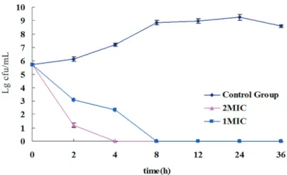
Time-kill curves of a-terpineol (Figure 1) showed
that the time-kill curve of control group ofa-terpineol had
the integral growth cycle (lag phase, logarithmic phase, sta-tionary phase and death phase). But at 1 x MIC and 2 x MIC concentration ofa-terpineol,Escherichia colidirectly
en-tered into decline phase without adjustment phase; loga-rithmic phase and stable phase. All the bacterial cells of
Escherichia coliwere killed bya-terpineol at 1 x MIC in
8 h and 2 x MIC in 4 h, the rate of killing increased by in-creasing the concentration of a-terpineol. The resulting
time-kill curve approach described the interaction between bacteria anda-terpineol in a dimensional way by a dynamic
Ultrastructural changes ofEscherichia coli
A transmission electron microscopic study of un-treated cells ofEscherichia colishowed a typical healthy bacilli-shape and clear integrated cell structure containing a continuous smooth cell wall, cell membrane, nuclear area (Figure 2 A). Besides,Escherichia colicytoplasm uniform distributed evenly and nuclear area stained slightly (Fig-ure 2 B).
Cells ofEscherichia coliexposed to MIC concentra-tion ofa-terpineol exhibited different changes during
dif-ferent time periods. Escherichia coli cell wall and cell membrane became thinner (Figure 3A, black arrows) and cytoplasmic material showed clumping (Figure 3A, white arrows) of after 1 h; The nuclear area was on the edge of cell (Figure 3B, arrows).
After 2 h, cells of Escherichia coli exhibited de-creased size, irregular shape, thin cell wall, some cells stained slightly(Figure 4A, black arrows) and some cells stained deepen (Figure 4A, white arrows); Cytoplasm of
Escherichia colihad lost its even distribution and cells un-equal division emerge (Figure 4B, arrows).
After 4 h,Escherichia colicells were forced to gather except the decreased cell size and irregular cell shape; Cy-toplasm had lost and the most cells stained slightly
(Figu-re 5A, arrows); Plasmolysis and bubble appea(Figu-red in the cell (Figure 5B, arrows).
The main morphostructural alterations inEscherichia coliinduced bya-terpineol performanced in decreased cell
size, irregular cell shape, cytoplasm condensed, cytoplasm lost, nuclear area edged, plasmolysis, unequal division and vacuolization of cell. The research results showed that, most likely, the antibacterial mechanism ofa-terpineol on
Escherichia coliwas realized through producing alterations on the structure ofEscherichia colidirectly. The
antibacte-Figure 1- The Time-kill curves ofa-terpineol againstEscherichia coli(each experiment in triplicate).
Figure 2- TEM graph (A, x 40000) ofEscherichia colikept normal mor-phology; TEM graph (B, x 25000) ofEscherichia colicytoplasm uniform distributed evenly and nuclear area stained slightly.
Figure 3- TEM graph ofEscherichia coli exposed to MIC value of a-terpineol for 1 h.Escherichia colicell wall and cell membrane became thinner (A, x 25000, black arrows) and cytoplasmic material showed clumping (A, x 25000, white arrows). The nuclear area was on the edge of cell (B, x 25000, arrows).
rial mechanism of many anti-bacterial herbal medicines was achieved by destroying the structure of cell directly and affecting the metabolism of cell (Sun and Wu, 2007). Berberine, achieves the antibacterial effect on Staphylococ-cus aureusby inhibition of, respiratory, glucose metabo-lism, synthesis of protein and nucleic acid; The mechanism of berberine onShigella flexneriis to influence the respira-tory process by inhibiting the oxidation process of aspartic acid and sodium succinate and Allicin, interfere with bacte-rial metabolism and achieve the effect of inhibition of bac-terial growth ultimately by linking with thiol in cysteine molecules that is necessary for bacterial growth (Sun and Wu, 2007). The marked action of oil components might have conferred lipophilic properties and the ability to pene-trate the plasma membrane (Knoblochet al., 1989), which can cause the changes of membrane permeability and loss of the important nutrients in the cell such as enzymes, phos-phoric acid, electrolytes, amino acids and nucleic acids, leading to cell death (Sangethaet al., 2009; Rasooliet al., 2006; Pothakamuryet al., 1997). As tea tree oil, carvacrol and thymol were all known to disrupt the cytoplasmic membrane and increase its permeability and depolarizes its potential (Xuet al., 2008). The results are comparable to the action ofa-terpineol. Besides, It was showed that the
presence of free hydroxyl group is essential for antimicrobial activity of carvacrol and that this compound could act as a protonophore (Ben Arfaet al., 2006), which is applicable fora-terpineol.
In conclusion, this study demonstrates that a
-ter-pineol has excellent antibacterial activitiy and the antibac-terial mechanism of a-terpineol against Escherichia coli
was realized through producing alterations on the structure ofEscherichia colidirectly. It might have good potential to be used for medical purposes.
Acknowledgments
This study was supported by Sichuan Youth Science and Technology Innovation Research Team for waterfowl disease prevention and control (2013TD0015), and Na-tional Natural Science Foundation of China (Grant No.31372477). The authors thank Department of Food
Sci-ence and Technology, Sichuan Agricultural University, Ya’an, China for supplying microorganism bacterium.
References
Ben Arfa A, Combes S, Preziosi-Belloy L, Gontard N, Chalier P (2006) Antimicrobial activity of carvacrol related to its chemical structure. Lett Appl Microbiol 43:149-154. Ghasemi PA, Jahanbazi P, Enteshari S, Malekpoor F, Hamedi
B(2010) Antimicrobial activity of some Iranian medicinal plants. Arch Biol Sci 62:633-641.
Hu HS, Hu HB, Zheng XD (2009) Study on chemical constituents and antimicrobial activity of the essential oil from
Acanthopanax brachypus. J Chinese Med Mater 32:67-70. Knobloch K, Pauli A, Iberl B, Weigand H, Weis N (1989)
Anti-bacterial and antifungal properties of essential oil compo-nents. J Essent Oil Res 1:119-128.
Levison KK, Takayama K, Okabe K, Nagai T (1994) Formulation optimization of indomethacin gels containing a combination of three kinds of cyclic monoterpenes as percutaneous pene-tration enhancers. J Pharm Pharmacol, 83:1367-1372. National Committee for Clinical Laboratory Standards (2008)
Performance standards for antimicrobial susceptibility test-ing. Ninth International Supplement M100-S9; Wayne, PA. Pothakamury U, Barbosa-Cánovas G, Swanson B, Spence K
(1997) Ultrastructural changes in Staphylococcus aureus
treated with pulsed electric fields. Food Sci Tech Int 3:113-121.
Pu ZH, Zhang YQ, Yin ZQ, Xu J, Jia RY, Lu Y, Yang F (2010) Antibacterial activity of 9-octadecanoic acid-hexadecanoic acid-tetrahydrofuran-3, 4-diyl ester from neem oil. Agr Sci China 9:1236-1240.
Park SN, Lim KY, Freire MO, Cho E, Jin DC, Kook JK (2012) Antimicrobial effect of linalool anda-terpineol against
pe-riodontopathic and cariogenic bacteria. Anaerobe 3:369-372.
Rasooli I, Rezaei MB, Allameh A (2006) Ultrastructural studies on antimicrobial efficacy of hyme essential oils onListeria monocytogenes. Int J Infect Dis 10:236-241.
Sun J, Wu GJ (2007) The research progress on Antibacterial mechanism of traditional hinese medicine. Chinese J Vet Med 43:42-43.
Sangetha S, Zuraini Z, Suryani S, Sasidharan S (2009) In situ TEM and SEM studies on the antimicrobial activity and pre-vention of Candida albicans biofilm by Cassia spectabilis extract. Micron 40:439-443.
Tao C, Wei Q, Yin ZQ (2011) Antifungal activity of the essential oil from Cinnamomum longepaniculatum leaves against three species of fungi. Chinese Vet Sci 41:89-93.
Tao GF, Ding JK, Sun HD (2002) The Chemical Constituents of the Essential Oil from Leaves of Cinnamomum longepaniculatum in Hubei, China. J Wuhan Bot Res 20:75-77.
Williams AC, Barry BW(1991) Terpenes and the lipid-protein-partitioning theory of skin penetration enhancement. Pharm Res 8:17-24.
Wei Q, Zhou YK, Zhou NJ, Yin LG, Zhang P (2009) Inhibitive Activity ofCinnamomum oil against bacteria. Chinese J Tropical Agr 1:5-7.
Xu J, Zhou F, Ji BP, Pei RS, Xu N (2008) The antibacterial mech-anism of carvacrol and thymol againstEscherichia coli. Lett Appl Microbiol 47:174-179.
Zewdu E, Cornelius P (2009) Antimicrobial resistance pattern of Salmonella serotypes isolated from food items and person-nel in Addis Ababa, Ethiopia. Trop Anim Health Pro 41:241-249.
Zhang YQ, Xu J, Yin ZQ, Jia RY, Lu Y, Yang F, Du YH, Zou P, Lv C, Hu TX (2010) Isolation and identification of the anti-bacterial active compound from petroleum ether extract of neem oil. Fitoterapia 81:747-750.