M
EDICAL
J
OURNAL
How should PCNA be assessed? Total of stained cells or only
the most intensely stained ones?
Departments of Gynecology and Pathology, Escola Paulista de Medicina, Universidade Federal de São Paulo - São Paulo, Brazil Claudio Kemp, Vânia Nosé Alberti, Geraldo Rodrigues de Lima, Filomena Marino de Carvalho
Address for correspondence: Claudio Kemp
Av. dos Carinás, 408 - Moema
São Paulo/SP - Brasil - CEP 04086-010
INTRODUCTION
R
ecent research has attempted to exploit data on the fractions of tumor growth and proliferating cells, thus obtaining a probable measure of tumor aggressiveness, with the purpose of establishing new prognostic indicators.From this information, various methods have been devised for measuring the proliferative activity of breast carcinomas: mitotic counts, index of tritiated thymidine, BrdU, computerized or flow cytometry.
Immunohistochemical methods involving monoclonal antibodies have been introduced to facilitate studies in this field.
In 1978, Miyachi et al(1) identified an antibody in the
serum of some patients with systemic erythomatous lupus that reacted with a nuclear antigen of proliferating cells (PCNA - Proliferating Cell Nuclear Antigen). This antigen was later characterized as a 36KDa polypeptide and described as a specific auxiliary protein of the DNA polymerase d, necessary for its catalytic activity(2,3).
A monoclonal antibody for this protein was obtained and retrospective analysis on tumor tissues was then conducted. These results were similar to the ones attained by flow cytometry of the same tumor(4,5).
In this way, several investigators reported on the significance of PCNA, using the mitotic index, grade of nuclear and histological differentiation, cancer-free survival and overall survival. They also observed substantial correlations with other markers of proliferative tumor activity such as: Ki67, incorporation of tritiated thymidine, incorporation of bromodeoxyuridine, flow cytometry and computerized static cytometry. Furthermore, correlations were also encountered with the protein c-erbB-2, the
Objective: This study aimed to analyse whether a marker of proliferative activity (PCNA) could provide a prognosis of tumor evolution and to determine whether different interpretation criteria could alter the results. Method: The presence of PCNA in 59 patients of state II (T2 NO,1 MO) mammary carcinoma was determined. Result: Numerical proportions of total and intensely stained cells were established. These data were compared with anatomopathological parameters. A significant association between higher cyclin values and worse histological and nuclear grading was encountered, particularly in patients with a “negative axilla” using the PCNA index. Cyclin values were not significant in relation to any parameters when indices from the intensely stained cells were considered exclusively. Conclusion: Higher nuclear (NG3) and histological (HGIII) grading, associated with a high PCNA index (>50), distinguish high-risk patients, and it is more appropriate considering all the stained cells as representative of PCNA indices, thus reflecting tumor aggressiveness.
expression of tumor suppressor gene p53, the receptor for EGF and the absence of hormonal receptors. These researchers emphasized that this method originated reproducible and reliable results, its technique was easily adaptable to laboratorial routines and, above all, involved low costs(5,6,7,8,9,10,11,12).
Nevertheless, other authors did not confirm these findings. Their results disagreed with the ones mentioned or even revealed a total absence of correlation with any parameter assessing the prognosis of breast cancer(13,14,15).
Since then studies have been published in the literature always emphasizing the correlation of PCNA-immunoreactivity with prognostic factors, discussing the controversies of the method and questioning the possible reasons for divergent results.
It is generally known that 50% of patients with T2 N0,1 M0 present non-compromised axilla, and, from these cases, 29% undergo unfavourable evolution within five years. However, this prognosis could be altered with the adoption of adjuvant chemotherapy(16).
Considering that the anatomopathological parameters are insufficient to support this indication, this study aimed to analyse whether the association with a marker of proliferative activity (PCNA) could provide a prognosis of the tumor evolution and possibly suggest the type of therapeutics needed following mastectomy. At the same time, the study also investigated whether different interpretation criteria could alter the results.
METHODS
Patients
A total of 59 patients with primary breast carcinoma in clinical state II treated in the Mastology section of the Gynecology Department of Escola Paulista de Medicina between July 1985 and July 1994 were selected to take part in the study.
The cases included corresponded to state II (T2 N0 M0,T2 N1 M0) according to the T.N.M. system as established by the International Union Against Cancer (1988).
The patients were classified into two groups. The first one (“Positive Axilla”; 27 patients) presented neoplastic compromise of the lymph nodes, determined by the anatomopathological examination, independent of the number of compromised lymph nodes. The second group (“Negative Axilla”; 32 patients), consisted of those who did not present metastasis in the axillary lymph nodes.
The patients’ average ages in the “Positive” and “Negative Axilla” groups were 53 and 56.6, respectively. The average tumor diameter in the “Positive Axilla” group was 3.2 cm and in the “Negative Axilla” group, 3.3 cm.
Anatomopathological Method
All material submitted to histological examination was previously fixed in a 10% solution of saturated liquid formaldehyde.
The slides were prepared in conformity with routine techniques of the Pathology Department of Escola Paulista de Medicina.
The slides corresponding to the primary tumor were examined under optical microscope to determine the following histological features: histological type, histological grade, nuclear grade, mitotic index, necrosis and vascular neoplastic embolization. Some of these parameters, histological grade, nuclear grade and mitosis were analysed quantitatively and semi-quantitatively(17,18).
The analysis of the histological variables was performed by two pathologists who had not been previously informed of the groups to which the patients belonged.
Immunohistochemical Method
The histological sections of the selected fragments were performed in the Pathology Department of Escola Paulista de Medicina and sent to the Immunopathology section for immunohistochemical processing by the Avidin-Biotin-peroxidase method (A.B.C.), as described by Hsu et al(19).
Each reaction included a positive and a negative control. The slides with positive controls were prepared with tissues known to be positive for the studied antigens, such as tonsil and intestinal mucosa. The negative-control slides were prepared from the blocks of the studied cases where, instead of using the primary antigen, a non-immune mouse serum was used.
The primary antibody employed was the anti-PCNA monoclonal antibody PC-10 from Dako (Denmark Dakopatts A/S) code M879, lot 121, previously tested and standardized for the dilution 1:80.
Immunohistochemical Interpretation
Table1
Percentages of the total of stained cells and the most intensely stained ones evaluated among 1000 cells
in patients with state II breast carcinoma with positive or negative axilla.
% Total of % The most stained cells intensely stained cells Axilla + Axilla - Axilla + Axilla
-75.9 76.5 35.0 33.6 64.4 50.7 23.6 14.3 66.0 40.1 23.4 15.0 50.5 34.2 18.6 3.0 43.3 71.5 4.3 24.7 48.0 10.8 4.9 2.6 73.0 75.0 36.5 16.4 51.8 50.6 12.9 12.5 65.1 39.9 19.4 3.91 41.9 33.4 9.5 8.6 49.8 59.1 2.2 17.5 56.5 38.0 6.4 1.4 36.7 27.8 0.8 7.1 91.6 70.8 73.2 6.7 33.9 58.9 3.6 6.6 60.3 10.1 5.7 0.9 45.5 29.1 6.0 3.7 74.6 36.3 11.0 1.9 60.5 10.0 18.4 0.0 38.4 70.5 8.2 14.1 44.4 73.6 4.0 12.1 79.5 40.0 11.8 3.6 37.8 51.7 4.5 11.2 69.9 50.4 27.4 2.6 73.3 54.8 22.0 7.3 69.2 63.0 19.0 1.0 62.6 30.4 32.3 4.5 61.7 18.8 28.0 8.5 48.6 16.0 48.6 12.1 65.8 18.9 MEAN 57.9 47.2 16.5 9.5 Mann-Whitney Test with approximation to the normal curve (axilla+ X axilla-)
critical Z = 1.96
total of stained cells the most intensely stained cells calculated Z = 2.01* calculated Z = 1.88 axilla + > axilla
-The staining intensity was subjectively analysed and the stained nuclei were quantified through the study of 1000 cells. This procedure was conducted with the help of a 100x immersion objective with a final magnification of 1000x .
The reaction was considered positive when nuclear staining occurred in a diffuse way (dot matrix of variable intensity) or granular (in clumps or homogeneously distributed)(2,3).
Nuclei counts were conducted without the author’s previous knowledge of the histological variables and the groups to which each case belonged.
The number of stained nuclei was counted to a total of 1000 cells, taking into account an area previously selected as the most representative, to give the PCNA index.
At the same time, the staining intensity was determined semi-quantitatively as poor, moderate or intense. The expression of the most intensely stained nuclei was as a percentage referring only to the most intensely stained ones of the 1000 cells.
From the initial 60 cases, only one presented a negative reaction and was then consequently excluded from the study. Thus, 59 patients were analysed.
Statistical Method
Non-parametric tests were utilized to assess the results, considering the nature of the studied variables. The following tests were applied: 1. Kruskal-Wallis rank variance analysis, 2. Mann-Whitney test to compare the “Positive” and “Negative Axilla” groups(20).
All the tests fixed the level for rejection of the null hypothesis at 0.05 or 5% (P < 0.05), marking significant values with an asterisk (*).
RESULTS
Correlation of patients’ ages and presence or absence of necrosis and vascular embolization with positive or negative axilla, evaluated by the Mann-Whitney or Kruskal-Wallis tests, revealed that the PCNA index did not show significant differences, neither for the total of stained cells nor for the most intensely stained ones.
DISCUSSION
The analysis of the 60 initial cases revealed that only in one did the reaction not occur. The corresponding slide of hematoxylin and eosin (H.E.) also exhibited poor tissue preservation, probably due to inadequate fixation.
Homogeneously attenuated reactions observed in another 7 cases may have been due to a prolonged fixation process, as the direct visualization of the reaction on the slides permitted the observation of good tissue conditions and our consequent trust in the stained cells counts.
Fifty-two (86.7%) of the studied specimens presented excellent results; and this figure reaches 98.4% when we consider those 7 less reactive cases.
The concomitant presence of poor, moderate and intensely-stained nuclei could be observed on analysis of the remaining 59 patients. The aspect encountered was diffuse or granular, or both. A heterogeneous distribution pattern with predominant reaction areas could also be observed.
The heterogeneous pattern found among the samples and within the same sample is inherent to the
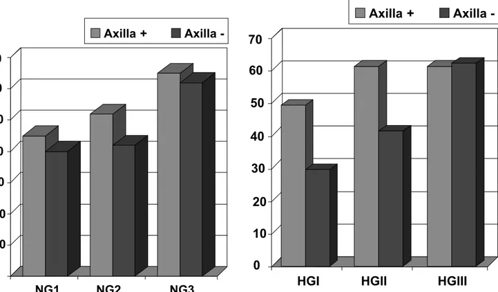
Figure 1 - Percentages of the total of stained cells (% PCNA) evaluated among 1000 cells in patients with state II breast carcinoma grouped according to nuclear grades NG1, NG2 and NG3 with positive or negative axilla.
The Kruskal-Wallis test demonstrated that among the nuclear grades NG1, NG2 and NG3 of the “Positive Axilla” group, the tumors of NG3 presented a significantly higher index of the total of stained cells (PCNA) when compared to the ones encountered in NG1 (Fig.1).
Concerning the nuclear grades NG1, NG2 and NG3 of the “Negative Axilla” group, the results suggested that the index of the total of stained cells (PCNA) in the NG3 were higher than the ones in NG2 and NG1.
Furthermore, correlations of the histological grades HGI, HGII and HGIII with positive or negative axilla (Fig.2) established by the Kruskal-Wallis test revealed that the index of the total of stained cells (PCNA) in the “Negative Axilla” group was significantly higher in the patients presenting tumors of histological grade HGIII than in patients with HGII and HGI tumors.
The same test applied among the patients of the “Positive Axilla” group demonstrated that the percentages of the total of stained cells (PCNA) in the histological grades HGI, HGII and HGIII were not significantly different.
The Mann-Whitney test and Kruskal-Wallis analysis did not show significant differences between the most intensely stained cells and histological and nuclear grades within either of the groups studied (“Positive Axilla” and “Negative Axilla”).
immunoreactivity with PCNA. This happens because tumor cells cycle asynchronously and cyclin expression in each cell relies on the phase of the cycle in which it is found(2).
Therefore, variability of reaction aspects or intensity among the samples or even within the same field is expected(5).
Observation of the above information provides support for establishing criteria to be applied in the various studies and from this, interpretation of the different results is enabled.
Most authors considered the most representative fields, ie those containing the highest numbers of positive nuclei. Total cell counts ranged from 200 to 1000, always evaluating the proportion of stained nuclei over total counts and then expressing the PCNA percentage. According to these authors, these figures would provide a measure of the maximum proliferative tumor activity(5,9,10,13,14,15,21).
At the same time, several authors considered all the stained nuclei as positive independently of staining intensity, thus aiming to widely represent the S-phase of the cellular cycle(2,4,5,9,10,13,15,21).
Other authors considered solely the most intensely stained nuclei or only those presenting granular aspect, eliminating weakly stained ones or those with diffuse staining(8,12,22).
Having found heterogeneous fields, some researchers made use of the mean of the are mas presenting higher and lower counts of stained nuclei(4,14,21).
In accordance with this procedure, the mean of the areas from a total of 1000 evaluated cells(7) or a random
observation of 10 fields(6) was applied in computerized
image analysis.
With the aim of analysing the resultant data, the authors made use of the PCNA percentages divided into quartiles: 0-25%, 26-50%, 51-75%, 76-100%(5,9); or with
a slight modification: < 19%, 20-39%, 40-61%, > 62%(15).
Other investigators reported cyclin expression as high or low in accordance with a cut-off value, which was statistically determined as 18(12), 25(14), 30(8,11), or expressing
high significance when over 50(11).
Two different monoclonal antibodies were used: 19A2 diluted in a proportion of 1/800 and 1/1000(4,6), and PC-10
in dilutions ranging from 1/15 to 1/800(5,7,8,9,10,12,13,15,21).
In the present study, we selected the most representative areas, successively counting nuclei in contiguous fields. Areas demonstrating necrosis or intense demoplasic reaction, where cell preservation or quantity were affected independently of staining, were discarded in an attempt to make the selected fields as homogeneous as possible.
Cell counts were performed and the cells were separated into 2 groups. The first group considered all the stained nuclei, independently of the aspect or intensity, whereas the second group comprised only the most intensely stained ones.
In order to assess the importance of the PCNA percentages encountered, these figures were compared with the available classic anatomopathological or clinical parameters.
In our series, 49 (83%) were invasive ductal carcinomas (47 pure and 2 mixed). Another 3 were “pure” infiltrative lobular carcinomas. The mean cyclin value for these cases (62.7) was higher than the one observed for the invasive ductal carcinomas (52.1), in agreement with Tahan’s findings(12). For
the remaining subtypes, the unitary sampling of each one did not allow a comparative analysis (Fig. 2).
In our material, the age, the presence or absence of necrosis, or vascular embolization did not present significant differences in the PCNA percentages among the “Positive” or “Negative” groups for the 2 analysed series of cells, ie total of stained cells or only the most intensely stained ones.
Evaluating the significant results relating to the first group (the percentage of the total cells stained by PCNA), PCNA values in the “Positive Axilla” group were significantly higher than the ones presented by the “Negative Axilla” (Table 1).
Haerslav and Jacobsen(23), analysing 509 patients with
cancers of different sizes, also encountered a higher PCNA-mean in tumors presenting metastases in axillary lymph nodes.
A comparison of the indices of total stained cells by PCNA and histological and nuclear grading shows that in the “Positive Axilla” group the mean PCNA values increased in association with the increase of nuclear grading. For the “Negative Axilla” group, a similar tendency was observed. Although the sample size did not allow the detection of a significant difference, the results suggest that NG3 is higher than NG1 and NG2 (Fig.1).
With regard to histological grading, an upward tendency of PCNA percentages in conformity with the grading was verified for the “Negative Axilla” group and significant differences were detected between HGIII and HGI, yet for the “Positive Axilla” group, mean values of this antigen in HGI, HGII and HGIII were more uniform and did not reveal significant disparities (Fig. 2).
The results obtained with histological and nuclear grades were not in agreement with other authors’ findings(13,14,15,21).
Thomas(14) and Gasparini(15) made use of PC-10
Sullivan(13) considered the mean value of fields with high
and low PCNA percentages to express the reaction value in each case. In addition to this, Leonardi(21) used the
PC-10 antibody diluted to 1/400 when fixed in formaldehyde and 1/800 when fixed in metacarnoy. Furthermore, the total count comprised 500 cells.
These various dilutions and criteria employed in the studies may interfere with the homogeneity of the results. On the other hand, similarities were encountered between our study and several other studies. Many of these studies confirmed the significant correlation of PCNA percentages with nuclear and histological grades, even showing evidence of this scaled proportion(6,7,8,9,10,12).
Moreover, a significant correlation was established between nuclear grade 3 and high indices of PCNA, Ki67 and aneuploidal tumors through computerized image analysis, indicating a worse prognosis. An inverse relation in these indices and a good prognosis was connected with nuclear grade 1(6). At the same time, a worsening of nuclear
grades was significantly associated with tumor recurrence(9).
The results of PCNA-positivity found exclusively through counts of the most intensely stained cells were not significant within the “Positive Axilla” or “Negative Axilla” groups, concerning the type of involvement of axillary lymph nodes.
Similarly, no significant differences were found between the groups “Positive Axilla” and “Negative Axilla” regarding nuclear and histological grading.
Thus, these data provide evidence that it is more appropriate to consider all the stained cells as a representation of PCNA indices for demonstrating the worsening of histological and nuclear grades, and more clearly demonstrating the differences in these indices in the “Positive Axilla” or “Negative Axilla”groups, thus better reflecting tumor aggressiveness.
From the prognostic viewpoint, higher nuclear and/ or histological grades, associated with increased PCNA values had already revealed the property of identifying groups of patients with a high risk of recurrence, particularly when a negative axilla was found(6,8,9,22,23).
In our material, from the 59 cases studied, 27 had a negative axilla. From these, PCNA values above 50 and
NG3 or HGIII determined 7(25.9%) and 8(29.6%) cases, respectively, considering the total of stained cells.
Based on the data analysed up until the present moment, we can conclude that the above-mentioned group of patients is characterized as a high-risk group for tumor recurrence and there is a necessity for systemic chemotherapy treatment.
Taking into account solely PCNA values over 50, we have the cases discussed above which were associated with HGIII and/or NG3, three associated with NG1 and six with NG2.
From those associated with NG1, two corresponded to the invasive lobular histological type, which are considered as having the worst prognosis(12).
From the other six classified as NG2 and HGII, five were infiltrative ductal carcinomas, and from these, three presented necrosis and vascular embolization, which are parameters regarded as unfavourable when analysed altogether(24). It is worth adding that tumors of heterogeneous
composition (distinct cellular clones) frequently run a course similar to more undifferentiated ones(24). Thus, they are also
connected with a bad prognosis. Narita(11) had already
reported that levels above 50 identified patients with a bad prognosis, equal to what was defined in our study.
It was also concluded that false judgements and disagreements concerning the evaluation of proliferative tumor activity may come from the sample selection, in the field chosen to count the cells or in the different aspects and intensities regarded as positive for the PCNA on a given smear.
Perhaps this has given rise to most of the disagreements among different observers. The most troublesome task after witnessing a selection criterion is transmitting the reproducibility threshold of this element to distant observers. For this method, it would be the threshold of aspects or staining intensity of the counted cells.
REFERENCES
1. Miyachi K, Fritzler MJ, Tan EM. Autoantibody to a nuclear antigen in proliferating cells. J Immunol 1978; 121: 2228-33.
2. Celis EJ, Celis A. Cell-cycle-dependent variations in the distribution of the nuclear protein cyclin proliferating cell nuclear antigen in cultured cells: Subdivision of S-phase. Proc Natl Acad Sci 1985; 82: 3262- 6.
3. Bravo R, Frank R, Blundell APP, Macdonad-Bravo H. Cyclin/PCNA is the auxiliary protein of DNA polymerase-d. Nature 1987; 326:515-17.
4. Garcia RL, Coltrera MD, Gown AM. Analysis of Proliferative Grade Using Anti-PCNA/Cyclin Monoclonal Antibodies in Fixed, Embedded Tissues. Am J Pathol 1989; 134: 733-39.
5. Hall AP, Levison AD, Woods LA, Yu WCC, Kellock BD, Watkins AJ, et al. Proliferating cell nuclear antigen (PCNA) immunolocalization in paraffin sections: an index of cell proliferation with evidence of deregulated expression in some neoplasms. J Pathol 1990; 162: 285-94.
6. Dawlson AE, Norton JA, Weinberg DS. Comparative assessment of proliferations and DNA content in breast carcinoma by image analysis and flow cytometry. AJP 1990; 136: 1115-24.
7. Shrestha P, Yamada K, Wada T, Maeda S, Watatani M, Yasutomi M, et al. Proliferating cell nuclear antigen in breast lesions: correlation of c-erb B-2 oncoprotein and EGF receptor and its clinicopathological significance in breast cancer. Virchows Archiv A Pathol Anat 1992; 421: 193-202.
8. Aaltomaa S, Lipponen P, Papinaho S, Syrjanen K. Proliferating cell nuclear antigen (PC10) immunolabelling and other proliferation indices as prognostic factors in breast cancer. J Cancer Rev Clin Oncol 1993; 119: 288-94. 9. Bianchi S, Paglierani M, Zampi G, Cardona G, Cataliotti L,
Bonardi R, et al. Prognostic value of proliferating cell nuclear antigen in lymph node - negative breast cancer patients. Cancer 1993; 72: 120-5.
10. Cummings CM, Furnival MC, Parsons GP, Townsend E. PCNA immunostaining in breast cancer. Aust NZJ Surg. 1993; 63: 630-6.
11. Narita T, Fünahashi H, Satoh Y, Takagi H. Proliferation cell nuclear antigen immunostaining in breast cancer and its relation to prognosis. Jpm J Clin Oncol 1993; 23(1): 20-5. 12. Tahan RS, Neuberg SD, Dieffenbach A, Yacoub L.
Prediction of early relapse and shortened survival in patients with breast cancer by proliferating cell nuclear antigen score. Cancer 1993; 71: 3552-9.
13. Sullivan RP, Mortimer G, Muircheartaigh IO. Cell proliferations in breast tumor: analysis of histological parameters Ki-67 and PCNA expression. IJMS 1993; 162: 343-7.
14. Thomas M, Noguchi M, Kitagawa H, Kinoshija K, Miyazaki I. Poor prognostic value of proliferating cell nuclear antigen labelling index in breast carcinoma. J Clin Pathol 1993; 46: 525-8.
15. Gasparini G, Boracchi P, Verderio P, Bevilacqua P. Cell kinetics in human breast cancer: comparison between the prognostic value of the cytofluorimetric S-phase fraction and that of the antibodies to Ki-67 and PCNA antigens detected by immunocytochemistry. Int J Cancer 1994; 57: 822-9.
R
ESUMO
16. Carter CL, Allen C, Henson DE. Relation of tumor size, lymph node status, and survival in 24,740 breast cancer cases. Cancer 1989; 63:181- 7.
17. World Health Organization. Histological typing of breast tumors. 2nd.ed.Geneva: WHO; 1981:106.
18. Elston CW, Ellis IO. Pathological prognostic factors in breast cancer. The value of histological grade in breast cancer: experience from a large study with long-term follow-up. Histopathology 1991; 19: 403-10.
19. Hsu S, Raine L, Fanger H. Use for Avidin-biotin-peroxidase comparison between ABC and unlabelled antibody PAP procedures. J Histochem Cytochem 1981; 29: 557-80.
20. Siegel S. Estadistica no parametrica. México: Trillas; 1975: 346.
21. Leonardi E, Girlando S, Serio G, Mauri AF, Perrone G, Scampini S, et al. PCNA and Ki67 expression in breast
carcinoma: correlations with clinical and biological variables. J Clin Pathol 1992; 45: 416-9.
22. Siitonen SM, Kallioniemi O, Isola JJ - Proliferating cell nuclear antigen immunohistochemistry using monoclonal antibody 19A2 and a new antigen retrieval technique has prognostic impact in archival paraffin-embedded node-negative breast cancer. AJP 1993; 142: 1081-9.
23. Haerslav T, Jacobsen GK. Proliferating cell nuclear antigen in breast carcinomas: an immunohistochemical study with correlation to histopathological features and prognostic factors. Virchows Archiv 1994; 424: 39-46.