In vitro antagonistic activity of
Lactobacillus casei
against
Helicobacter pylori
Shymaa Enany, Salah Abdalla
Department of Microbiology and Immunology, Suez Canal University, Ismailia, Egypt.
Submitted: August 8, 2014; Approved: February 2, 2015.
Abstract
Helicobacter pyloriis one of the most common causes of chronic infections in humans. CuringH. pyloriinfection is difficult because of the habitat of the organism below the mucus adherent layer of gastric mucosa. Lactobacilli are known as acid-resistant bacteria and can remain in stomach for a long time than any other organism, we aimed in this study to examine the efficacy ofLactobacillus caseias a probiotic againstH. pyloriin humans. Particularly,L. caseiwas opted as it is considered to be one of the widely used probiotics in dairy products. One hundred and seven strains ofH. pylori were isolated from dyspeptic patients and were tested for their antibiotic susceptibility to metro-nidazole (MTZ), clarithromycin (CLR), tetracycline (TET), and amoxicillin (AMX) by the disc dif-fusion method. The strains were examined for their susceptibility toward L. casei - present in fermented milk products - by well diffusion method. It was found that 74.7% strains were resistant to MTZ; 1.8% to MTZ, TET, and CLR; 3.7% to MTZ and CLR; 4.6% to MTZ and TET; and 0.9% were resistant to MTZ, TET, and AMX. The antibacterial activity ofL. caseiagainstH. pyloriwas deter-mined on all the testedH. pyloriisolates including antibiotic resistant strains with different patterns. Our study proposed the use of probiotics for the treatment ofH. pyloriinfection as an effective ap-proach.
Key words:Helicobacter pylori,Lactobacillus casei, probiotics.
Introduction
Helicobacter pyloriis a Gram-negative, microaero-philic, and spiral-shaped rod bacterium. It has infected over 50% of the population around the world and is considered to be the most important etiological agent for gastric cancer and mucosa-associated lymphoid tissue (Dubois and Bo-ren, 2007; Wroblewskiet al., 2010). It is well known that early childhood is an important period whereH. pylori in-fection is usually acquired. The colonization of this bacte-rium occurs very early in life, and is often persistent throughout the life without inducing any symptoms (Dunn et al., 1997a). In developing countries, the percentage of population carryingH. pylorican reach 94%, while in de-veloped countries, the prevalence of infection is lower, ranging from 1.2% to 30% (Huntet al., 2011).H. pylori in-fection is still a challenge for many researchers and physi-cians particularly when it is related to the treatment of this infection. The Maastricht IV Consensus Report recom-mended the choice of antibiotic combination for treatment
according to localH. pyloriantibiotic resistance patterns, and it recommended bismuth-containing quadruple thera-pies as the first-line empirical treatment in areas of high clarithromycin resistance (Malfertheiner et al., 2012). If first-line clarithromycin-based regimen has already used, a second-line metronidazole-based quadruple therapy may be used, and then a levofloxacin-based combination would be a third-line option (Gisbert, 2009). However, eradication of these bacteria is still difficult for many reasons. First, these bacteria develop sophisticated strategies to colonize epithelial cells that line the antrum of the stomach and to survive in the acidic environment (Goodwin and Arms-trong, 1990). The access of the antimicrobial agent from the lumen of stomach to this site is limited. Second, many antimicrobial agents are poorly secreted in gastric mucosa, and are inactivated in the stomach environment, or reach the stomach in a low concentration (Dunnet al., 1997b). Third, in 10%-35% of the cases, the presence of resistance in H. pylori has been observed as in other species
DOI: http://dx.doi.org/10.1590/S1517-838246420140675
Send correspondence to Dr. S. Enany. Department of Microbiology and Immunology, Faculty of Pharmacy, Suez Canal University, 41522 Ismailia, Egypt. E-mail: [email protected].
(Gotteland and Cruchet, 2003). Fourth, the clinical therapy is often accompanied by unwanted side effects of using multiantibiotics (Megraud and Lehours, 2007) as well as their high cost for families from the low socioeconomic level.
For these reasons, it is an essential demand to develop low-cost and large-scale alternative solutions to preventH. pylori colonization and to search for new or additional treatment agents. Consequently, much attention has been paid to the health-promoting probiotics (Sgouras et al., 2004). FAO-WHO has defined probiotics as nonpatho-genic live microorganisms, which when administered in adequate amounts confer a health benefit on the host (WHO, 2001). Preoperative oral administration of sym-biotics can enhance immune responses, attenuate systemic postoperative inflammatory responses, and improve the in-testinal microbial environment (Sugawara et al., 2006). Most commonly used probiotics are lactic acid-producing bacteria such as Lactobacillus, Bifidobacterium, Enterococcus, and Bacillus (Sullivan and Nord, 2005). Lactobacilli have been successfully used in prophylaxis and in the treatment of gastrointestinal disorders and infec-tions (Servin, 2004). They are acid resistant and can persist in the stomach for a long time than any other bacteria (Bhatiaet al., 1989). Thus,H. pyloricould be a good target for lactobacilli probiotic prophylaxis and therapy. Lacto-bacilli are noninvasive and induce various epithelial cell re-sponses by competing with pathogenic bacteria for host adhesion-binding sites, thereby improving the epithelial cell barrier function and stimulating the host immune re-sponse (Forestier et al., 2001). Several authors have re-ported the production of antimicrobial substances from lactobacilli (Bernet et al., 1994; Bernet-Camard et al., 1997; Silva et al., 1987). Certain lactobacilli synthesize antimicrobial compounds that are related to the bacteriocin family (Jacket al., 1995), while others are well-known met-abolic end products of lactic acid fermentation such as lac-tic and acelac-tic acids, and hydrogen peroxide (Vandenbergh, 1993). Recently, studies have reported that various Lactobacillusstrains can inhibit the growth ofH. pyloriin vitro and in vivo such asLactobacillus gasseri(Fujimuraet al., 2012), Lactobacillus johnsonii (Hsieh et al., 2012), Lactobacillus salivarius (Lionetti et al., 2010), and Lactobacillus acidophilusNAS (Lionettiet al., 2010).
The aim of this study was to find out the potential, synergistic, and additive inhibitory activity developed by the spent culture supernatants ofL. caseiagainstH. pylori clinical isolates with mixed antibiotic resistance patterns in vitro by an agar-well diffusion method. The basis for the se-lection ofL. casei was its known activity against Gram-negative pathogens, particularly,H. pyloriand their pro-biotic properties. As reported in previous clinical studies, it has the ability to survive transit through stomach and resist the bile salts (Midoloet al., 1995; Sykoraet al., 2005).
Materials and Methods
H. pyloristrains
A total of 150H. pylorisuspected strains were iso-lated from antral biopsy specimens of patients with dyspep-tic symptoms and clinically suggested for upper gastroin-testinal endoscopy. All endoscopic examinations were carried out using a video endoscope, PENTAX EPM-3500. Three biopsy specimens (2-3 mm in diameter) were taken from the antrum region of the stomach along the greater curvature from each patient (Matsukuraet al., 2004). Con-sent was obtained from all patients to use their data in the current research work. The first biopsy specimen was pushed onto the reaction pad of the Pylori Tek test® (Serim, USA) to examine the urease production. The sec-ond one was examined by direct Gram’s strain. The third was rapidly homogenized and streaked on fresh brain-heart infusion agar supplemented with 10% sterile horse serum and on Columbia blood agar with a selective supplement (Dent, Oxoid). The plates were incubated under micro-aerophilic conditions using the Gas Generating Kits Campylobacter system BR 056A (Oxoid, Hampshire, Eng-land) at 37 °C for 3-5 days. The suspected colonies ofH. pyloriwere subjected to the following identification: mor-phological identification, microscopical identification, and biochemical reactions including testing for the presence of oxidase, catalase, and urease. A total of 107 isolates were confirmed as H. pylori. The antibiotic susceptibility test against metronidazole (MTZ), clarithromycin (CLR), tetra-cycline (TET), and amoxicillin (AMX) for the positive iso-lates ofH. pyloriwas carried out using the disc diffusion method (Elvisset al., 2004; McNultyet al., 2002). Isolated colonies from 3-day culture were suspended in 1 mL sterile saline to a density equivalent to McFarland number 4 stan-dard (12 108 colony forming units; Elvisset al., 2004). One hundred microliter from this suspension was streaked on Müller-Hinton agar with 10% sheep blood using cotton swab (Ducket al., 2004). Disks of MTZ (5 mg, Oxoid), CLR (15 mg, Oxoid), TET (30 mg, Oxoid), and AMX (25mg, Oxoid) were kept on a plate (Ducket al., 2004). The plates were incubated in a microaerophilic atmosphere at 37 °C for 3 days before examination. Plates used were of size as large as 16 cm in diameter. The strains were consid-ered resistant to MTZ, CLR, TET, and AMX when the inhi-bition zone was < 15 mm,£40 mm, < 19 mm, and < 17 mm, respectively (Wolleet al., 2002; Ducket al., 2004). Poly-merase chain reaction (PCR) for detecting the subunit ofH. pyloriurease gene (ureA) was carried out according to the procedure of Heet al., and Enanyet al.(He et al., 2002; Enany, 2005, 2011). All these tests were performed in du-plicates.
England) was used in the subsequent agar diffusion method (Sgouraset al., 2004).
L. casei Imunitassstrains
L. casei ImunitassDN-114001 strains were isolated from a fermented milk product (ACTIMEL, DANONE, Spain)® and cultured in DeMan-Rogosa-Sharpe (MRS) broth (Oxoid, England) for 24 h at 30 °C and a pH around 5.5.
The growingL. caseicolonies were subjected to the following identification: morphological identification, di-rect Gram strain, and biochemical reactions including the glucose fermentation test and oxidase test, and catalase pro-duction. It was stored in MRS broth at -80 °C till use. Iso-latedL. caseiwere subcultured twice in MRS broth for 24 h at 30 °C prior to the viable count.Lactobacilliwere counted by serial dilutions and subcultured onto MRS agar plates under the same incubation conditions. The pH for each cul-ture was measured after 0, 1, 2, 3, 4, 5, 6, and 24 h of incu-bation.
Agar-well diffusion assay
SubculturedH. pyloriisolates were inoculated with McFarland turbidity equal to 2 in saline and were plated on MRS agar plates without antibiotics (Boyanova et al., 2009). Wells were drilled into the agar using sterile Cork pourers of 7 mm diameter. Colonies ofL. caseistrain resus-pended in fresh MRS broth were susresus-pended in the agar wells at a concentration of 4 108cfu/mL (Gotteland and Cruchet, 2003; Sgouraset al., 2004). Plates were incubated for 48-72 h under microaerophilic conditions at 37 °C. The diameters of inhibition zones around the wells were mea-sured in millimeters. The assay was performed in triplicates for each H. pyloriisolate, and the result expressed as a mean diameter.
Results
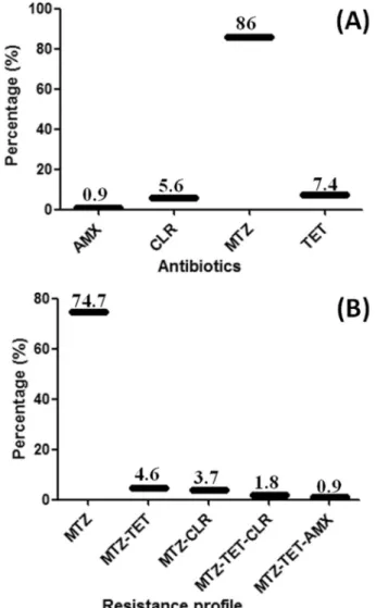
Among 150 examined patients, 20.7% (31of 150) were normal, 20% (30 of 150) were suffering from gastric ulcer, 24% (36 of 150) from duodenal ulcer, 33.3% (50 of 150) from gastritis, and 2% (3 of 150) from gastric cancer. H. pyloriwas cultured, and the urease test was positive in 71.33% (107) of the total 150 gastric biopsy specimens. The confirmedH. pylori isolates were oxidase, catalase, and urease positive. Overall, the resistance rate for the 107 confirmedH. pyloriwas 86% (92 of 107) for MTZ, 7.4% (8 of 107) for TET, 5.6% (6 of 107) for CLR, and 0.9% (1 of 107) for AMX (Fig. 1A), and the antibiotic resistance pat-tern based on the combined resistance to antibiotics is shown in Figure 1B. Most strains (74.7%) were only resis-tant to MTZ, while 1.8% strains were resisresis-tant to MTZ, TET, and CLR. Four isolates (3.7%) were resistant to MTZ and CLR, and five isolates (4.6%) were resistant to MTZ and TET, while only one isolate (0.9%) was resistant to MTZ, TET, and AMX.
Thelactobacillistrains used in our study were cata-lase and oxidase negative and were able to ferment glucose. Apparent reduction in pH of theLactobacillusculture was detected through time points. The antibacterial activity of L. caseistrains againstH.pyloriwas observed on the 107 tested H.pylori isolates, thereby showing the inhibition zones from 11.0 to 15.0 mm. All the clinical isolates ofH. pyloriwere completely inhibited byLactobacillusculture. Mean inhibition zones in millimeters are shown in Table 1.
Discussion
market. Different lactobacilli could be used for inhibition of H. pylori colonization through different mechanisms. Oral intake ofL. johnsoniiLa1 was reported in many clini-cal studies and confirmed by different bacteriologiclini-cal and pathological examinations to attenuateH. pyloriby reduc-ing pro-inflammatory chemotactic signals, which are responsible for the recruitment of lymphocytes and neutro-phils in the lamina propria (Gotteland and Cruchet, 2003; Sgouraset al., 2005). Moreover,L. gasseriOLL2716 was proved to follow the mechanism of induction. The coccoid conversion ofH. pyloriby lactic acid secretion (Canducci et al., 2002) andL. acidophilusexert an antagonistic effect againstH. pylori through the production of a heat stable protein that exerts the antimicrobial effect other than lactic acid, which is only sensitive to enzymatic treatments (Coconnieret al., 1998). In this study,L. caseiproduces a large amount of lactic acid that has been confirmed by the reduction in pH; lactate has been implicated as an inhibi-tory factor forH. pyloridue to its antimicrobial effect re-sulting from lowering the pH and its ability to inhibit the urease enzyme (Midoloet al., 1995; Sgouraset al., 2004). Although some studies have reported that only viable cells ofL. caseican inhibitH. pylorigrowth (Coconnieret al., 1998; Sgouraset al., 2004), other studies have investigated the importance of other factors such as pH and lactic acid production (Bhatiaet al., 1989; Lorcaet al., 2001). In our study, we adjusted the pH to 5.5 as it is known that the low pH environment was found to increase the activity of lactobacilli (Boyanovaet al., 2009), while lactic acid pro-duction was found to inhibit urease enzyme secretion from H. pylori, which plays a role in counteracting the stomach acidity (Servin, 2004; Sgouraset al., 2004). Recently, a striking mechanism forL. caseihas been reported, which gleaned its power in making irreversible inhibition of the swimming motility ofH. pylori through changes in cell morphology, replacing the helical cells by “c”-shaped and coccoid cells, and losing of FlaA and FlaB flagellins (Le Moalet al., 2013). Another in vivo study showed thatL. caseicould prevent enteric infections and stimulate secre-tory IgA in malnourished animals by inducing the in-creased systemic immune response, and it is also proved in the same study thatL. caseiwas the most effective among other lactobacilli (Perdigonet al., 1995). Proteomic analy-sis of bacteria usually revealed plenty of information about its proteins and their role in the cell (Enanyet al., 2012;
Enanyet al., 2013; Enanyet al., 2014). Two-dimensional gel electrophoresis, one of the gel-based proteomic meth-ods, is used to fractionate, identify, and quantify proteins (Wuet al., 2009; Magdeldinet al., 2014). Proteomic analy-sis ofL. caseiusing two-dimensional gel electrophoresis expressed different proteins, which act as stress response proteins and are also involved in central and intermediary metabolism. These proteins have a potential role for the ad-aptation to the surroundings, particularly the accumulation of lactic acid in the course of growth and the physiological processes in bacterial cells (Wuet al., 2009).
It was interesting to find all antibiotic resistance pat-terns of ourH. pyloriisolates responded by 100% to the an-tagonistic effect of theL. caseistrains. Human and animal clinical studies showed that the administration ofL. casei probiotics alone decreases but does not clearH. pylori in-fection, suggesting that it should be taken together with the antibiotic therapy to improve the eradication rates (Sgouras et al., 2004; Catset al., 2003). Many studies have reported that the concurrent combination of yogurt probiotic prod-ucts containingL. caseitogether with the antibiotic treat-ment could significantly reduce their side effects such as reduction in vomiting and nausea, decreasing the taste dis-turbance, and correcting antibiotic-induced intestinal dys-biosis and consequently reduction in diarrhea (Lesbros-Pantoflickovaet al., 2007). A recent meta analysis suggests that supplementation with probiotics compared with eradi-cation therapy may be considered an option to increase eradication rates, particularly when antibiotic therapies are relatively ineffective (Danget al., 2014).
Our results in the present study can be used as a base for the additional assessment of the inhibitory effects ofL. casei strains. More in vivo trials for L. casei and other lactobacilli are still under investigation.
In conclusion, the data presented here clearly show thatL. caseistrains isolated from probiotic preparation of dairy products inhibited the growth ofH. pyloristrains and even the antibiotic-resistant strains in vitro. Further, in vivo clinical studies are still required to assessL. caseiability to be utilized in the treatment ofH. pyloriinfection.
Acknowledgments
We thank “Sci-Edit Publications (Language Editing Services) (www.sci-edit.net)” for editing the manuscript.
References
Bernet-Camard MF, Lievin V, Brassart Det al.(1997) The human Lactobacillus acidophilus strain LA1 secretes a non-bacteriocin antibacterial substance(s) active in vitro and in vivo. Appl Environ Microbiol 63:2747-2753.
Bernet MF, Brassart D, Neeser JRet al. (1994) Lactobacillus acidophilus LA 1 binds to cultured human intestinal cell lines and inhibits cell attachment and cell invasion by enterovirulent bacteria. Gut 35:483-489.
Table 1- Inhibition of clinical isolates ofH. pyloribyL. caseistrain cul-ture.
Mean diameter of inhibition zone (mm)
Number (%) ofH. pyloristrains inhibited byL. casei
11:12 9 (8.4%)
12:13 4 (3.7%)
13:14 66 (61.7%)
Bhatia SJ, Kochar N, Abraham P et al. (1989) Lactobacillus acidophilus inhibits growth of Campylobacter pylori in vi-tro. J Clin Microbiol 27:2328-2330.
Boyanova L, Stephanova-Kondratenko M, Mitov I (2009) Anti-Helicobacter pylori activity of Lactobacillus delbrueckii subsp. bulgaricus strains: preliminary report. Lett Appl Microbiol 48:579-584.
Canducci F, Cremonini F, Armuzzi Aet al.(2002) Probiotics and Helicobacter pylori eradication. Digestive and liver disease: official journal of the Italian Society of Gastroenterology and the Italian Association for the Study of the Liver 34:S81-S83.
Cats A, Kuipers EJ, Bosschaert MAet al.(2003) Effect of fre-quent consumption of a Lactobacillus casei-containing milk drink in Helicobacter pylori-colonized subjects. Aliment Pharmacol Ther 17:429-435.
Coconnier MH, Lievin V, Hemery Eet al.(1998) Antagonistic ac-tivity against Helicobacter infection in vitro and in vivo by the human Lactobacillus acidophilus strain LB. Appl Envi-ron Microbiol 64: 4573-4580.
Dang Y, Reinhardt JD, Zhou X et al. (2014) The Effect of Pobiotics Supplementation on Helicobacter pylori Eradica-tion Rates and Side Effects during EradicaEradica-tion Therapy: A Meta-Analysis. PloS one 9:e111030.
Dubois A, Boren T (2007) Helicobacter pylori is invasive and it may be a facultative intracellular organism. Cell Microbiol 9:1108-1116.
Duck WM, Sobel J, Pruckler JMet al.(2004) Antimicrobial resis-tance incidence and risk factors among Helicobacter pylori-infected persons, United States. Emerg Infect Dis 10:1088-1094.
Dunn BE, Cohen H, Blaser MJ (1997a) Helicobacter pylori. Clin Microbiol Rev 10:720-741.
Dunn BE, Vakil NB, Schneider BGet al.(1997b) Localization of Helicobacter pylori urease and heat shock protein in human gastric biopsies. Infect Immun 65:1181-1188.
Elviss NC, Owen RJ, Xerry Jet al.(2004) Helicobacter pylori an-tibiotic resistance patterns and genotypes in adult dyspeptic patients from a regional population in North Wales. J Anti-microb Chemother 54:435-440.
Enany S (2011)Helicobacter pylori myths vs. truth. Lambert Aca-demic Publishing, Usikker, Leveringstid, 863 pp.
Enany S, Yoshida Y, Magdeldin Set al.(2013) Two dimensional electrophoresis of the exo-proteome produced from commu-nity acquired methicillin resistant Staphylococcus aureus belonging to clonal complex 80. Microbiol Res 168:504-511.
Enany S, Yoshida Y, Magdeldin S et al. (2012) Extensive proteomic profiling of the secretome of European commu-nity acquired methicillin resistant Staphylococcus aureus clone. Peptides 37:128-137.
Enany S, Yoshida Y, Yamamoto T (2014) Exploring extra-cellu-lar proteins in methicillin susceptible and methicillin resis-tant Staphylococcus aureus by liquid chromatography-tan-dem mass spectrometry. World J Microbiol Biotechnol 30:1269-1283.
Enany SAS, Ali K (2005) The prevalence of h.pylori and resis-tance patterns in dyspeptic patients from ismailia, Egypt. Suez Canal University Medical Journal 8:87-92.
Forestier C, De Champs C, Vatoux Cet al.(2001) Probiotic activ-ities of Lactobacillus casei rhamnosus: in vitro adherence to
intestinal cells and antimicrobial properties. Res Microbiol 152:167-173.
Fujimura S, Watanabe A, Kimura Ket al.(2012) Probiotic mech-anism of Lactobacillus gasseri OLL2716 strain against Helicobacter pylori. J Clin Microbiol 50:1134-1136. Gisbert JP (2009) Second-line rescue therapy of helicobacter
pylori infection. Therap Adv Gastroenterol 2:331-356. Goodwin CS, Armstrong JA (1990) Microbiological aspects of
Helicobacter pylori (Campylobacter pylori). European jour-nal of clinical microbiology & infectious diseases: official publication of the European Society of Clinical Microbiol-ogy 9:1-13.
Gotteland M, Cruchet S (2003) Suppressive effect of frequent in-gestion of Lactobacillus johnsonii La1 on Helicobacter pylori colonization in asymptomatic volunteers. J Anti-microb Chemother 51:1317-1319.
He Q, Wang JP, Osato Met al.(2002) Real-time quantitative PCR for detection of Helicobacter pylori. J Clin Microbiol 40:3720-3728.
Hsieh PS, Tsai YC, Chen YCet al.(2012) Eradication of Helico-bacter pylori infection by the probiotic strains Lactobacillus johnsonii MH-68 and L. salivarius ssp. salicinius AP-32. Helicobacter 17:466-477.
Hunt RH, Xiao SD, Megraud Fet al.(2011) Helicobacter pylori in developing countries. World Gastroenterology Organisation Global Guideline. J Gastrointestin Liver Dis 20: 299-304. Jack RW, Tagg JR, Ray B (1995) Bacteriocins of gram-positive
bacteria. Microbiol Rev 59:171-200.
Le Moal VL, Fayol-Messaoudi D, Servin AL (2013) Com-pound(s) secreted by Lactobacillus casei strain Shirota YIT9029 irreversibly and reversibly impair the swimming motility of Helicobacter pylori and Salmonella enterica serovar Typhimurium, respectively. Microbiology 159:1956-1971.
Lesbros-Pantoflickova D, Corthesy-Theulaz I, Blum AL (2007) Helicobacter pylori and probiotics. J Nutr 137:812S-818S. Lionetti E, Indrio F, Pavone Let al.(2010) Role of probiotics in
pediatric patients with Helicobacter pylori infection: a com-prehensive review of the literature. Helicobacter 15:79-87. Lorca GL, Wadstrom T, Valdez GFet al.(2001) Lactobacillus
acidophilus autolysins inhibit Helicobacter pylori in vitro. Curr Microbiol 42:39-44.
Magdeldin S, Enany S, Yoshida Yet al.(2014) Basics and recent advances of two dimensional- polyacrylamide gel electro-phoresis. Clin Proteomics 11:16.
Malfertheiner P, Megraud F, O’Morain CAet al.(2012) Manage-ment of Helicobacter pylori infection - the Maastricht IV/ Florence Consensus Report. Gut 61:646-664.
Matsukura N, Tajiri T, Kato Set al.(2004) Diagnostic value of culture, histology and PCR for Helicobacter pylori in the remnant stomach after surgery. Aliment Pharmacol Ther 20:33-38.
McNulty C, Owen R, Tompkins D et al. (2002) Helicobacter pylori susceptibility testing by disc diffusion. J Antimicrob Chemother 49:601-609.
Megraud F, Lehours P (2007) Helicobacter pylori detection and antimicrobial susceptibility testing. Clin Microbiol Rev 20:280-322.
Perdigon G, Alvarez S, Rachid Met al.(1995) Immune system stimulation by probiotics. J Dairy Sci 78:1597-1606. Servin AL (2004) Antagonistic activities of lactobacilli and
bifi-dobacteria against microbial pathogens. FEMS Microbiol Rev 28:405-440.
Sgouras D, Maragkoudakis P, Petraki Ket al.(2004) In vitro and in vivo inhibition of Helicobacter pylori by Lactobacillus casei strain Shirota. Appl Environ Microbiol 70:518-526. Sgouras DN, Panayotopoulou EG, Martinez-Gonzalez Bet al.
(2005) Lactobacillus johnsonii La1 attenuates Helicobacter pylori-associated gastritis and reduces levels of proinflam-matory chemokines in C57BL/6 mice. Clin Diagn Lab Immunol 12:1378-1386.
Silva M, Jacobus NV, Deneke Cet al.(1987) Antimicrobial sub-stance from a human Lactobacillus strain. Antimicrob Agents Chemother 31:1231-1233.
Sugawara G, Nagino M, Nishio Het al. (2006) Perioperative synbiotic treatment to prevent postoperative infectious com-plications in biliary cancer surgery: a randomized controlled trial. Ann Surg 244:706-714.
Sullivan A, Nord CE (2005) Probiotics and gastrointestinal dis-eases. J Intern Med 257:78-92.
Sykora J, Valeckova K, Amlerova Jet al.(20050 Effects of a spe-cially designed fermented milk product containing probiotic Lactobacillus casei DN-114 001 and the eradication of H.
pylori in children: a prospective randomized double-blind study. J Clin Gastroenterol 39:692-698.
Vandenbergh PA (1993) Lactic acid bacteria, their metabolic products and interference with microbial growth. FEMS Microbiol Rev 12:221-237.
WHO F (2001) Report on Joint FAO/WHO Expert Consultation on Evaluation of Health and Nutritional Properties of Pro-biotics in Food Including Powder Milk with Live Lactic Acid Bacteria. ftp://ftpfaoorg/es/esn/food/probio_re-port_enpdf.
Wolle K, Leodolter A, Malfertheiner Pet al.(2002) Antibiotic susceptibility of Helicobacter pylori in Germany: stable pri-mary resistance from 1995 to 2000. J Med Microbiol 51:705-709.
Wroblewski LE, Peek RMJr, Wilson KT (2010) Helicobacter pylori and gastric cancer: factors that modulate disease risk. Clin Microbiol Rev 23:713-739.
Wu R, Wang W, Yu D et al. (2009) Proteomics analysis of Lactobacillus casei Zhang, a new probiotic bacterium iso-lated from traditional home-made koumiss in Inner Mongo-lia of China. Mol Cell Proteomics 8:2321-2338.
Associate Editor: Maricê Nogueira de Oliveira