Effect of an aqueous extract of
Phaseolus vulgaris
on the properties
of tail tendon collagen of rats with
streptozotocin-induced diabetes
Department of Biochemistry, Faculty of Science,
Annamalai University, Annamalainagar, Tamil Nadu, India L. Pari and
S. Venkateswaran
Abstract
Changes in the structural and functional properties of collagen caused by advanced glycation might be of importance for the etiology of late complications in diabetes. The present study was undertaken to inves-tigate the influence of oral administration of aqueous pod extract (200 mg/kg body weight) of Phaseolus vulgaris, an indigenous plant used in Ayurvedic Medicine in India, on collagen content and characteris-tics in the tail tendon of streptozotocin-diabetic rats. In diabetic rats, collagen content (117.01 ± 6.84 mg/100 mg tissue) as well as its degree of cross-linking was increased, as shown by increased extent of glycation (21.70 ± 0.90 µg glucose/mg collagen), collagen-linked fluorescence (52.8 ± 3.0 AU/µmol hydroxyproline), shrinkage tem-perature (71.50 ± 2.50ºC) and decreased acid (1.878 ± 0.062 mg hydroxyproline/100 mg tissue) and pepsin solubility (1.77 ± 0.080 mg hydroxyproline/100 mg tissue). The α/ß ratio of acid- (1.69) and pepsin-soluble (2.00) collagen was significantly decreased in strepto-zotocin-diabetic rats. Administration of P. vulgaris for 45 days to streptozotocin-diabetic rats significantly reduced the accumulation and cross-linking of collagen. The effect of P. vulgaris was compared with that of glibenclamide, a reference drug administered to streptozo-tocin-diabetic rats at the dose of 600 µg/kg body weight for 45 days by gavage. The effects of P. vulgaris (collagen content, 64.18 ± 1.97; extent of glycation, 12.00 ± 0.53; collagen-linked fluorescence, 33.6 ± 1.9; shrinkage temperature, 57.0 ± 1.0; extent of cross-linking - acid-soluble collagen, 2.572 ± 0.080, and pepsin-acid-soluble collagen, 2.28 ± 0.112) were comparable with those of glibenclamide (collagen con-tent, 71.5 ± 2.04; extent of glycation, 13.00 ± 0.60; collagen-linked fluorescence, 38.9 ± 2.0; shrinkage temperature, 59.0 ± 1.5; extent of cross-linking - acid-soluble collagen, 2.463 ± 0.078, and pepsin-soluble collagen, 2.17 ± 0.104). In conclusion, administration of P. vulgaris pods had a positive influence on the content of collagen and its properties in streptozotocin-diabetic rats.
Correspondence
L. Pari
Department of Biochemistry Faculty of Science Annamalai University Annamalainagar-608 002 Tamil Nadu
India
Fax: +91-4144-23-8145 E-mail: [email protected] Research supported by the Indian Council of Medical Research, New Delhi (No. 45/29/99 - BMS) in the form of a senior research fellowship to S. Venkateswaran.
Received November 13, 2002 Accepted April 14, 2003
Key words
•Collagen
•Diabetes
•Phaseolus vulgaris •Cross-linking
•Glibenclamide
Introduction
Collagen is a major component of mam-malian connective tissue and is present in all major tissues that require strength and flex-ibility such as skin, bone and tendon (1). Type I collagen is the principal extracellular matrix protein (2). Collagens are character-ized by the presence of one or more triple helix domains. The helical structure consists of polypeptide chains containing Gly-x-y repeats, where x and y typically represent proline or hydroxyproline residues. Collagen molecules, after being secreted by the cells, assemble into characteristic fibers respon-sible for the functional integrity of tissues such as bone, cartilage, skin and tendon. They provide a structural framework for other tissues, such as blood vessels, aorta and most organs. Cross-links between adjacent molecules are a prerequisite for the collagen fibers to withstand the physical stresses to which they are exposed (3,4).
Nonenzymatic glycation of collagen lead-ing to the formation of advanced glycation end products is increased in patients with diabetes mellitus and in animals with exper-imental diabetes (5). Changes in the struc-tural and functional properties of proteins caused by advanced glycation might be of importance for the etiology of late complica-tions in diabetes. The process of nonenzy-matic glycation is initiated by covalent bind-ing of the reactive aldehyde moiety of glu-cose to free amino groups of protein (6). Collagenous proteins are especially exposed to glycation because they contain several lysine, hydroxyl lysine and arginine residues with free amino groups, have a very slow turnover rate and are exposed to ambient levels of glucose (7,8). The content of ad-vanced glycation end products in collagen increases with age and diabetes (8).
In recent years the popularity of alterna-tive medicine has increased for various rea-sons. Dietary measures and traditional plant therapies as prescribed by Ayurvedic and
other indigenous medicine systems are com-monly used in India (9).The beneficial ac-tions of these diets on the amelioration of diabetic symptoms are well documented (10,11).
Phaseolus vulgaris L. (Leguminosae), commonly known as kidney bean, is a food item of mass consumption in Asia and East-ern countries. The various parts of the plant have been extensively used in Ayurvedic and Unani practice on the Indian subconti-nent for the treatment of diabetes mellitus (12).In 1995, Roman-Ramos et al. (13) showed that the aqueous extract of P. vul-garis pods possessed antihyperglycemic ac-tivity. P. vulgaris has also been reported to contain nearly 50 mg of flavonoids per 100 g (14). Recently we have demonstrated the antioxidant (15) and hypolipidemic (16) ef-fects of P. vulgaris pods in diabetic rats.
To our knowledge, the present investiga-tion is the first report on the effects of P. vulgaris extracts on the collagen content and its physical and chemical characteristics in the tail tendon of streptozotocin-diabetic rats.
Material and Methods
Experimental animals
Male albino Wistar rats weighing 170-200 g bred in the Central Animal House, Rajah Muthiah Medical College, were used. The animals received a normal laboratory pellet diet (Hindustan Lever Ltd., Mumbai, India) and water ad libitum. The animals used in the present study were maintained in accordance with the guidelines of the Na-tional Institute of Nutrition, Indian Council of Medical Research, Hyderabad, India. The study was approved by the Ethics Commit-tee of Annamalai University.
Drugs and chemicals
MO, USA). The chemicals were of analyti-cal grade or equivalent.
Plant material
P. vulgaris was purchased on the local market in Chidambaram, Cuddalore District, Tamil Nadu, India. The plant was identified in the herbarium of the Botany Directorate of Annamalai University. A voucher speci-men (No. 2387) was deposited in the Botany Department of Annamalai University.
Preparation of plant extract
One hundred and thirty-two grams of dried P. vulgaris pods was extracted with 1.0 liter of water for 2 h at 60-70ºC by continu-ous hot extraction and evaporated to dryness in a rotary evaporator at 40-50ºC under re-duced pressure.A semisolid material was obtained (20 g) and stored at 0-4ºC until the time for use. When needed, the residual extract was suspended in distilled water and used in the study (13).
Induction of experimental diabetes
A freshly prepared solution of streptozo-tocin (45 mg/kg) in 0.1 M citrate buffer, pH 4.5, was injected intraperitoneally in a vol-ume of 1 ml/kg (17). Forty-eight hours after streptozotocin administration, rats with mod-erate diabetes having glycosuria and hyper-glycemia (i.e., with blood glucose of 200-300 mg/dl) were used for the experiment.
Experimental procedure
A total of 40 rats (30 surviving diabetic rats and 10 normal rats) were used. The rats were divided into 4 groups of 10 rats each. Group 1: normal untreated rats; group 2: diabetic control rats; group 3: diabetic rats receiving the P. vulgaris pod extract (200 mg/kg body weight) in 1 ml aqueous solu-tion daily by gavage for 45 days (13); group
4: diabetic rats receiving glibenclamide (600 µg/kg body weight) in aqueous solution daily by gavage for 45 days (18).
After 45 days, the animals were deprived of food overnight and sacrificed by decapita-tion. Blood was collected with potassium oxalate and sodium fluoride as anticoagu-lants for the determination of blood glucose. Plasma was separated by centrifugation for insulin assay. The tail was removed and stored frozen at -80ºC and tail tendon tissue was prepared as described below.
Blood glucose and plasma insulin
Fasting blood glucose was measured by the O-toluidine method (19) and plasma in-sulin was assayed with an ELISA kit (Boeh-ringer-Mannheim, Mannheim, Germany).
Preparation and purification of type I collagen
precipitated from the supernatant by slow addition with stirring of 0.2 volumes 30% NaCl in 0.5 M acetic acid. The washed pellet was suspended in two volumes of 0.5 M acetic acid and dialyzed overnight against several changes of 20 mM Na2HPO4. The precipitate was collected by centrifugation at 17,000 g for 30 min, washed once with 20 mM Na2HPO4 and redissolved in two vol-umes of 0.5 M acetic acid. The solution was centrifuged at 28,330 g for 60 min and dia-lyzed for 2 days against 0.1 M acetic acid with three changes. The purified collagen was lyophilized and stored in a freezer in containers sealed under vacuum.
Estimation of collagen content
Weighed tail tendon tissue was hydro-lyzed in 6.0 N HCl for 18 h at 110ºC. The collagen content was determined by measur-ing hydroxyproline, as described by Woess-ner (21).
Extent of glycation
The extent of glycation was determined by the method described by Rao and Pattabiraman (22) in which 1.0 ml of puri-fied collagen (containing 1.0 mg collagen) was mixed with 3.0 ml of concentrated H2SO4, vortexed, cooled on ice, mixed with 0.5 ml of 80% phenol, and left to stand at room temperature for 30 min. Absorbance was measured at 485 nm using glucose as standard.
Collagen-linked fluorescence
Collagen-linked fluorescence was meas-ured by the method of Monnier et al. (23). Approximately 3.0 mg tissue was finely minced in PBS and centrifuged at 3,330 g for 10 min. The pellet was washed with distilled water and the lipids were extracted with 5.0 ml of chloroform:methanol (2:1, v/v) over-night. The samples were rehydrated by the
addition of 2.0 ml methanol and 0.5 ml distilled water and centrifuged at 3,330 g for 10 min and the pellet was washed twice with methanol, three times with distilled water, twice with 20 mM N-[2-hydroxyethyl] piperazine-N’-[2-ethanesulfonic acid], pH 7.5, containing 0.12 M CaCl2 (buffer H) and stored overnight at 4ºC in buffer H. The buffer was then removed by centrifugation at 3,330 g for 10 min and the pellet resus-pended in 3.5 ml of buffer H containing 120 units of type VII collagenase. Four drops of toluene were added to prevent bacterial growth and the material was digested for 48 h at 37ºC. A blank containing collagenase in buffer H was included. The digest was cen-trifuged at 3,330 g for 30 min and the clear supernatant containing digested collagen was used for the fluorescence assay. Fluores-cence was measured with a Hitachi spectro-fluorometer (Hitachi, Tokyo, Japan) against distilled water at 440 nm after excitation at 370 nm and was corrected for the collagen-ase blank.
Solubility pattern of tail tendon collagen
The solubility pattern of tail tendon col-lagen was determined as described by Miller and Rhodes (24).
Neutral salt-soluble collagen
Tail tendon tissue was thoroughly minced, homogenized in 10 volumes of neutral salt solvent (1.0 M NaCl, 50 mM Tris, pH 7.5) containing 20 mM EDTA and 2.0 mM N-ethyl maleimide and stirred for 24 h. The suspension was then centrifuged at 35,000 g
for 1 h at 4ºC and the extraction was repeated with the pellet. The supernatants were pooled and an aliquot was used for the assay of hydroxyproline (21).
Acid-soluble collagen
10 volumes of 0.5 M acetic acid and extract-ed for 24 h with constant stirring, after which the contents were centrifuged. The pellet was re-extracted with acetic acid, the super-natants were pooled and an aliquot was used for the determination of hydroxyproline.
Pepsin-soluble collagen
The residue obtained after acid extrac-tion was resuspended in 0.5 M acetic acid containing 100 mg pepsin per g wet tissue. Digestion was carried out for 24 h, followed by centrifugation and re-extraction. Aliquots of pooled supernatant were used for hy-droxyproline measurement.
Shrinkage temperature
The shrinkage temperature of tail tendon collagen was determined as described by Nutting and Borasky (25). Small strips of collagen fibers were cut from the tail tendon and placed on microscope slides mounted on a holder for viewing under the microscope. An electric bulb with heat-producing capac-ity was placed under the holder. A thermom-eter was inserted into the hole available in the holder to monitor the temperature. The electric bulb was then switched on and the heating rate was set at 3ºC/min. The shrink-age temperature of collshrink-agen fibers was moni-tored through the microscope and the exact shrinkage temperature was recorded during the shrinkage process (26).
SDS-PAGE
Acid- and pepsin-soluble collagen was prepared from tail tendon as described by Miller and Rhodes (24). Collagen samples were investigated by SDS-PAGE using a 3% stacking gel with a 5% running gel and Coo-massie brilliant blue staining. The gels were scanned with a densitometer and the α/ß
ratio of acid- and pepsin-soluble collagen was calculated.
Statistical analysis
The data for the various biochemical pa-rameters were analyzed by analysis of vari-ance (ANOVA) and the group means were compared by Duncan’s multiple range test (27). Values were considered statistically significant when P < 0.05.
Results
Blood glucose and plasma insulin
Blood glucose and plasma insulin levels of normal and experimental rats are given in Table 1. The diabetic rats showed a cant increase in blood glucose and a signifi-cant decrease in plasma insulin levels. The administration of Phaseolus pod extract and glibenclamide to diabetic rats caused a sig-nificant decrease in blood glucose levels and a significant increase in plasma insulin. How-ever, neither the plant extract nor glibencla-mide normalized glucose or insulin levels completely.
The dose of P. vulgaris (200 mg/kg body weight) was selected based on our previous studies in which a dose-dependent effect of
P. vulgaris on blood glucose was obtained.
P. vulgaris was studied with three different doses - 50, 100 and 200 mg/kg body weight. Among the three different doses, P. vulgaris
showed a highly significant effect at 200 mg/ kg body weight (15,16). So we have chosen
Table 1. Effect of Phaseolus vulgaris pod extract (PPEt) on blood glucose and plasma insulin levels in normal and diabetic rats.
Group Blood glucose (mg/dl) Plasma insulin (µU/ml)
Normal 79.12 ± 5.42a 14.41 ± 0.70a
Diabetic control 278.40 ± 22.00b 4.13 ± 0.26b
Diabetic + PPEt 91.20 ± 4.65c 7.78 ± 0.40c
Diabetic + glibenclamide 98.20 ± 6.50d 7.50 ± 0.34c
the dose for our present study. The effect of the dose was compared with glibenclamide at a dose of 600 µg/kg body weight, which is a well-accepted standardized dose in exper-imental animals. Glibenclamide is also known to cause severe side effects at doses higher than 600 µg/kg (18).
Hydroxyproline, total collagen, extent of glycation, collagen-linked fluorescence and shrinkage temperature
The data in Table 2 show that the levels of hydroxyproline and total collagen and the extent of glycation and collagen-linked fluo-rescence were significantly increased in dia-betic animals. The shrinkage temperature was also increased in the diabetic group
compared to the normal group. Administra-tion of both Phaseolus pod extract and gli-benclamide significantly reduced collagen levels, extent of glycation and collagen-linked fluorescence in diabetic rats. The increased shrinkage temperature was also significantly prevented in diabetic rats treated with Pha-seolus pod extract and glibenclamide. The effect of the extract was almost similar to that of glibenclamide.
Pattern of collagen solubility
Data concerning the solubility pattern of tail tendon collagen of normal rats and of the experimental groups after extraction with neutral salt and acid solution and by pepsin digestion are presented in Table 3. The
per-Table 2. Effect of Phaseolus vulgaris pod extract (PPEt) on hydroxyproline, total collagen, extent of glycation, fluorescence and shrinkage temperature of rat tail collagen from normal and diabetic rats.
Group Hydroxyproline Total collagen Extent of glycation Fluorescence Shrinkage (mg/100 mg tissue) (mg/100 mg tissue) (µg glucose/mg (AU/µmol temperature
collagen) hydroxyproline) (oC)
Normal 8.87 ± 0.233a 66.13 ± 1.14a 10.30 ± 0.42a 27.7 ± 1.3a 60.5 ± 1.0a
Diabetic 15.69 ± 0.917b 117.01 ± 6.84b 21.70 ± 0.90b 52.8 ± 3.0b 71.5 ± 2.5b
control
Diabetic + 9.68 ± 0.265c 64.18 ± 1.97c 12.00 ± 0.53c 33.6 ± 1.9c 57.0 ± 1.0c
PPEt
Diabetic + 9.59 ± 0.254d 71.5 ± 2.04d 13.00 ± 0.60d 38.9 ± 2.0d 59.0 ± 1.5d
glibenclamide
For drug administration see Table 1 legend. AU = arbitrary units. Data are reported as means ± SD for 6 rats in each group. Values not sharing a common superscript letter differ significantly at P < 0.05 (Duncan multiple range test).
Table 3. Effect of Phaseolus vulgaris pod extract (PPEt) on neutral salt-, acid- and pepsin-soluble collagen content (hydroxyproline/100 mg tissue) of tail tendon in normal and diabetic rats.
Group Neutral salt-soluble collagen Acid-soluble collagen Pepsin-soluble collagen (µg/100 mg) (mg/100 mg) (mg/100 mg)
Normal 137.56 ± 9.2a 2.895 ± 0.091a 2.96 ± 0.100a
Diabetic control 71.99 ± 4.2b 1.878 ± 0.062b 1.77 ± 0.080b
Diabetic + PPEt 113.60 ± 7.8c 2.572 ± 0.080c 2.28 ± 0.112c
Diabetic + glibenclamide 101.40 ± 7.4d 2.463 ± 0.078d 2.17 ± 0.104d
centage of neutral salt-, acid- and pepsin-soluble collagen was significantly decreased in diabetic animals. Administration of the
Phaseolus pod extract and glibenclamide significantly increased collagen solubility in diabetic rats.
SDS-PAGE
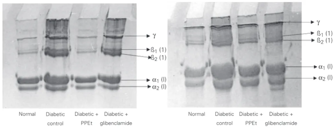
The gel patterns obtained by SDS-PAGE of acid- and pepsin-soluble collagen from the tail tendon of normal and experimental rats are shown in Figure 1. The α/ß ratio of
both acid- and pepsin-soluble collagen was decreased significantly in diabetic rats (Table 4). Administration of Phaseolus pod extract and glibenclamide significantly increased the
α/ß ratio to near normal values.
Discussion
We determined the influence of an
ex-tract of P. vulgaris on collagen content and characteristics in diabetic rats.
The capacity of the Phaseolus pod ex-tract to decrease the elevated blood sugar to normal levels is an essential trigger for the liver to revert to its normal homeostasis dur-ing experimental diabetes. The possible mechanism by which the plant extract exerts its hypoglycemic action in diabetic rats may be by potentiating the plasma insulin effect by increasing either the pancreatic secretion of insulin from the existing ß-cells or its release from the bound form, as demon-strated by the significant increase in insulin levels induced by the plant extract in dia-betic rats (Table 1).
Streptozotocin-induced diabetes mellitus characterized by hyperglycemia caused a sig-nificant increase in hydroxyproline levels and collagen content. The correlation be-tween collagen and intracellular degradation is of interest and may have a role in the
α2 (I) γ
ß1 (1)
ß2 (1)
α1 (I) α2 (I)
γ
ß1 (1)
ß2 (1)
α1 (I)
Normal Diabetic control
Diabetic + PPEt
Diabetic + glibenclamide
Normal Diabetic control
Diabetic + PPEt
Diabetic + glibenclamide
Figure 1. Effect of Phaseolus vulgaris pod extract (PPEt) on SDS-PAGE pattern of acid-soluble (left) and pepsin-acid-soluble (right) collagen of tail tendon in normal and diabetic rats.
Table 4. Effect of Phaseolus vulgaris pod extract (PPEt) on the α/ß ratio of acid- and pepsin-soluble collagen in
tail tendon of normal and diabetic rats.
Group Acid-soluble collagen component Pepsin-soluble collagen component
α ß α/ß ratio α ß α/ß ratio
Normal 69.5 30.3 2.27a 73.1 26.9 2.71a
Diabetic control 62.9 37.1 1.69b 66.7 33.3 2.00b
Diabetic + PPEt 66.8 33.2 2.00c 69.4 30.6 2.26c
Diabetic + glibenclamide 65.5 34.5 1.89d 66.5 33.5 1.98d
regulation of collagen content. Golub et al. (28) have suggested that increased degrada-tion of newly synthesized collagen during streptozotocin-induced diabetes might con-tribute to collagen deposition in the early stages. Diabetic rats treated with the plant extract and glibenclamide showed a signifi-cant decrease in total collagen content when compared to untreated diabetic rats. This decrease may be attributed to the significant decrease in blood glucose and consequent decrease in nonenzymatic glycation and deposition of collagen in diabetic rats treated with the Phaseolus pod extract and gliben-clamide. The extract exhibited effects simi-lar to those of glibenclamide.
In addition, prolyl hydroxylase, an ascor-bic acid-dependent enzyme, is required to maintain the normal properties of collagen. The activity of prolyl hydroxylase has been reported to change in diabetic rats (29). This alteration is mainly due to the reduction in the concentration of ascorbic acid in diabe-tes (29). In a previous study, we also ob-served a significant reduction in the concen-tration of ascorbic acid in streptozotocin-diabetic rats (16). We also observed a sig-nificant increase in the concentration of ascor-bic acid in diabetic rats treated with the plant extract and glibenclamide (16).The decrease in the ascorbic acid concentration and con-sequently altered prolyl hydroxylase could also be responsible for the alteration of col-lagen observed in streptozotocin-diabetic rats. A significant increase in the concentration of ascorbic acid in diabetic rats treated with the plant extract and glibenclamide (16) may also be responsible for the significant reduc-tion in collagen content.
In the present study, an increase in the extent of glycation was observed in the tail tendon of diabetic rats, probably due to ex-posure of the tissues to glucose in the dia-betic state. Earlier studies have also reported that glucose is directly involved in the accel-erated cross-linking of collagen in the dia-betic state. Several studies have also
estab-lished that collagen glycation is increased during exposure to high glucose levels in vitro and in vivo (30). Flavonoids were re-ported to have antiglycating activity (31). The decrease in the extent of glycation in diabetic rats treated with the plant extract could be due to the antiglycating property of the flavonoids present in the extract.
The cross-linking of tail tendon collagen was assessed by measuring the shrinkage temperature. The shrinkage temperature of collagen fibers is related to the number of covalent cross-links present in collagen and to the content of imino acids such as proline and hydroxyproline (32). The shrinkage tem-perature of collagen was reported to increase with age and in diabetes and was explained in terms of intermolecular cross-links (32). Therefore, the measurement of collagen shrinkage temperature may be used to deter-mine the gross tissue changes at the molecu-lar level.
We observed a significant increase in the collagen shrinkage temperature in diabetic rats, clearly indicating the increase in the cross-links of tail tendon collagen in these animals. Administration of the plant extract and glibenclamide induced a significant re-duction in the cross-linking of tail tendon collagen.
neutral, acid and pepsin digestion. This is an indication of decreased levels of cross-link-ing in the collagen of the treated groups. The reduction in the advanced glycation and cross-linking of collagen in diabetic rats treated with the plant extract may be due to its antiperoxidative activity (15), since lipid peroxidation products have been shown to directly influence collagen cross-linking and advanced glycation end product formation (36,37). In addition, advanced glycation end products were also reported to induce the upregulation of the expression of type I col-lagen genes that could result in excess depo-sition of collagen in diabetes (38). The in-crease in advanced glycation end product levels in diabetic rats observed in the present study could be responsible for the upregula-tion of collagen gene expression which re-sults in the increased deposition of collagen and consequent increased cross-linking in streptozotocin-diabetic rats. The Phaseolus
pod extract is reported to be rich in fla-vonoids. These flavonoids may contribute by their protective action to the reduction of collagen cross-linking in treated diabetic rats. Collagen obtained from the tail tendon of diabetic animals showed increased fluores-cence, which is a strong indication of in-creased advanced glycation. Previous stud-ies have also documented an overall increase in the fluorescence of diabetic tissue col-lagen (39,40).The 370/440 nm fluorescence is usually due to Maillard reaction-related fluorescence (6).
It has been shown that, in addition to glucose, free radicals and lipid peroxides also play an important role in the develop-ment of collagen-linked fluorescence (41). It
appears that the reactive radicals formed during glycation and oxidation reactions can also have an influence on the development of fluorescence.
Administration of the Phaseolus pod ex-tract and glibenclamide significantly reduced the intensity of fluorescence in diabetic rats. This may be due to the significant reduction in blood glucose and consequent decreased glycation, and to the significant scavenging of free radicals generated during diabetes by flavonoids present in Phaseolus pod extract. The increased band size of ß-components in diabetic collagen clearly indicates the in-creased cross-linking. Golub et al. (28) have also reported that the acid-soluble collagen from streptozotocin-diabetic rats contains higher than normal amounts of ß-component and hence exhibits increased cross-linking. The increased intensity of the ß-component observed here in diabetic rats suggests that collagen chains are capable of enhanced in-tramolecular cross-linking since the ß-com-ponent is a dimer of α-chains. In diabetic
rats treated with the Phaseolus pod extract, collagen content in the ß-region as well as in the high molecular weight region was near normal when compared to diabetic control groups.
On the basis of these observations, it is clear that the P. vulgaris pod extract had a positive influence on the content of collagen and its characteristics in streptozotocin-dia-betic rats. Further work is currently under-way to analyze the components of the aque-ous extract of P. vulgaris for their beneficial effects on collagen content in streptozoto-cin-diabetic rats.
References
1. Prockop DJ & Kivirikko KI (1995). Collagens. Molecular biology, diseases and potentials for therapy. Annual Review of Biochemis-try, 64: 403-434.
2. George A, Malone JP & Veis A (1999). The secondary structure of type I collagen N-telopeptide as demonstrated by Fourier transform IR spectroscopy and molecular modeling. Proceedings of the Indian
Academy of Sciences (Chemical Sciences), 111: 121-131. 3. Nimni ME & Han BO (1999). Collagen and
collagen-glycosaminogly-can matrices as carriers for growth factors. Proceedings of the Indian Academy of Sciences (Chemical Sciences), 111: 283-289. 4. Brown JC & Timpl R (1995). The collagen superfamily. International
5. Vlassara H, Bucala R & Striker L (1994). Pathogenic effects of advanced glycosylation: biochemical, biologic, and clinical implica-tions for diabetes and ageing. Laboratory Investigation, 70: 138-151. 6. Njoroge FG & Monnier VM (1989). The chemistry of the Maillard
reaction under physiological conditions: a review. In: Baynes JW & Monnier VM (Editors), The Maillard Reaction and Aging, Diabetes and Nutrition. AR Liss, New York.
7. Brownlee M, Cerami A & Vlassara H (1988). Advanced glycosylation end products in tissue and the biochemical basis of diabetic compli-cations. New England Journal of Medicine, 318: 1315-1321. 8. Reiser KM (1998). Non-enzymatic glycation of collagen in aging and
diabetes. Proceedings of the Society for Experimental Biology and Medicine, 218: 23-37.
9. Warier PK (1995). Eugenia jambolana Linn. In: Warier PK, Nambiar VPK & Ramankutty C (Editors), Indian Medicinal Plants. Orient Longman, Madras, India.
10. Bordia A, Verma SK & Srivastava KC (1997). Effect of ginger (Zingiber officinale Rosc.) and fenugreek (Trigonella foenumgraecum L.) on blood lipids, blood sugar and platelet aggregation in patients with coronary artery disease. Prostaglandins, Leukotrienes, and Essential Fatty Acids, 56: 379-384.
11. Khan A & Safdar M (2003). Role of diet, nutrients, spices and natural products in diabetes mellitus. Pakistan Journal of Nutrition, 2: 1-12. 12. Chopra RN, Chopra IC, Handa KI & Kapur LD (1958). Medicinal plants in diabetes. In: Gupta P (Editor), Indigenous Drugs of India. 2nd edn. U.N. Dhar & Sons Ltd., Calcutta, India.
13. Roman-Ramos R, Flores-Sanoz JL & Alarcon-Aguilar FJ (1995). Antihyperglycemic effect of some edible plants. Journal of Ethno-pharmacology, 48: 25-32.
14. Sushmita N & Ranjana N (1997). Anti-oxidant flavonoids in common Indian foods. South Asian Journal of Preventive Cardiology, 1: 33-35. 15. Venkateswaran S & Pari L (2002). Antioxidant effect of Phaseolus vulgaris in streptozotocin induced diabetic rats. Asia Pacific Journal of Clinical Nutrition, 11: 206-209.
16. Venkateswaran S, Pari L & Saravanan G (2002). Effect of Phaseolus vulgaris on circulatory antioxidants and lipids in streptozotocin in-duced diabetic rats. Journal of Medicinal Food, 5: 97-103. 17. Siddique O, Sun Y, Lin JC & Chien YW (1987). Facilitated
transder-mal transport of insulin. Journal of Pharmaceutical Sciences, 76: 341-345.
18. Pari L & Umamaheswari J (2000). Antihyperglycaemic activity of Musa sapientum flowers: Effect on lipid peroxidation in alloxan diabetic rats. Phytotherapy Research, 14: 136-138.
19. Sasaki T, Matsy S & Sonae A (1972). Effect of acetic acid concentra-tion on the colour reacconcentra-tion in the O-toluidine boric acid method for blood glucose. Rinshbo Kagaku, 1: 346-353.
20. Chandrakasan G, Torchia DA & Piez KA (1976). Preparation of intact monomeric collagen from tail tendon and skin and the structure of the nonhelical ends in solution. Journal of Biological Chemistry, 251: 6062-6067.
21. Woessner JF (1961). The determination of hydroxyproline in tissue and protein samples containing small portions of this imino acid. Archives of Biochemistry and Biophysics, 93: 440-447.
22. Rao P & Pattabiraman TN (1989). Reevaluation of the phenol sulphuric acid reaction for the estimation of hexoses and pentoses. Analytical Biochemistry, 181: 18-22.
23. Monnier VM, Vishwanath V, Frank KE, Elmets CA, Dauchot P & Kohn RR (1986). Relation between complications of type I diabetes mellitus and collagen-linked fluorescence. New England Journal of Medicine, 314: 403-408.
24. Miller EJ & Rhodes RK (1982). Preparation and characterization of
different types of collagen. Methods in Enzymology, 82: 33-64. 25. Nutting GC & Borasky R (1949). Microscopic methods for
determin-ing shrinkage temperature of collagen and leather. Journal of the American Leather Chemists Association, 44: 831-839.
26. Vangsness CT, Mitchell W, Nimni M, Erlich M, Saadat V & Schmotzer H (1997). Collagen shortening. An experimental approach with heat. Clinical Orthopaedics, 337: 267-271.
27. Duncan BD (1957). Multiple range tests for correlated and hetero-scedastic means. Biometrics,13: 359-364.
28. Golub IM, Greenwald RA, Zebrowski EJ & Ramamurthy K (1978). The effect of experimental diabetes on the molecular characteristics of soluble rat collagen. Biochimica et Biophysica Acta, 534: 73-81. 29. McLennan S, Yue DK, Fisher E, Capogreco C, Heffernan S, Ross GR
& Turtle JR (1988). Deficiency of ascorbic acid in experimental diabetes. Relationship with collagen and polyol abnormalities. Dia-betes, 37: 359-361.
30. Bensusan HB (1965). A novel hypothesis for the mechanism of cross-linking in collagen: Structure and chemistry of collagen. In: Harkness ND, Partridge SM & Tristiam GR (Editors), Structure and Function of Connective and Skeletal Tissue. Butterworth, London, UK.
31. Yamaguchi F, Ariga T, Yoshimura Y & Nakazawa H (2000). Antioxi-dative and anti-glycation activity of garcinol from Garcinia indica fruit rind. Journal of Agricultural and Food Chemistry, 48: 180-185. 32. Rao CN, Rao VH & Sanjeevi R (1981). Effect of Biflavonoids on the
mechanical and thermal properties of skin and tendon. Indian Jour-nal of Biochemistry and Biophysics, 18: 224-228.
33. Meng J, Sakata N, Takebayashi S, Asano T, Futata T, Araki N & Horiuchi S (1996). Advanced glycation end products of the Maillard reaction in aortic pepsin-insoluble and pepsin-soluble collagen from diabetic rats. Diabetes, 45: 1037-1043.
34. Wolff SP & Dean RT (1987). Glucose autoxidation and protein modi-fication. Biochemical Journal, 245: 243-250.
35. Ahmed MU, Thorpe SR & Baynes JW (1986). Identification of N epsilon-carboxymethyllysine as a degradation product of fructose lysine in glycated protein. Journal of Biological Chemistry, 261: 4889-4894.
36. Hicks M, Delbridge L & Yue DK (1988). Increase in cross-linking of nonenzymatically glycosylated collagen induced by products of lipid peroxidation. Archives of Biochemistry and Biophysics, 268: 249-254.
37. Fu MX, Requena JR, Jenkins AJ, Lyons TJ, Baynes JW & Thorpe SR (1996). The advanced glycation end product, N epsilon-(carboxy-methyl)lysine, is a product of both lipid peroxidation and glycoxida-tion reacglycoxida-tions. Journal of Biological Chemistry, 271: 9982-9986. 38. Kim YS, Kim BC, Song CY, Hong HK, Moon KC & Lee HS (2001).
Advanced glycosylation end products stimulate collagen mRNA synthesis in mesangial cells mediated by protein kinase C and transforming growth factor-beta. Journal of Laboratory and Clinical Medicine, 38: 59-68.
39. Odetti P, Pronzato MA, Noberasco G, Cosso L, Traverso N, Cottalasso D & Marinari UM (1994). Relationship between glycation and oxidation related fluorescence in rat collagen during aging. An in vivo and in vitro study. Laboratory Investigation, 70: 61-67. 40. Sakata N, Meng J, Jimi S & Takebayashi S (1995). Nonenzymatic
glycation and extractability of collagen in human atherosclerotic plaques. Atherosclerosis, 116: 63-75.