Objective: To report a case of severe dystrophic calciication in maxillary sinus of a child with liver transplantation and dental organs pigmented by hyperbilirubinemia.
Case description: female patient, 12 years old, with liver transplantation performed at the age of 7 due to extrahepatic biliary atresia (EHBA). The patient was receiving the immunosuppressant tacrolimus (2 mg daily). Intraoral clinical exam showed tooth green pigmentation by bilirubin. Cone-beam volumetric computed tomography (CT) was performed to verify radiographic density of pigmented dental elements. Hounsield scale measurement did not show changes in radiographic density of dental structures. However, CT scan showed intense dystrophic calciication in the maxillary sinus region.
Comments: CT scan indicated relevant radiographic findings, with radiopacity of the maxillary sinus due to fungal or non-fungal sinusitis. This case report highlights the presence of radiographic image associated with acute infectious processes that could compromise the systemic state of immunosuppressed patients.
Keywords: Adolescent; Liver transplantation; Maxillary sinus; Pigmentation; Dentition, permanent; Tomography, X-ray computed.
Objetivo: Relatar um caso de calciicação distróica intensa no interior do seio maxilar em uma criança com transplante hepático e órgãos dentais pigmentados por hiperbilirrubinemia.
Descrição do caso: Paciente do sexo feminino, 12 anos de idade, com transplante hepático efetuado aos 7 anos de vida devido à atresia de vias biliares extra-hepática, uso de tacrolimus imunossupressor (2 mg diários). No exame clínico intrabucal, observou-se a presença de pigmentação esverdeada no órgão dental por bilirrubina. Efetuou-se um exame de tomograia computadorizada volumétrica de feixe cônico para análise da densidade radiográfica dos elementos dentais pigmentados. Mediante interpretação da imagem pela escala de Hounsield, não foi constatada nenhuma alteração na densidade radiográica das estruturas do órgão dental. No entanto, a tomograia computadorizada evidenciou a presença de calciicação distróica intensa em região de seio maxilar.
Comentários: A alteração de imagem observada no exame de tomograia computadorizada demonstrou achado radiográico relevante, com presença de radiopacidades no interior do seio maxilar decorrentes de sinusites fúngicas ou não fúngicas. O relato desse caso é relevante por apresentar alteração de imagem radiográica exacerbada associada a quadros infecciosos agudos que podem comprometer o estado sistêmico do paciente imunossuprimido.
Palavras-chave: Adolescente; Transplante de fígado; Seio maxilar; Pigmentação; Dentição permanente; Tomograia computadorizada por raios X.
ABSTRACT
RESUMO
*Correspoding author. Email: [email protected] (A. F. Macedo).
aEscola Paulista de Medicina, Universidade Federal de São Paulo, São Paulo, SP, Brazil.
bUniversidade Cruzeiro do Sul, São Paulo, SP, Brazil.
cUniversidade de São Paulo, São Paulo, SP, Brazil.
Received on December 8, 2016; approved on April 11, 2017; available online on November 13, 2017.
DYSTROPHIC CALCIFICATION OF MAXILLARY SINUS IN
PEDIATRIC PATIENTS WITH LIVER TRANSPLANTATION
AND PIGMENTATION OF DENTAL ORGAN
Calcificação distrófica em seio maxilar de paciente pediátrico
com transplante hepático e pigmentação do órgão dental
Adriana Furtado de Macedo
a,b,*, Claudio Costa
c, Regina Helena Guedes da Motta Mattar
a,
Ramiro Anthero de Azevedo
aDystrophic calcification of maxillary sinus
118
Rev Paul Pediatr. 2018;36(1):117-120
INTRODUCTION
Calciication is a biochemical process in which deposition of calcium salts occurs; however, it may happen in unusual sites of human body.1 Pathological calciications are classiied
as idiopathic, metastatic, dystrophic or intrasinus. hese are called idiopathic when calcium builds up in healthy tissues but blood calcium level is normal. However, when blood tests positive calcium elevation with consequent ion deposition, metastatic calciication will be present. In dystrophic calcii-cation, there is poor vascularization where calcium deposits, that is, not suicient blood supply; in addition, necrotic tis-sues and ischemia may be seen on the site.1 It usually occurs
in the core of growing tumors, where there is carbon dioxide decrease and extracellular luid alkalinity increase, resulting in a microenvironment in which calcium is easily deposited. Intrinsic calciication derives from inlammatory and infec-tious conditions.2
he liver is the main organ for intermediate metabolism of proteins, carbohydrates, and fats as it metabolizes and excretes toxic substances. Chronic liver disease may alter these func-tions, especially in the presence of a perinatal inlammatory process initiated in bile ducts, resulting in progressive ibro-sclerosis and intra- and extrahepatic obstruction.3-5 Hepatic
transplantation is often the preferred therapy for a wide range of chronic liver diseases.6 After transplantation, calcineurin
inhibitors such as cyclosporine and tacrolimus are initiated, which dramatically increases the transplanted organ’s lifetime.6
Some oral manifestations are relevant and speciic to pediat-ric patients with this systemic disease. Color change in den-tal enamel and soft tissues is one of them, in both cases pre-senting greenish pigmentation, as well as enamel hypoplasias, eruption delay, and increased volume of pulp chamber and root canals.3 In order to analyze such alterations,
volumet-ric computed tomography is often required, as it is classiied as the best available method to evaluate hard-tissue lesions, especially in the mandible regions.7-8
hus, the aim of this paper is to describe the case of a pediatric patient with dystrophic calciications in max-illary sinus and bilirubin dental pigmentations after liver transplantation.
CASE DESCRIPTION
A female patient, 12 years and 9 months old, presented for den-tal treatment at Universidade Federal de São Paulo (Unifesp), complaining of color change in dental enamel. he patient had been born of 40 weeks, by C-section, with intense neonatal jaundice. Even after phototherapy for three days, the condition showed no remission. At three years of age, she was diagnosed
with biliary atresia, and liver transplantation was performed at the age of seven years and 11 months. he immunosuppres-sive medication administered was tacrolimus, with 1 mg in the morning and 1 mg in the evening.
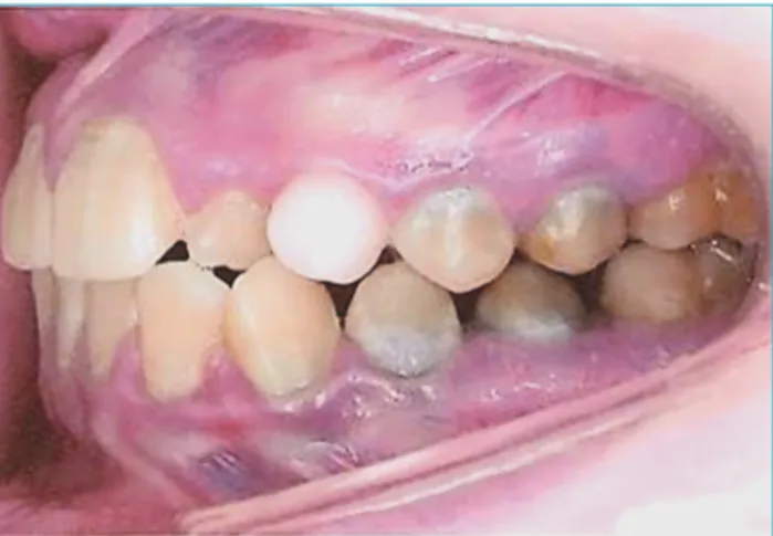
Mixed dentures with greenish pigmentation in dental elements, as well as dental bioilm and prolonged retention of the left superior deciduous canine were observed upon intraoral examination (Figure 1). Cone beam CT scan of the maxilla was performed at Centro de Tomograia Avançada
(CTA). he equipment used was an I-CAT (Kavo•) with
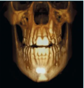
cone-beam X-ray system, focal point of 0.5 mm, voxel of 0.125 mm, 14-bit gray scale, 17x23 cm ield of view (FOV), automatic collimation with pulsed exposure, efective dose of 36 to 74 μSv and cylindrical reconstruction. he method consisted of a single exposure using cone-beam X-ray, cap-turing an image of the whole volume with a single exposure and 360° rotation of x-ray source around the patient’s head. To perform the tomographic report on the diferent radio-graphic densities of pigmented teeth, the Hounsield scale was used in 16 shades, from light gray to black. CT scan showed prolonged retention of the left superior deciduous canine (Figure 2) in frontal three-dimensional view, and sagittal sections showed peripheral hyperdense images of maxillary sinus (Figure 3). Upon panoramic examination, peripheral dystrophic calciications of maxillary sinus were seen (Figure 3).
DISCUSSION
Dystrophic calcification in maxillary sinus originates from an inflammatory picture with chronicity character-istics and may be related to fungal or non-fungal sinusitis.
Figure 1 Intraoral view with prolonged retention of left
Macedo AF et al.
119
Rev Paul Pediatr. 2018;36(1):117-120
Intrasinus calcium deposits may arise along with non-fun-gal inflammatory processes such as presence of mucocele and bacterial sinusitis.2 Few reports in literature mention
non-fungal sinusitis, and differential diagnosis between both conditions is relevant.
he present report shows absence of diferences in radio-graphic density of pigmented teeth in relation to enamel organ, without color change after cone-beam volumetric CT scan. his method allows examining the human body in segments with few millimeters of thickness, which helps to diagnose pathologies that afect bone tissues, besides
being non-invasive, fast and of high diagnostic accuracy, being able to identify and delineate pathological processes.9
Computed tomography is currently used for oral rehabilita-tion, delimitarehabilita-tion, and visualization of maxillofacial pathol-ogies, but it is still little used to diagnose head and neck systemic pathologies.9
At images, the pigmented dental structure did not show radiographic density diference, suggesting that dental organs with bilirubin chromatic alterations in their structure can be submitted to restorative treatment. his pigmentation is a consequence of the high concentration of bilirubin in the dentinal tubules.3 Tomographic imaging also showed
dystro-phic calciications inside the maxillary sinus, with peripheral areas of hyperdensity and hypodense center. he images sug-gest non-fungal sinusitis, in which calciication is close to the thickened submucosal layer of the maxillary sinus, constantly afected by chronic inlammatory conditions.2 Other etiologies
cited in relation to this radiographic inding are inlamma-tory diseases, malignant tumors and benign lesions, mucoce-les, and bacterial sinusitis.2 Calciications in fungal sinusitis
occur in the core of maxillary sinus, with hyperdensity orig-inating from well-delineated nodular masses arising from the calcium depositions within mycelial mass.2 here is no
con-sensus on the level of thickening of the sinus mucosa that is considered abnormal, ranging from 2 to 6 mm.10 he
radio-graphic pattern of nonfungal sinusitis difers from fungal sinusitis, which is characterized by high density in maxillary sinus, bone destruction, and iniltration of adjacent soft tis-sue, allowing aggravating processes that may lead to death in immunosuppressed patients.2 In this case, a patient with liver
transplantation may present serious clinical implications, once the adequate treatment of infections and sinusitis requires reduction or complete elimination of immunosuppression. Figure 2 Frontal three-dimensional image.
Figura 3 (A) Sagittal view showing peripheral dystrophic calciication of maxillary sinus; (B) Sagittal view showing
peripheral dystrophic calciication of maxillary sinus and onset of new calciication areas in its center; (C) Panoramic view showing dystrophic calciications of maxillary sinus.
Dystrophic calcification of maxillary sinus
120
Rev Paul Pediatr. 2018;36(1):117-120
If low immunosuppression continuation is needed, transplant rejection may occur.
Another factor to be highlighted in this case is the absence of symptoms, although dental pathological processes may poten-tially induce inlammation in maxillary sinus because of the proximity of dental loor to the radicular portion.10 he patient
had healthy teeth, which shows no relationship between calci-ication and pathological dental processes. Patients with acute sinusitis usually report unilateral headache and maxillary algesia in dental region, with sensitive and painful teeth; facial edema and thick purulent secretion may occur in chronic sinusitis.11
his inding underlines the importance of conducting a cone-beam CT scan to diagnose orofacial pathologies.10
Along with results obtained by imaging evaluation, a tai-lored treatment plan was developed based on weekly dental
prophylaxis with oral hygiene guidance and dental bioilm dis-closure in order to avoid the installation of a gingival inlamma-tory process and the onset of incipient caries lesions. Later on, the deciduous canine was extracted.
herefore, it can be inferred that imaging was fundamen-tal for diagnosis of non-fungal sinusitis in immunosuppressed and asymptomatic patient, which allowed the medical team to start the treatment for a disease that can aggravate the overall condition of liver transplantation recipient.
Funding
his study did not receive funding.
Conflict of interests
he authors declare no conlict of interests.
REFERENCES
1. Jácome AM, Abdo EN. Aspectos radiográicos das calciicações em tecidos moles da região bucomaxilofacial. Odontol Clín-Cient. 2010;9:25-32.
2. Yoon JH, Na DG, Byun HS, Koh Yh, Chung SK, Dong HJ. Calcification in Chronic Maxillary Sinusitis: Comparison of CT Findings with Histopathologic Results. AJNR Am J Neuroradiol. 1999;20:571-4.
3. Macedo AF, Azevedo RA, Zanin FA, Duarte DA. Manifestações bucais e sistêmicas em crianças com doença hepática crônica. Rev Gaúcha Odont. 2007;55:403-6.
4. Cauduro SM. Atresia biliar extra-hepática: métodos diagnósticos. J Pediatr (Rio J). 2003;79:107-14.
5. Shen QL, Chen YJ, Wang ZM, Zhang TC, Pang WB, Shu J, et al. Assessment of liver ibrosis by Fibroscan as compared to liver biopsy in biliary atresia. World J Gastroenterol. 2015;21:6931-6.
6. Isa HM, Mohamed AM, Alderazi AE. Efect of pediatric liver transplantation on renal function. Saudi J Kidney Dis Transpl. 2016;27:1-8.
7. Sumida AE, Oliveira FA, Oliveira HW. Uso da tomograia computadorizada (TC) na odontologia: estudo comparativo entre 2 métodos de reformatação da imagem tomográica na avaliação de retenções dentárias na região anterior da maxila. Rev Gaúcha Odontol. 2002;50:192-6.
8. Perella A, Borsatti MA, Tortamano IP, Rocha RG, Cavalcanti MG. Validation of computed tomography protocols for simulated mandibular lesions: a comparison study. Braz Oral Res. 2007;21:165-9.
9. Bramante AS, Bramante CM, Bernadineli N, Moraes IG, Garcia RB. Diagnóstico de defeitos ósseos por meio da radiografia convencional, digital e tomografia helicoidal. Rev Port Estomatol Med Dent Cir Maxilofac. 2007; 48:15-21.
10. Rege IC, Sousa TO, Leles CR, Mendonça EF. Occurrence of maxillary sinus abnormalities detected by cone beam CT in asymptomatic patients. BMC Oral Health. 2012;12:30.
11. Oliveira RA, Pedrazini MC, Wassall T. Relative area measurement of maxillary sinus by computed tomography. Rev Gaúcha Odontol. 2014;62:111-6.