BrazJOtorhinolaryngol.2014;80(4):359---361
Brazilian
Journal
of
OTORHINOLARYNGOLOGY
www.bjorl.org
CASE
REPORT
Late
displacement
of
a
dental
implant
into
maxillary
sinus
夽
Deslocamento
tardio
de
um
implante
dentário
para
o
seio
maxilar
Rodrigo
Nunes
Tavares
a,
Alexandre
Simões
Nogueira
a,b,
Marcelo
Bonifácio
da
Silva
Sampieri
a,∗,
Marcelo
Ferraro
Bezerra
a,
Eduardo
Sanches
Gonc
¸ales
baFaculdadedeFarmácia,OdontologiaeEnfermagem,UniversidadeFederaldoCeará(UFC),Sobral,CE,Brazil bFaculdadedeOdontologiadeBauru,UniversidadedeSãoPaulo(FOB-USP),SãoPaulo,SP,Brazil
Received17July2012;accepted24November2012 Availableonline23May2014
Introduction
Dental implants can be accidentally introduced into the maxillarysinus bothduring andaftersurgical installation. Bothsituationsareuncommon andusuallyinducesinusitis orothersimportantcomplications.1Theaimofthepresent
study was to report a rare clinical case of late displace-mentofadentalimplantintothemaxillarysinus,inwhich aCaldwell-Luc(CL)approachwasperformed, followedby thereconstructionoftheanteriormaxillarysinuswallusing atitaniummesh.
Case
presentation
A49-year-oldwomanwastreatedsixmonthspreviouslywith dental implants, and received three in the left posterior
夽 Pleasecitethisarticleas:TavaresRN,NogueiraAS,SampieriMB,
BezerraMF,Gonc¸alesES.Latedisplacementofadentalimplantinto maxillarysinus.BrazJOtorhinolaryngol.2014;80:359---61.
∗Correspondingauthor.
E-mail:marsampieri@hotmail.com(M.B.S.Sampieri).
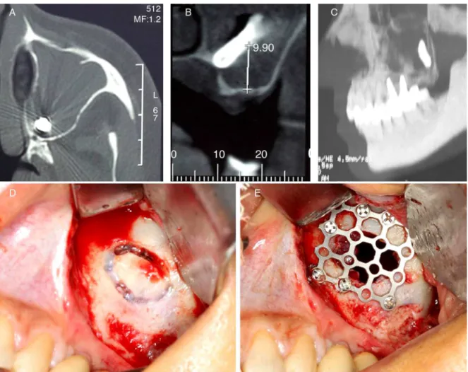
maxilla. She was referred to this clinic since one of the threeimplantsdisappearedduringcasting.CTscanshowed whatappeared tobeadentalimplant locatedposteriorly andinferiorlyinsideofthemaxillarysinus(Fig.1A---C).The mucosafromthesinusdemonstratedimportantalterations; itsdensitywascompatiblewithacutesinusitis, corroborat-ingtheclinicalfindings,whichincludedairwayobstruction andmoderate facialpain. Therewas noevidence of oro-antralfistula. After antibiotic therapy for acutesinusitis, the dental implant was removed through a CL approach. Underlocalanesthesia, asmallsurgicalincisionwasmade onthebuccalsulcus.Theanteriorbonywallofthesinuswas exposed,andanaccesswasmadewithacarbideroundbur (Fig.1D).Thesinusmucosaaroundthedentalimplantwas resected,followedbyintensecleaning.Finally,theanterior wallofmaxillarysinuswasreconstructedusingatitanium mesh,whichwasfixatedbymonocorticalscrews(Fig.1E). Thepatient hasbeen followed upfor 32months, withno complications.
Discussion
Poor bone quality and quantity of the posterior max-illa,inadditiontoalveolar pneumatizationfrommaxillary
http://dx.doi.org/10.1016/j.bjorl.2012.11.001
360 TavaresRNetal.
Figure1 (A)TCaxial,(B)TCcoronal,(C)TCsagittal,(D)Calwell-Lucapproachand(E)reconstructionofthesinusanteriorwall withatitaniummesh.
sinus,arepredisposingfactorsforthedisplacementof den-tal implants.The majorrisk factor is inadequatesurgical technique, which includes overtreatment of the implant preparation,sinusfloorperforation,andpoorprimary sta-bility.Latedisplacementisrareandusuallyhappensduring thefirstsixmonthsafterimplantation.2Inthepresentcase,
thedisplacementoccurredaftersixmonths,during manip-ulationoftheimplantforprostheticrehabilitation.
Thereareseveralmethods toremoveadentalimplant from the maxillary sinus, such as suction through bone alveolar defect, CL approach, functional endoscopy sinus surgery(FESS),andtransoralendoscopyapproachviacanine fossa.3,4Inthepast fewdecades,FESShasbeen replacing
theCLapproachforthetreatmentofparanasalpathologies, since it has been more effective. Besides all its advan-tages, isolated FESS is not effective in removing larger materials,especiallythoselocatedintheposteriorand infe-rioraspectsofthesinus.5,6Thepresenceofanoral---antral
communication,inflammatoryalterationsofsinusmucosa, and ostium patency also must be taken into account to choose the correct plan of treatment. The anterior max-illarysinus wallreconstructionbecomesimportant due to potentialcomplicationsfromtheCLapproach,suchas per-sistentbonedefectandretractionsofthesofttissuesofthe cheek.5,6Bonegrafts,guidedtissueregeneration,and
buc-calfatpadhavebeenusedtodecreaseCLfailure.1Forthis
purpose,atitaniummeshwasusedinthepresentcase.
Final
remarks
TheCLapproachisstillindicatedtoremoveobjectslocated posteriorly/inferiorly inside of the maxillary sinus, with additional care to reconstruct the bone defect created. Endoscopy procedures isolated or in association with the CL approacharealso proven tobe effective. The profes-sionalmusttake accountthesethreealternativesinorder tochoosethemostsuitableprocedure.
Conflicts
of
interest
Theauthorsdeclarenoconflictsofinterest.
References
1.González-GarcíaA,González-GarcíaJ,Diniz-FreitasM, García-García A, Bullón P. Accidental displacement and migration of endosseous implants into adjacent craniofacial structures: a review and update. Med Oral Patol Oral Cir Bucal. 2012, http://dx.doi.org/10.4317/medoral.18032.
Latedisplacementofadentalimplantintomaxillarysinus 361
3.BarzilaiG,GreenbergE,UriN.IndicationsfortheCaldwell-Luc approach in theendoscopicera. OtolaryngolHead NeckSurg. 2005;132:219---20.
4.ChiapascoM,FelisatiG,MaccariA,BorloniR,GattiF,DiLeoF. Themanagementofcomplicationsfollowingdisplacementoforal implantsintheparanasalsinuses:amulticenterclinicalreport and proposedtreatmentprotocols. IntJOralMaxillofacSurg. 2009;38:1273---8.
5.NakamuraN,MitsuyasuT,OhishiM.Endoscopicremovalofa den-talimplantdisplacedintothemaxillarysinus:technicalnote.Int JOralMaxillofacSurg.2004;33:195---7.