Congenital heart defects and extracardiac malformations
Cardiopatias congênitas e malformações extracardíacas
Cardiopatías congénitas y malformaciones extracardiacas
Rosana Cardoso M. Rosa1, Rafael Fabiano M. Rosa2, Paulo Ricardo G. Zen3, Giorgio Adriano Paskulin4
Instituição: Universidade Federal de Ciências da Saúde de Porto Alegre (UFCSPA) e Complexo Hospitalar Santa Casa de Porto Alegre (CHSCPA), Porto Alegre, RS, Brasil
1Médica Pediatra e Aluna de Mestrado pelo Programa de Pós-Graduação
em Patologia da UFCSPA, Porto Alegre, RS, Brasil
2Doutor pelo Programa de Pós-Graduação em Patologia da UFCSPA;
Geneticista Clínico do Hospital Materno-Infantil Presidente Vargas, Porto Alegre, RS, Brasil
3Doutor pelo Programa de Pós-Graduação em Patologia da UFCSPA;
Professor Adjunto da Disciplina de Genética Clínica da UFCSPA, Porto Alegre, RS, Brasil
4Doutor pelo Programa de Pós-Graduação em Genética e Biologia Molecular
da Universidade Federal do Rio Grande do Sul; Professor-Associado da Disciplina de Genética Clínica da UFCSPA, Porto Alegre, RS, Brasil AbstrACt
Objective: To review the association between congenital heart defects and extracardiac malformations.
Data sources: Scientific articles were searched in the Medline, Lilacs, and SciELO databases, using the descriptors “congenital heart disease,” “congenital heart defects,” “con-genital cardiac malformations,” “extracardiac defects,” and “extracardiac malformations.” All case series that specifically explored the association between congenital heart defects and extracardiac malformations were included.
Data synthesis: Congenital heart diseases are responsible for about 40% of birth defects, being one of the most common and severe malformations. Extracardiac malformations are observed in 7 to 50% of the patients with congenital heart disease, bring-ing a greater risk of comorbidity and mortality and increasbring-ing the risks related to heart surgery. Different studies have at-tempted to assess the presence of extracardiac abnormalities in patients with congenital heart disease. Among the changes described, those of the urinary tract are more often reported. However, no study has evaluated all patients in the same way. Conclusions: Extracardiac abnormalities are frequent among patients with congenital heart disease, and patients with these alterations may present an increased risk of mor-bimortality. Therefore, some authors have been discussing the importance and cost-effectiveness of screening these children for other malformations by complementary exams.
Key-words: congenital abnormalities; heart defects, congenital; urinary tract; ultrasonography.
resumo
Objetivo: Revisar a associação entre cardiopatias congêni-tas e malformações extracardíacas.
Fontes de dados: A pesquisa incluiu artigos cientí-ficos presentes nos portais Medline, Lilacs e SciELO, utilizando-se os descritores “congenital heart disease”, “congenital heart defects”, “congenital cardiac malformations”, “extracardiac defects” e “extracardiac malformations”. Foram incluídos os artigos de séries de casos que exploravam especificamente a associação entre cardiopatias congênitas e malformações extracardíacas.
Síntese dos dados: A cardiopatia congênita é responsável por cerca de 40% dos defeitos congênitos, sendo uma das malformações mais frequentes e a de maior morbimortali-dade. Malformações extracardíacas são observadas em 7 a 50% dos pacientes com cardiopatia congênita, trazendo um risco ainda maior de comorbidade e mortalidade e tornando a cirurgia cardíaca mais arriscada. Diferentes estudos têm tentado avaliar a presença de anormalidades extracardíacas em pacientes portadores de cardiopatia congênita. Dentre as alterações descritas, destacam-se aquelas do trato urinário. Contudo, não houve um estudo que tenha avaliado do mesmo modo todos os pacientes.
Endereço para correspondência: Paulo Ricardo G. Zen
Rua Sarmento Leite, 245, sala 403 – Centro CEP 90050-170 – Porto Alegre/RS E-mail: [email protected]
Fonte financiadora: Bolsa de Estudos cedida pela Coordenação de Aper-feiçoamento de Pessoal de Nível Superior (Capes)
Conflito de interesse: nada a declarar
Conclusões: Anormalidades extracardíacas são frequentes em pacientes com cardiopatia congênita, sendo que os por-tadores de tais alterações podem apresentar um risco maior de morbimortalidade. Consequentemente, alguns autores vêm discutindo a importância e o custo-benefício da triagem destas crianças à procura de outras malformações por meio de exames complementares.
Palavras-chave: anormalidades congênitas;cardiopatias congênitas; sistema urinário; ultrassonografia.
resumeN
Objetivo: Revisar la asociación entre cardiopatías congé-nitas y malformaciones extracardiacas.
Fuentes de datos: Se investigaron artículos científicos presentes en los portales Medline, Lilacs y SciELO, utili-zándose los descriptores «congenital heart disease», «congenital heart defects», «congenital cardiac malformations», «extracardiac defects» y «extracardiac malformations». Se incluyeron todos los artículos de casos que exploraban específicamente la asociación entre cardiopatías congénitas y malformaciones extracardiacas.
Síntesis de los datos: La cardiopatía congénita es res-ponsable por un 40% de los defectos congénitos, siendo una de las malformaciones más frecuentes y la de mayor morbimortalidad. Malformaciones extracardiacas se obser-van en 7 a 50% de los pacientes con cardiopatía congénita, trayendo un riesgo todavía más grande de comorbilidad y mortalidad y haciendo la cirugía cardíaca más arriesgada. Distintos estudios vienen intentando evaluar la presencia de anormalidades extracardiacas en pacientes portadores de cardiopatía congénita. Entre las alteraciones descriptas, se destacan aquellas del sistema urinario. Sin embargo, no hubo estudio que haya evaluado del mismo modo a todos los pacientes.
Conclusiones: Anormalidades extracardiacas son fre-cuentes en pacientes con cardiopatía congénita, siendo que los portadores de estas alteraciones pueden presentar un riesgo mayor de morbimortalidad. Por consiguiente, algunos autores vienen discutiendo la importancia y el costo-beneficio de la selección de estos niños en bús-queda de otras malformaciones por medio de exámenes complementares.
Palabras clave: anormalidades congénitas; cardiopatías congénitas; sistema urinario; ultrasonografía.
Introduction
Congenital heart diseases (CHD) are detected among 3 to 5% newborns(1), and are classiied as severe in one out of every 33 livebirths(2). In developed countries they repre-sent the main cause of mortality in early childhood and are responsible for one ifth of the total deaths(3). In Brazil, congenital malformations were responsible for nearly 19% of the mortality in children under one year old in 2008, and represented the second most frequent cause of death among this age group(4).
According to the deinition proposed by Mitchell et al(5), CHD consists of a macroscopic structural abnormality of the heart or the intra-thoracic great vessels, which have signiicant or potentially signiicant functional consequen-ces(6). They comprise about 40% of the congenital defects and represent one of the most frequent malformations(7-9). The prevalence of CHD range from 4 to 19/1,000 livebir-ths(6,10-13). However, according to Bosi et al(14), the prevalence of CHD has been increasing due to the greater detection of minor defects by the Doppler echocardiography, which has been widely used. In addition, the advances in intensive, surgical and anesthetic care have allowed a greater survival and, consequently, a greater number of adults with this condition(13,15).
CHD are the congenital malformations with greater impact on children’s morbidity and mortality, as well as on the health system’s costs(14). They represent the main cause of death among the congenital malformations(9). Extra-cardiac malformations (EM), such as intra-abdominal organs defects and/or associated with genetic syndromes, are observed from 7 to 50% of the patients with CHD, and impose a greater risk of morbidity and mortality to these patients, in addi-tion to increasing the risks of surgical correcaddi-tion(16-20). Also, such patients may need surgical procedures or intensive care regardless of the heart condition(16).
For these reasons, some authors have been discussing the importance and the cost-effectiveness of screening all chil-dren with CHD to detect EM using ancillary tests, such as abdominal ultrasound(20,21).
papers retrieved in these databases and was not limited to a pre-speciied period of time. We included all the case series that speciically analyzed the association between CHD and EM. Case reports were not included.
studies that evaluated em in patients with
congenital heart defects
Different studies have evaluated the presence of EM in patients with CHD. Studies conducted before the 80’s, howe-ver, show important limitations, since ultrasound tests were not available yet. The diagnosis of CHD, for instance, was performed according to the physical examination, surgery and catheterism indings, or even autopsy indings(5,22-24).
In the study by Julian and Farrú(25), published in 1986, the diagnostic method used to evaluate the EM was not re-ported. These authors conducted a retrospective evaluation of 207 children with CHD in a cardiology hospital in Chile and found EM in 31.9% of the patients, 22.7% of which constituted part of some syndrome. The authors did not iden-tify any speciic association between the different EM and CHD, except for the classic syndromes. The most frequent EM were observed in the gastrointestinal, musculoskeletal and genitourinary tracts.
In 1987, Ferencz et al(26) conducted a case control study of CHD among livebirths in the US. Individuals with CHD (n=1,494) were compared to a sample of neonates without CHD born in the same area (n=1,572). CHD were diagno-sed by echocardiography, cardiac catheterization, surgery or autopsy. EM were observed among 26.8% of the individuals with CHD, 8.3% of which were not associated with chro-mosomal anomalies or any other syndromes. Central nervous system malformations, eye disorders, major defects of the abdominal wall, gastrointestinal and urinary tract defects were more frequent among the patients with CHD.
In 1987, Kramer et al (27) prospectively evaluated 1,016 children with CHD up to 16 years old in Germany. The diagnosis was conirmed by echocardiography or cardiac catheterization. In 13.3% of the cases, the heart defect was part of a syndrome, embriopathy, association or sequence. Major EM occurred in 7.7% of the patients without any speciic syndrome, embriopathy, association or sequence (the results of the urography were not taken into consideration in these results). The malformations affected mainly the musculoskeletal and central nervous systems, eyes, ears, gas-trointestinal, respiratory and urinary tracts. A greater num-ber of EM was identiied among individuals with tetralogy
of Fallot (p=0.01). A urogram following angiography was performed in 302 individuals for screening of malforma-tions, and revealed abnormalities of the upper urinary tract in 8.9%. Only one of these individuals was symptomatic. The most frequent anomalies of the urinary tract were the total kidney duplication and the duplication of the ureter or renal pelvis. There were no differences in the frequency of the urinary tract malformations in association with any speciic heart defect. In the other hand, they found an association (p<0.01) between minor EM and CHD in comparison with healthy children.
In 1898, Stoll et al(28) prospectively studied 801 newborns with CHD and a control group in France. The study inclu-ded fetuses and stillbirths. The diagnosis of heart defect was conirmed by echocardiogram, cardiac catheterization, surgery or autopsy reports. Among the reported cases, 25.7% showed at least one EM, and such abnormalities were ten times more frequent in this group than in the control group. Among individuals with heart defects, 11.5% exhibited some type of syndrome. The most frequent EM affected the kidneys (21.4%) and the digestive system (19.6%). The most frequent abnormalities of the upper renal system were the urethral anomalies, hydronephrosis and unilateral renal agenesis. The presence of olygohydramnios and polyhydra-mnios and threatened miscarriage (the occurrence of vaginal bleeding before fetal viability) were more frequent among children with CHD and concomitant EM or syndromes.
Following this publication, Murugasu et al(21) conducted a prospective study in a Singapore hospital in 1990, including 109 children with CHD who underwent cardiac catheteriza-tion and ultrasound of the urinary tract. Important urologic malformations were found in 11.9% of the children, inclu-ding hydronephrosis, duplication, ectopy, agenesis and renal dysplasia. Five of the 13 patients with renal malformations had some syndrome, four of which were chromosomal. None of the children had any signs or symptoms suggestive of urinary tract disease. The authors concluded that the early identiication of the urologic anomalies in these patients would only have been possible using a screening test, such as renal ultrasound or urography.
these, 27 had a well-recognized syndrome or a well-deined association of malformations. The authors did not identify any association between speciic CHD and the main groups of EM, except for a possible relation between spleen abnor-malities and endocardic wall defects.
In 1999 Grech and Gatt(16) retrospectively studied 231 patients with CHD in Malta. The diagnosis was conirmed by echocardiography, catheterization, surgery or autopsy. The authors observed that 39 (17%) of the children had EM, 9% of them being chromosomal anomalies, 2% well recognized syndromes and 6% other non-cardiac. The most common EM were the musculoskeletal abnormalities.
In the study of Bosi et al in 2003(14), 2,442 livebirths with CHD born from 1980 to 2000 were retrospectively evaluated in Italy. The diagnosis of cardiac malformation was establi-shed by physical examination, echocardiography, cardiac catheterization, surgery and/or necropsy indings. Twenty four percent of these patients had EM. Chromosomal ano-malies were identiied in 9.1% of the newborns. In addition, 4.4% of the patients with CHD were diagnosed with some syndrome and/or sequence. EM were found among 10% of the children with CHD who did not have any syndrome, sequence or chromosomal anomalies. The genitourinary system was the most frequently affected (25.8%).
In 2003, Calzolari et al(30) conducted a retrospective study of 1,549 livebirths and stillbirths with CHD, also in Italy. The diagnosis of the cardiac malformation was conirmed by echocardiography, heart surgery, cardiac ca-theterism and/or necropsy. EM were found in 26% of the cases. Interventricular communication, ostium secundum atrial septal defects and complex heart defects were the cardiac malformations more frequently associated with EM. The most common EM were found in the musculoskele-tal (25.3%), genitourinary (22.9%) and gastrointestinal (11.5%) systems. Karyotypical analysis was performed in 19.4% of the cases, and chromosomopathies were detected in 152 patients.
In 2004, Eskedal et al(31) studied the prevalence of major EM anomalies in children with CHD and its impact on the survival. The registers of 3527 children with CHD who were born from 1990 to 1999 in Norway were analyzed. All the patients had the diagnosis of the heart defect conirmed by echocardiography, surgery, cardiac catheterization or necrop-sy. The authors reported the presence of EM in 20% of the children, with the gastrointestinal malformations, mainly intestinal anomalies and esophageal atresia, being the most frequent ones. The authors observed an improvement in the
survival of children born from 1995 to 1999 in comparison with those born from 1990 to 1994, except for those with EM, whose survival did not differ between the periods. However, patients with Down’s syndrome with CHD and no major EM had a better survival in the second period of the study. Also, children with right atrial isomerism were observed to have a higher risk for asplenia.
Still in 2004, Stephensen et al(32) retrospectively evaluated 740 livebirths with CHD from 1990 to 1999 in Iceland. The diagnosis of the heart defect was conirmed by echo-cardiography, cardiac catheterization or autopsy. EM were observed in 89 individuals (12%), including those with chromosomal anomalies, syndromes and/or other congenital anomalies. Chromosomopathies were observed in 4.9%. Among the patients with chromosomopathies and syndro-mes, genitourinary and gastrointestinal malformations were the most frequently observed EM. Among those with major heart defects, 18.2% had other congenital anomalies.
Güçer et al(33) conducted a retrospective study of 305 autopsies of children who had been born alive and had been diagnosed with a CHD in a Turkey hospital, from a total of 3320 autopsies performed in the study period. The prevalence of CHD was 9.1%. In addition, one or more EM were observed in 45.9% of the cases, with the craniofacial (19.7%) and genitourinary (15.1%) anomalies being the most common ones. The gastrointestinal and spleen malformations comprised 11.1% and 4.6% of the EM, respectively. Intra-atrial and interventricular commu-nication, coarctation of aorta, single ventricle, pulmonary stenosis, hypoplastic right heart syndrome, double outlet right ventricle, atrial and ventricular septal defects, aortic arch anomalies, right and left atrial isomerism were fre-quently accompanied by EM (>50%). Spleen malforma-tions were signiicantly more common among the cases of single ventricle (p<0.002). Furthermore, gastrointestinal and genitourinary anomalies were signiicantly associated with conotruncal defects (p<0.001).
not ind any correlation between CHD and concurrent EM. In children with multiple organs malformations, the mul-tivariate logistic regression analysis detected a signiicant association of age, the primary heart surgery and the type of CHD with mortality. Patients with digestive system malformations had a signiicant greater number of emer-gency surgeries (p=0.001). The mortality rate of the total sample was 8.9%, while the mortality of children with EM was 19%, increasing to 50% among newborns in this last group. Therefore, the treatment of children with concomi-tant CHD and EM is related to high mortality. EM were more frequently observed among children with ventricular septal defects (7.6%, p=0.0012).
In the prospective observational study by Meberg et al(19) conducted in Norway in 2007, which evaluated 57,027 births from 1982 to 2005, 662 CHD conirmed by echo-cardiography or autopsy were observed. Twenty two percent of the individuals with CHD had associated EM. Forty six percent of the EM malformations were not related to any chromosomal anomaly, genetic syndrome, microdeletion or teratogenic syndromes. Atrioventricular septal defects, intra-atrial communication, tetralogy of Fallot and single ventricle were the heart defects most frequently associated with EM. Associated anomalies were observed in 31% of the patients with perimembranous ventricular septal defect. Mortality was signiicantly higher among patients with CHD associated with other disorders (29%) in comparison with those with isolated heart defects (6%).
In the case control study by Amorim et al(2) published in 2008, 277 newborns and 75 stillbirths with CHD diagnosed from 1990 to 2003 in a University Hospital in Minas Gerais, Brazil, were evaluated. The livebirths had a control group, composed by the next newborn of the same gender, with no malformations, born in the same hospital. The CHD was con-irmed by postnatal echocardiography or necropsy. Noncardiac anomalies were detected in 31.4% of the livebirths and 48% of the stillbirths in whom no syndromes could be detected. EM constituted part of some syndrome in 23.1% of the livebirthis and 32% of the stillbirths. The most common EM among the newborns were those of the genitourinary system (48.3%). Among the stillbirths with no diagnosed syndromes, EM were more frequently observed in the genitourinary (52.8%) and musculoskeletal systems (52.8%).
In 2009, Gonzalez et al(20) conducted a retrospective analy-sis of the medical charts of 223 neonates with a prenatal diag-nosis of structural CHD from 1998 to 2007, in a general and
a cardiologic hospital in the US. The CHD was conirmed by postnatal echocardiography. The EM detected by cranial and abdomen ultra-sonogram were classiied as signiicant, mo-derately signiicant and nonsigniicant (variants of normal). Abdominal ultrasound (performed in 58.7% of the cases) detected some abnormality in 41.2% of the cases, and 36.6% were clinically signiicant (most of them consisting of renal malformations or heterotaxy disorders). Patients with septal defects were 3.7 times more likely to have abnormal indings in the abdominal ultra-sonogram than those without septal defects. Nearly 50% of the individuals had one or more extra-cardiac or genetic disorders identiied by abdominal ultrasound, cranial ultrasound (performed in 134 patients) or karyotype analysis (performed in 158 cases), resulting in a cost-yield ratio of 4,508 USD/patients with extracardiac or genetic abnormalities (the costs of performing the three screening tests was 2,254 USD/patient). The authors con-cluded, thus, that a universal screening program using the three tests represent a reasonable strategy for the neonates who need a cardiac surgery.
In 2010, Dilber and Malcić(35) retrospectively analyzed 1480 newborns with CHD in the period from 2002 to 2007 in pediatric cardiologic hospitals in Croatia. The diagnosis of CHD was established by physical examination, electrocardiogram, X-Ray, echocardiography, cardiac cathe-terization or autopsy. Congenital rhythm disturbances and cardiomyopathies were also included. During the ive years period, 57 (3.85%) children died due to the CHD or other related problems. Chromosomal defects, syndromes and/or major congenital abnormalities were identiied in 14.5% of the patients. Among the individuals who did not have any chromosomal disorder or syndrome, the most common EM were the gastrointestinal defects (8.4%). The CHD most frequently associated with extra cardiac malformations were the major heart defects (33.3%).
communication (18.5%), malrotation of the heart (17.2%) and conotruncal defects (16%). Therefore, 27.8% of the infants with CHD had major EM, including those with syndromes or laterality defects. The most common noncardiac defects were those of the skeleton (35%), gas-trointestinal (25.2%) and urinary (23.1%) systems. The clinical information was reviewed by a clinical geneticist, who classiied the patients as having isolated CHD, CHD associated with EM or with syndromes. The authors ob-served a higher frequency of speciic combinations, such as hydronephrosis or urethral atresia with cardiac malrotation,
right ventricular outlet obstruction, intra-atrial and inter-ventricular communication. When the analysis was limited to the cases with EM who underwent necropsy, the skeleton (46.1%) and urinary system (35.8%) defects were the most frequently observed.
Nevertheless, we must emphasize that none of the studies described herein used the same method to evaluate all the patients. In the present review, we did not ind any study that used the dysmorphological physical examination per-formed by a clinical geneticist, karyotype tests, luorescent
in situ hybridization (FISH) test for detection of the 22q11
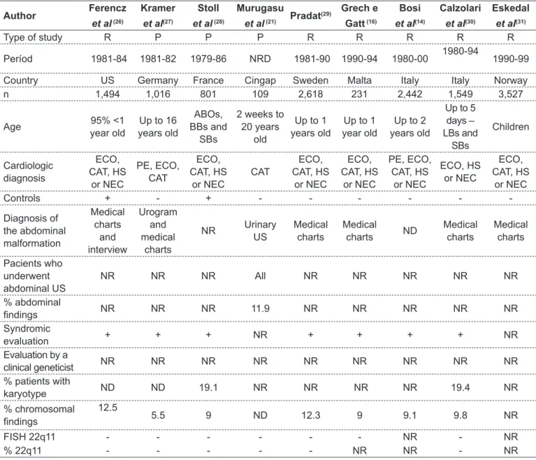
table 1 - Studies of extra-cardiac malformations in children with congenital heart diseases published in the literature
Author Ferencz
et al (26)
Kramer
et al(27)
stoll
et al (28)
murugasu
et al (21) Pradat
(29) Grech e
Gatt (16)
bosi
et al(14)
Calzolari
et al(30)
eskedal
et al(31)
Type of study R P P P R R R R R
Períod 1981-84 1981-82 1979-86 NRD 1981-90 1990-94 1980-00 1980-94 1990-99
Country US Germany France Cingap Sweden Malta Italy Italy Norway
n 1,494 1,016 801 109 2,618 231 2,442 1,549 3,527
Age 95% <1
year old
Up to 16 years old
ABOs, BBs and
SBs
2 weeks to 20 years
old
Up to 1 years old
Up to 1 year old
Up to 2 years old
Up to 5 days – LBs and
SBs
Children
Cardiologic diagnosis
ECO, CAT, HS
or NEC
PE, ECO, CAT
ECO, CAT, HS
or NEC
CAT
ECO, CAT, HS
or NEC
ECO, CAT, HS
or NEC
PE, ECO, CAT, HS
or NEC
ECO, HS or NEC
ECO, CAT, HS
or NEC
Controls + - + - - -
-Diagnosis of the abdominal malformation
Medical charts
and interview
Urogram and medical
charts
NR Urinary
US
Medical charts
Medical
charts ND
Medical charts
Medical charts
Pacients who underwent abdominal US
NR NR NR All NR NR NR NR NR
% abdominal
indings NR NR NR 11.9 NR NR NR NR NR
Syndromic
evaluation + + + NR + + + + NR
Evaluation by a
clinical geneticist NR NR NR NR NR NR NR NR NR
% patients with
karyotype ND ND 19.1 NR NR NR NR 19.4 NR
% chromosomal
indings
12.5
5.5 9 ND 12.3 9 9.1 9.8 NR
FISH 22q11 - - - NR - NR
% 22q11 - - - NR NR - NR
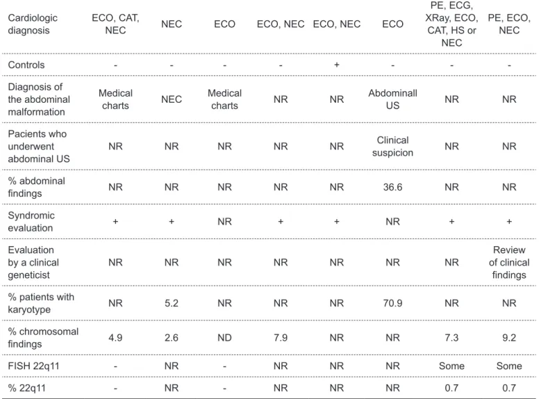
table 1 - Continuation
Author stephensen
et al(32)
Güçer
et al(33)
Wojtalik
et al (34)
meberg
et al(19)
Amorim
et al (2)
Gonzalez
et al(20)
Dilber e
Malčić(35)
miller
et al (36)
Type of study R R R P R R R R
Períod 1990-99 1977-02 1997-02 1982-05 NR 1998-07 2002-07 1968-05
Country Iceland Turkey Ploand Norway Brazil US Croatia US
n 740 305 1,856 662 352 223 1,480 7,984
Age Children
1 day to 16 years old -
NEC
Children Neonates
Neonates – LBs and
SBs
Neonates Neonates
ABOs, fetuses, LBs and
SBs
Cardiologic diagnosis
ECO, CAT,
NEC NEC ECO ECO, NEC ECO, NEC ECO
PE, ECG, XRay, ECO,
CAT, HS or NEC
PE, ECO, NEC
Controls - - - - + - -
-Diagnosis of the abdominal malformation
Medical
charts NEC
Medical
charts NR NR
Abdominall
US NR NR
Pacients who underwent abdominal US
NR NR NR NR NR Clinical
suspicion NR NR
% abdominal
indings NR NR NR NR NR 36.6 NR NR
Syndromic
evaluation + + NR + + NR + +
Evaluation by a clinical geneticist
NR NR NR NR NR NR NR
Review of clinical
indings
% patients with
karyotype NR 5.2 NR NR NR 70.9 NR NR
% chromosomal
indings 4.9 2.6 ND 7.9 NR NR 7.3 9.2
FISH 22q11 - NR - NR NR NR Some Some
% 22q11 - NR - NR NR NR 0.7 0.7
R: retrospective; P: prospective; NR: not reported; US: United States; Cingap: Cingapure; n: number of patients; ABOs: abortions; LBs: livebirths; SBs: stillbirths; NEC: necropsies; ECO: echocardiography; CAT: catheterization; HS: heart surgery; PE: physical examination; ECG:
deletion syndrome and abdominal ultrasound in all children with CHD. Table 1 summarizes the main features of these studies. Chart 1 lists the main EM reported among patients with CHD.
Conclusions
In summary, EM, including abdominal abnormalities, are frequent in patients with CHD, and those who have both conditions may present higher risk of morbidity and mortality. Consequently some authors have been discussing the importance and cost-effectiveness of the screening of children with CHD for EM using ancillary tests such as abdominal ultra-sonogram. Only a few studies have eva-luated this approach using a universal screening program; however, these studies have indicated that this represents a reasonable strategy, not only for the frequency of EM, but also because many of them, specially the abdominal malformations, may be assymptomatic. Such approach could be helpful to prevent further complications, such as chronic kidney disease. However, almost all the published studies are retrospective and not uniform (i.e., did not use a standardized evaluation method, by using ultrasound in all the studied individuals) and are based mainly in me-dical charts and population registers. Therefore, further studies are still necessary to evaluate the real importance of performing such a screening.
Acknowledgements
We thank the Coordination for the Improvement of Higher Level Personnel (CAPES) for the scholarship given to this study.
references
1. Robinson A, Linden MG. Clinical genetics handbook. Boston, USA: Blackwell Science; 1993.
2. Amorim LF, Pires CA, Lana AM, Campos AS, Aguiar RA, Tibúrcio JD et al. Presentation of congenital heart disease diagnosed at birth: analysis of 29,770 newborn infants. J Pediatr (Rio J) 2008;84:83-90.
3. Anderson RN, Smith BL. Deaths: leading causes for 2002. Natl Vital Stat Rep 2005;53:1-89.
4. Brasil. Ministério da Saúde. DATASUS [homepage on the Internet]. Informações de Saúde: Nascidos Vivos Brasil [cited 2007 Feb 10]. Available from: http://tabnet.datasus.gov.br/cgi/deftohtm.exe?sinasc/cnv/nvuf.def 5. Mitchell SC, Korones SB, Berendes HW. Congenital heart disease in 56,109
births. Incidence and natural history. Circulation 1971;43:323-32.
6. Hoffman JI, Kaplan S. The incidence of congenital heart disease. J Am Coll Cardiol 2002;39:1890-900.
7. Grech V. The evolution of diagnostic trends in congenital heart disease: a population-based study. J Paediatr Child Health 1999;35:387-91. 8. Acharya G, Sitras V, Maltau JM, Dahl LB, Kaaresen PI, Hanssen TA et al.
Major congenital heart disease in Northern Norway: shortcomings of pre- and postnatal diagnosis. Acta Obstet Gynecol Scand 2004;83:1124-9.
9. Jenkins KJ, Correa A, Feinstein JA, Botto L, Britt AE, Daniels SR et al. Noninherited risk factors and congenital cardiovascular defects: current
knowledge: a scientiic statement from the American Heart Association Council
on Cardiovascular Disease in the Young: endorsed by the American Academy of Pediatrics. Circulation 2007;115:2995-3014.
10. Ferencz C, Rubin JD, McCarter RJ, Brenner JI, Neill CA, Perry LW et al. Congenital heart disease: prevalence at livebirth. The Baltimore-Washington infant study. Am J Epidemiol 1985;121:31-6.
11. Gill HK, Splitt M, Sharland GK, Simpson JM. Patterns of recurrence of Chart 1 - Extracardiac malformations most frequently reported
among patients with congenital heart diseases(2,14,16,19, 20, 26-36) extracardiac malformations
Central nervous system Hydrocephalus
Corpus callosum agenesis Defects of the neural tube closure Craniofacial
Cleft lip/palate eyes
Microphtalmia/anophtalmia respiratoty
Diaphragmatic hernia
Pulmonary hypoplasia/ agenesis
Tracheoesophageal istula
Pulmonary segmentation anomalies Digestive
Esophageal atresia/stenosis Duodenal atresia/stenosis Omphalocele
Anal atresia/stenosis musculoskeletal
Upper limbs deiciency
Polydactyly/syndactyly Costovertebral anomalies Dislocation of the hip Clubfoot
Genitourinary Renal duplication
Uretheral/renal pelvis duplication Hydronephrosis
Renal agenesis/hypoplasia Cystic kidney disease Ectopic kidney
Vesicouretheral relux
Hypospadia spleen anomalies
congenital heart disease: an analysis of 6,640 consecutive pregnancies evaluated by detailed fetal echocardiography. J Am Coll Cardiol 2003;42:923-9.
12. van Beynum IM, Kapusta L, Bakker MK, den Heijer M, Blom HJ, de Walle HE. Protective effect of periconcepcional folic acid supplements on the risk of congenital heart defects: a registry-based case-control study in the northern Netherlands. Eur Heart J 2010;31:464-71.
13. Dolk H, Loane M, Garne E; European Surveillance of Congenital Anomalies (EUROCAT) Working Group. Congenital heart defects in Europe: prevalence and perinatal mortality, 2000 to 2005. Circulation 2011;123:841-9.
14. Bosi G, Garani G, Scorrano M, Calzolari E; IMER Working Party. Temporal variability in birth prevalence of congenital heart defects as recorded by a general birth defects registry. J Pediatr 2003;142:690-8.
15. Winlaw D. Congenital heart disease in the 21st century. Crit Care Resusc 2007;9:270-4.
16. Grech V, Gatt M. Syndromes and malformations associated with congenital heart disease in a population-based study. Int J Cardiol 1999;68:151-6. 17. Marino B, Digilio MC. Congenital heart disease and genetic syndromes:
speciic correlation between cardiac phenotype and genotype. Cardiovasc
Pathol 2000;9:303-15.
18. Begić H, Tahirović H, Mesihović-Dinarević S, Ferković V, Atić N, Latifagić A.
Epidemiological and clinical aspects of congenital heart disease in children in Tuzla Canton, Bosnia-Herzegovina. Eur J Pediatr 2003;162:191-3. 19. Meberg A, Hals J, Thaulow E. Congenital heart defects – chromosomal
anomalies, syndromes and extracardiac malformations. Acta Paediatr 2007;96:1142-5.
20. Gonzalez JH, Shirali GS, Atz AM, Taylor SN, Forbus GA, Zyblewski SC et al. Universal screening for extracardiac abnormalities in neonates with congenital heart disease. Pediatr Cardiol 2009;30:269-73.
21. Murugasu B, Yip WC, Tay JS, Chan KY, Yap HK, Wong HB. Sonographic screening for renal tract anomalies associated with congenital heart disease. J Clin Ultrasound 1990;18:79-83.
22. Greenwood RD, Rosenthal A, Parisi L, Fyler DC, Nadas AS. Extracardiac abnormalities in infants with congenital heart disease. Pediatrics 1975;55:485-92.
23. Kenna AP, Smithells RW, Fielding DW. Congenital heart disease in Liverpool: 1960-69. Q J Med 1975;44:17-44.
24. Hoffman JI, Christianson R. Congenital heart disease in a cohort of 19,502 births with long-term follow-up. Am J Cardiol 1978;42:641-7.
25. Jullian PM, Farrú AO. Extra cardic abnormalities in congenital heart defects. Rev Chil Pediatr 1986;57:430-3.
26. Ferencz C, Rubin JD, McCarter RJ, Boughman JA, Wilson PD, Brenner JI et al. Cardiac and noncardiac malformations: observations in a population-based study. Teratology 1987;35:367-78.
27. Kramer HH, Majewski F, Trampisch HJ, Rammos S, Bourgeois M. Malformation patterns in children with congenital heart disease. Am J Dis Child 1987;141:789-95. 28. Stoll C, Alembik Y, Roth MP, Dott B, De Geeter B. Risk factors in congenital
heart disease. Eur J Epidemiol 1989;5:382-91.
29. Pradat P. Noncardiac malformations at major congenital heart defects. Pediatr Cardiol 1997;18:11-8.
30. Calzolari E, Garani G, Cocchi G, Magnani C, Rivieri F, Neville A et al. Congenital heart defects: 15 years of experience of the Emilia-Romagna Registry (Italy). Eur J Epidemiol 2003;18:773-80.
31. Eskedal L, Hagemo P, Eskild A, Aamodt G, Seiler KS, Thaulow E. A population-based study of extra-cardiac anomalies in children with congenital cardiac malformations. Cardiol Young 2004;14:600-7.
32. Stephensen SS, Sigfusson G, Eiriksson H, Sverrisson JT, Torfason B, Haraldsson A et al. Congenital cardiac malformations in Iceland from 1990 through 1999. Cardiol Young 2004;14:396-401.
33. Güçer S, Іnce T, Kale G, Akçören Z, Ozkutlu S, Talim B et al. Noncardiac malformations in congenital heart disease: a retrospective analysis of 305 pediatric autopsies. Turk J Pediatr 2005;47:159-66.
34. Wojtalik M, Mrówczyński W, Henschke J, Wronecki K, Siwińska A, Piaszczyński
M et al. Congenital heart defect with associated malformations in children. J Pediatr Surg 2005;40:1675-80.
35. Dilber D, Malcić I. Sprectrum of congenital heart defects in Croatia. Eur J
Pediatr 2010;169:543-50.