309
Rev Bras Oftalmol. 2015; 74 (5): 309-11
C
ASER
EPORTReceived for publication 27/08/2012 - Accepted for publication 02/05/2013
The authors declare no conflicts of interest
1,2 Ophthalmology Resident of the Instituto de Olhos de Goiânia, Goiânia (GO), Brazil. 3 Department of Retina and Vitreous, Instituto de Olhos de Goiânia, Goiânia (GO), Brazil.
Catarata em árvore de Natal
Christmas tree cataract
Larissa Rossana Souza Stival
1, Ricardo Henrique Goulart Bittar
2, Anelise Medeiros Lago
2, João Jorge Nassaralla
Junior
3R
ESUMOA catarata em árvore de Natal é um tipo raro de opacificação do cristalino caracterizado por depósitos policromáticos em forma de agulhas no córtex profundo e no núcleo do mesmo, que podem ser isolados ou associados a outras opacidades. Neste estudo relata-mos e registrarelata-mos, por meio de fotografias, dois casos deste tipo de opacidade cristaliniana.
Descritores: Catarata; Cristalização; Opacidades; Cristalino/metabolismo; Relatos de casos
A
BSTRACTThe Christmas tree cataract is a rare type of lens opacification characterized by deposits of needle-shaped polychromatic cortex deep in the core, that can be isolated or associated with other opacities. We report and record, through photographs, two cases of this type of lens opacity.
Keywords: Cataract; Crystallization; Opacities; Lens, crystalline/metabolism; Case reports
310 StivalLRS, BittarRHG, LagoAM, Junior JJN
I
NTRODUCTIONC
hristmas tree cataract, also called starry cataract, is a rare type of opacity in the crystalline characterized by polychromatic needle-shaped deposits in the deep cortex and nucleus thereof which may be isolated or associated to other opacities(1). It is presented unilaterally or asymmetricallybilaterally(2).
There are controversies regarding the composition of said opacities. Hayes and Fisher(3) state that the images formed are
the result of light diffraction when reflected in the cells of the crystalline. Pau and Förster(4) suggest that the composition of
the opacities would mostly be cholesterol. Shun-Shin et al.(5)
suggested cystine as the substance related to the formation of the Christmas tree opacities typical of cataract.
The objective of this study was to describe two cases of cataract in Christmas tree seen at the Eye Institute of Goiânia in January 2012.
C
ASER
EPORTTwo cases of cataract in Christmas tree seen at the Eye Institute of Goiânia were selected.
Case 1 – A.M.G., female, 81 years old, complaining of low visual acuity associated to tearing. AVCC: RE: 20/40-1, LE: 20/
80. Dynamic refraction: RE = + 1.50 / -1.00 100 = 20 / 40-1. LE =
+ 1.75 / -2.50 55 = 20/80. Biomicroscopy: RE: nuclear cortical cataract + 2 / + 4. LE: thin and diffuse opacities in the cortex of the crystalline in which multicolored refractive bodies are scattered.
Case 2 – A.P.P., male, 69 years old, complaining of low vi-sual acuity. AVCC RE: 20/60-1, LE:20/60. Dynamic refraction:
RE +0.75esf -1.50cil x 20° = 20/80-1 LE = +1.00esf -0.75cil x
100° = 20/60. Biomicroscopy: RE: nuclear cortical cataract +2/ +4, crystalline cortex with iridescent multicolored crystals suggestive of cataract in Christmas tree. LE: Corticonuclear cataract +2/+4.
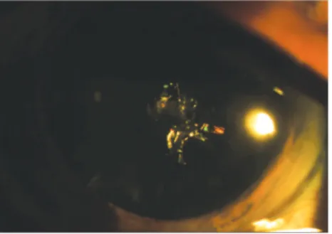
In March 2012 pictures of the eye of patient in case 2 were obtained in mydriasis with tropicamide 10% (Mydriacyl® - Alcon Brazil) and phenylephrine 10% (Fenilefrina® Allergan -Brazil) with a digital camera Pro-Pix DC-200 in slit lamp Shin-NIPPON (Japan), which documented the veracity of the exam described. Figure 1 without zoom: polychromatic crystalline opacity in needle shape. Figure 2, in slit lamp with zoom 6.5X: presence of iridescent crystals in the crystalline cortex. Figure 3, in slit lamp with zoom 16X: greater clarity of the multicolored refractive bodies, with a predominance of colors red and green.
D
ISCUSSIONCataracts in Christmas tree is a rare form of opacity in the crystalline in which images similar to highly refringent multicolored needles can be seen crossing the fibers in the deep layers of the cortex.
This type of change has already been the subject of several clinical discussions, and the composition of said opacities is still controversial(3-5). The colors vary according to the angle of
incident light and the colors red and green are predominant, which explains its nomenclature, since the brightness and variety of colors refer to the ornaments that commonly decorate the Christmas tree(3).
There are case reports of the association vetween cataract in Christmas tree and myotonic dystrophy, also called Steinert’s disease, which is characterized by a delay in muscle relaxation after a volunteer effort (myotomy). It is a dominant autosomal inheritance, and is presented as a common ophthalmologic finding, as well as the early onset of starry cataract, ptosis. Other unusual ophthalmologic findings include external ophthalmoplegia, dissociation of combined pupillary reflexes, light pigmentary retinopathy and reduced intraocular pressure(6). The two patients
reported did not present any systemic or ophthalmic symptom of myotonic dystrophy.
There are other rare forms of opacity in the crystalline, such as spear cataract and staghorn crystalline. As well as other types of polychromatic cataract, such as ceruelan cataract, nu-clear cataract, traumatic cataract(7), drug-induced cataract(8),
classic cataract of the diabetic patient and of the patient with uveitis.In the eyes studied, the appearance matches what is described in the literature in relation to cataract in Christmas tree.
It becomes difficult to register with pictures the true color of cataract in the Christmas tree, as well as most of polychromatic cataracts, due to reflection and association to other opacities. The flash traversing the optical system eliminates the most obvious colors, and makes the image less real than what it seems
Rev Bras Oftalmol. 2015; 74 (5): 309-11
Figure 1: Crystalline with polychromatic deposits.
Figure 2: Iridescent crystals in the cortex of the crystalline.
Figure 3: Multicolored refractive bodies, with a predominance of colors
311
Rev Bras Oftalmol. 2015; 74 (5): 309-11
to be to the naked eye. That is why the pictures were taken with no flash, in order to document in most realistic way what was found on the clinical examination (Figures 1, 2 and 3).
This study reported 2 cases with typical changes of cataract in Christmas tree in which the frequency increases with age, and isolated is of little interest in the reduction of visual acuity(9). It is
a rare type of polychromatic cataract that must be known not to surprise in the clinical examination or during the surgical procedure. Consequently, an increased time of surgery is avoided, since the crystals are highly refringent and change significantly the viewing of the procedure by the surgeon.
R
EFERENCES1. Brown NP, Bron AJ. Lens disorders: a clinical manual of cataract diag-nosis. 3rd ed. Oxford: Butterworth-Heinemann; 1996.
2. Kobayashi Y, Suzuki T. The aging lens: ultrastructural changes in cata-ract. In: Bellows JG, editor. Cataract and abnormalities of the lens. New York: Grune & Stratton; 1975. p. 313-43.
3. Hayes BP, Fisher RF. Ultrastructural appearances of a lens with marked polychromatic lustre: evidence for diffraction as a cause. Br J Ophthalmol.1984;68(12):850-8.
4. Pau H, Förster H. [Double refraction of crystals in the lens (spheroliths, ‘Christmas tree ornaments’) and in the vitreous body (scintillatio nivea)]. Graefes Arch Clin Exp Ophthalmol.1982;219(6):295-7. Ger-man.
5. Shun-Shin GA, Vrensen GF, Brown NP, Willekens B, Smeets MH, Bron AJ. Morphologic characteristics and chemical composition of Christ-mas tree cataract. Invest Ophthalmol Vis Sci.1993;34(13):3489-96. 6. Kanski JJ. Oftalmologia clínica: uma abordagem sistemática. 5a ed.
Rio de Janeiro: Elsevier; 2004. p. 161-91.
7. Reggi JR, Dantas MC, Dantas PE, Borges MJ. Catarata traumática: estudo de 60 casos. Arq Bras Oftalmol. 1997;60(5):489-92. 8. Paranhos FR. Estudo da incidência de catarata estelar em pacientes
em uso de clorpromazina. Arq Bras Oftalmol. 1991;54(2):63-8. 9. Stevens P, Swann PG. Christmas tree cataract. Clin Exp Optom.