! " #
#
$
, - (.
TABLE OF CONTENTS
Acknowledgements v
Resumo 1
Abstract 7
Chapter I – Introduction 11
I.I Learning to fear: auditory fear conditioning 12
I.II The Auditory System 22
a) The ascending flow of auditory information 23
b) The descending flow of auditory information 38
I.III Goals 45
Chapter II - The role of the auditory cortex in auditory fear conditioning 47
Acknowledgements 47
Summary 48
Material and Methods 50
Results 55
Chapter III - Discriminative auditory fear learning requires both
tuned and non-tuned auditory pathways to the amygdala 63
Acknowledgements 63
Summary 64
Material and Methods 66
Results 74
Discussion 91
Chapter IV - Conclusions and Perspectives 95
ACKNOWLEDGMENTS
First of all, I would like to thank my supervisor, Dr. Marta
Moita, for giving me the opportunity to develop my project in her
laboratory, for accepting such a peculiar student and embarking on
the challenge that these past few years have become. It was an
enriching experience watching the lab grow and taking part in its
scientific diversity. And we couldn’t be more different, but it definitively
taught me a lot. So thank you Marta for all the trust in me and for
helping me to grow as a scientist and as a person.
Also my sincere thanks to my thesis committee, Dr. Jorge
Carneiro and Dr. Monica Dias, who guided me in my first steps along
this road, and Dr. Rui Costa and Dr. Rui Oliveira, who kindly advised
me through the final years. For all the input, all the fruitful
discussions, all the support and guidance, I owe you my sincere
acknowledgments. And to Dr. Rui Oliveira, brave commander of the
team were I first set foot on the world of science, thank you for
showing me how joyful and heartfelt science can be.
I would also like to thank Marta Guimarãis, my PhD buddy in
Moita’s lab. Together we went through all the ups and downs a PhD
and a growing lab can have. So Marta, thank you for being there, for
the complicity, for all the help, for all the laughs and all the tears we
shared. Those memories will last forever.
And a special thank to Rosalina Fonseca, always willing to
lend a helping hand when electronic stuff were puzzling my head, but
has always been an inspiration to me, and I really cherish all your
support and friendship.
Thank you to my dearest colleagues at INPI, in particular to
the amazing Cluster of Biochemistry and Genetics, for all the laughs
together and for always being understanding and helpful when my
mind was lost somewhere in my experiments, even during working
hours... Also my gratitude to Eng Ana Bandeira for all those times in
which I was spared from “heavy” work so that I could take my PhD to
the end.
I must also not forget my friends and family, for all the loving
patience and support along this sometimes bumpy road. Paulinha,
Andreia, Ana, Cátia, Joana, Iris, Candy, Joaninha…sorry for all the
absences, and thank you for always being there! And to Aninhas, for
the precious help with the cover design, but especially for all the
crazyness and for never letting me loose my smile.
And my deepest gratefulness to my parents António and
Teresa, they really made this possible. Thank you for always
believing in me throughout the years, for all the right words in the right
moments, for all the unconditional support, for all the love and care
and for always making home the cosiest place to be. I really hope
you’re proud because this is your achievement too! And for last, a
special thank to my grandparents. You left me the willing to make you
proud, and I will always honour that. And especially to you my
gorducha, whose sweet memory always brought me comfort in the
I must also thank the IGC and IGC PhD Programme for giving
me the opportunity to develop my work in such a tremendously
brilliant scientific institution, and to all the PhD colleagues and all the
people in the institute who took part in my life there and made it such
an enriching experience.
This work was supported by Fundação para a Ciência e a
Resumo
O medo é uma resposta fisiológica com um forte impacto na
sobrevivência e adaptação. Grandes progressos têm vindo a ser
alcançados na compreensão dos mecanismos de aprendizagem de
medo, fundamentalmente através do condicionamento da resposta de
medo a estímulos auditivos. Neste paradigma comportamental um
som inicialmente neutro (estímulo condicionado, EC) adquire
propriedades aversivas após a sua associação a um estímulo
aversivo (e.g. choque; estímulo não condicionado, ENC). Assim, após
o condicionamento, o ENC desencadeia respostas condicionadas
originalmente despoletadas pelo estímulo aversivo.
A amígdala tem sido identificada como um substracto neuronal
relevante para a aprendizagem associativa da resposta de medo,
sendo apontada como o local onde ocorre a associação EC-ENC. A
informação auditiva converge na amígdala através de duas vias:
directamente através do tálamo auditivo, ou indirectamente através de
projecções tálamo-cortex-amígdala. Em virtude desta segregação de
inputs foi proposta uma hipótese que defende a existência de uma
“via superior” e uma “via inferior”. Segundo esta hipótese, a via
cortical (“via superior”) é essencial para a discriminação perceptiva
entre sons, enquanto que a via talâmica (“via inferior”) transmite
informação auditiva de uma forma mais rápida mas menos precisa.
O presente trabalho visava fundamentalmente testar esta
hipótese para a qual, apesar de largamente aceite, poucas evidências
têm sido apresentadas. O condicionamento da resposta de medo a
estímulos auditivos foi usado como paradigma comportamental, e a
identificaçao dos substratos neuronais da discriminaçao auditiva na
aprendizagem das respostas de medo.
Vários autores têm vindo a demonstrar que ambas as vias
auditivas intervêm na aprendizagem da resposta de medo a estímulos
auditivos, sendo que cada uma das vias é suficiente, por si só, para
suportar a aquisição da resposta condicionada. Contudo, segundo um
estudo recente, o córtex auditivo parece ser necessário para a
expressão da memória após a aprendizagem de medo, o que, a
confirmar-se, inviabilizaria experiencias destinadas a testar o papel da
via cortical na aprendizagem discriminativa. Por esse motivo, o
presente trabalho foi precedido de um estudo preliminar destinado a
clarificar o contributo da via cortical para a aprendizagem no cérebro
intacto, através da realização de lesões pós-treino do córtex auditivo.
Não obstante, importa também considerar que as lesões do
córtex auditivo, subjacentes às dificuldades de aprendizagem
anteriormente reportadas, afectaram quer o córtex auditivo primário,
quer o cortex secundário e associativo. Face ao efeito destas lesões
de grande extensão, e uma vez que os núcleos talâmicos que
projectam para a amígdala estão também reciprocamente interligados
ao cortex auditivo (sobretudo cortex secundário e perirrinal), as lesões
efectuadas no âmbito do presente trabalho foram limitadas ao córtex
auditivo primário (A1), cuja conectividade com os núcleos talâmicos
que projectam para a amígdala é reduzida. Procurou-se, assim,
minimizar a interferência de possíveis efeitos resultantes de lesões
corticais que afectassem simultaneamente ambas as vias auditivas,
quer por interferirem com a modulação corticofugal quer por induzirem
Resultados preliminares mostram a normal expressão da
memória de medo em ratos treinados num protocolo standard de AFC
e aos quais foram efectuadas lesões pós-treino do A1. Estes
resultados permitiram então estabelecer a base dos estudos
realizados subsequentemente com vista a avaliar a contribuição do
cortex auditivo para a especificidade da informação auditiva
transmitida à amígdala.
O facto de ter sido observado um resultado contrastante ao
das lesões corticais de maior extensão anteriormente reportadas
sugere uma contribuição diferencial dos inputs auditivos, a qual se
encontra, provavelmente, segregada ao nível das vias lemniscal
(tonotópica) e não-lemniscal (não tonotópica) que constituem a
segmentação funcional base do sistema auditivo. Portanto, no
presente trabalho foi levantada a hipótese de que a via tonotópica
(mas não a via não-tonotópica), suporta a resposta discriminativa de
medo a estímulos auditivos.
O córtex primário e secundário diferem principalmente na
selectividade da resposta auditiva. O córtex auditivo primário,
tonotopicamente organizado e cujos neurónios são caracterizados por
uma resposta altamente selectiva, constitui o último elemento da via
lemniscal e recebe projecções talâmicas originadas principalmente na
igualmente tonotópica divisão ventral do tálamo auditivo (MGv), o
único núcleo do tálamo que não possui projecções directas para a
amígdala. Pelo contrário, na via não-lemniscal, a divisão média do
tálamo auditivo (MGm) constitui o principal input directo para a
amígdala (apesar de possuir também eferentes difusamente
distribuídos no córtex auditivo), e apresenta respostas multisensoriais
Assim, a divergência funcional e conectiva verificada entre os
inputs talâmicos foi utilizada como base teórica para testar a hipótese
da “via superior/via inferior”. Para o efeito, foram realizadas lesões
electrolíticas do MGv ou do MGm e testado o seu efeito na aquisição,
expressão e extinção da memória adquirida através de um protocolo
de condicionamento diferencial da resposta de medo. Neste protocolo
foram utilizados duas frequências sonoras diferentes, sendo que um
dos sons foi associado ao choque (EC+) e o outro não (EC-).
Os resultados presentemente reportados mostram que
através de condicionamento diferencial com uma só apresentação de
cada um dos estímulos (single-trial training), todos os grupos testados
(controlo, lesão do MGv e lesão do MGm) adquiriram respostas
generalizadas de medo a ambos os sons. Por sua vez, utilizando um
treino com múltiplas apresentações dos estímulos (multiple-trial
training) os controlos expressam uma resposta diferencial de medo ao
CS+ e ao CS-, enquanto que os animais com qualquer uma das
lesões não discriminam os dois sons.
Por outro lado, quando as lesões foram realizadas após o
treino de condicionamento, apenas os ratos com lesão do MGm
revelaram incapacidade de discriminação entre os dois sons, sendo
que este grupo de animais demonstrou igualmente elevados níveis de
imobilidade (freezing), quer ao CS+ quer ao CS-, mesmo após uma
sessão de extinção. Portanto, apesar de ambas as vias auditivas
serem necessárias para a aquisição de respostas discriminativas de
medo, a expressão destas respostas depende unicamente da via
talâmica, sendo que esta via parece ser importante para a
discriminação através da supressão da resposta de medo a estímulos
De um modo geral, os resultados apresentados sugerem o
papel do MGv como modulador da aquisição de respostas
discriminativas a estímulos auditivos aversivos, enquanto que o MGm
parece sustentar continuamente a discriminação auditiva através de
uma regulação negativa das respostas de medo. Face aos resultados
obtidos, novas hipóteses são presentemente levantadas e discutidas
no âmbito da contribuição das vias lemniscal e não-lemniscal para a
aprendizagem discriminativa. O MGv poderá ser importante para a
aprendizagem discriminativa por facilitação da selectividade no córtex,
aumentando o contraste entre o EC+ e o EC-. O MGm, por sua vez,
poderá agir como facilitador da plasticidade no cortex, ou através de
convergência com o input cortical na amígdala. Por outro lado, o
papel do MGm poderá estar relacionado com a manutenção do tónus
inibitório ou na modulação inibitória dos neurónios da amigdala. Os
mecanismos propostos, não sendo mutuamente exclusivos, podem
em conjunto contribuir para a normal aprendizagem e expressão
Abstract
Fear is a physiological trait with a strong weight on survival and
adaptation. Great progress has been made to understand the
mechanisms of fear learning, mainly using auditory fear conditioning
(AFC). In this behavioral paradigm, an initial neutral tone (conditioned
stimulus, CS) acquires aversive predictive properties after successive
pairings with a footshock (unconditioned stimulus, US) and comes to
elicit responses characteristically elicited by threatening stimuli. In this
behavioral paradigm, the amygdala has been identified has a key
neural substrate for associative fear learning, and the site where
unconditioned stimuli (US) and conditioned (CS) auditory stimuli come
to be associated.
Auditory information may reach the amygdala either directly
from the auditory thalamus or indirectly via thalamo-cortico-amygdala
projections. The “high route/low route” hypothesis has thus been
proposed, which claims that the cortical pathway (“high route”) is
crucial for discrimination between fearful and neutral sounds, while the
direct thalamic pathway (“low route”) provides a rapid but less
accurate relay of auditory information to the amygdala. This
hypothesis relies on the assumption that more complex processing
requires cortical activity and that thalamic relay is faster then cortical
transmission to the amygdala. The present work essentially aims at
putting to test this largely accepted hypothesis. Auditory fear
conditioning was used as the behavioral paradigm to unravel the
possible functional explanation for the coexistence of two parallel
route/low route hypothesis was the working model for the identification
of neuronal substrates of auditory discrimination.
Accumulating evidence has been showing that each one of the
pathways alone is sufficient to support auditory fear conditioning.
However, according to a recent study, the auditory cortex might be
necessary for the recall of auditory fear learning, which would render
impossible the task of testing the role of the cortical pathway in the
recall of discriminative fear. Therefore, the present work was preceded
by a preliminary task aimed at clarifying the involvement of the cortical
pathway in AFC in the intact brain, by performing post-training lesions
of the auditory cortex.
Moreover, as lesions underlying the previously reported
learning impairments encompassed both primary, secondary and
association cortices, and because thalamic nuclei projecting to the
amygdala are reciprocally connected to the auditory cortex, we
selectively lesioned the primary auditory cortex (A1) which has limited
connectivity with thalamic nuclei projecting to the amygdala. Through
this selective cortical lesion we hoped to minimize effects resulting
from lesions simultaneously affecting both pathways to the amygdala,
either due to interference with corticofugal modulation or due to
induced neuronal degeneration of thalamic nuclei projecting to the
amygdala. Preliminary data shows normal expression of fear memory
in animals with post-training lesions of A1 and trained in a standard
AFC protocol. These results thus settled the basis for the following
studies aimed at testing the role of the auditory cortex in the accuracy
of conveyed auditory information during fear learning.
Because primary and secondary auditory cortex mostly differ
inputs to the amygdala essentially relies on the segregation of inputs
in the leminscal (tonotopic) and non-lemniscal (non-tonotopic)
systems, we further hypothesized that the tonotopic, but not the
non-tonotopic, pathway supports discriminative fear to auditory cues. In the
lemniscal pathway, sharply tuned neurons in primary auditory cortex
receive their main input from the ventral division of the medial
geniculate nucleus (MGv), which is tonotopically organized, has
narrowly tuned neurons and does not project directly to the amygdala.
In contrast, in the non-lemniscal pathway, the medial division of medial
geniculate nucleus (MGm) shows multisensory and non-tuned auditory
responses and is the main direct input to the amygdala, although it
also sends diffuse projections to auditory cortex.
We thus went further on testing the high/low route hypothesis
by assessing the effect of electrolytic lesions of the MGv or MGm on
the acquisition, expression and extinction of fear responses. A
discriminative auditory fear conditioning protocol was used, where one
tone was followed by shock (CS+) and another was not (CS-). Here
we show that with single-trial conditioning all the tested groups
(control, MGv- and MGm-lesioned rats) acquire non-discriminative fear
of both the CS+ and the CS-. This redundancy in neuronal pathways
involved in the acquisition of fear may guarantee self-preservation,
even though in single trial learning the learned fear responses
generalize to other auditory stimuli.
After multiple-trial conditioning, control rats discriminate
between the CS+ and CS-, whereas MGv- and MGm-lesioned rats do
not, meaning that discriminative fear learning requires the activity of
the two co-existing pathways. On the other hand, post-training lesions
fear. Thus, although for the acquisition of discriminative fear both the
lemniscal and non-lemniscal auditory pathways seem to be necessary,
the recall of discriminative fear memory seems to rely solely on the
latter. Interestingly, MGm-lesioned rats display high levels of freezing
to both the CS+ and CS- even after an extinction session to the CS+,
suggesting that this pathway might be important for discriminative fear
by suppressing freezing to the neutral cues.
Altogether the present findings point out a role for the MGv as
a modulator of the acquisition of discriminative fear responses, while
the MGm continuously holds up for auditory discrimination by
negatively regulating fear responses. New testable hypothesis are
presently put forward concerning the contribution of the lemniscal and
non-lemniscal routes for the mechanisms of auditory discrimination
learning. MGv may be important for discriminative learning by
facilitating cortical re-tuning, which might enhance the contrast
between CS+ and CS- evoked activity in AC. The MGm may impact
on discrimination learning by induction or facilitation of plasticity in
cortex or through convergence with cortical input onto the amygdala
neurons. Alternatively, the role of MGm may rely on sustaining
inhibitory tone in the amygdala, or in the inhibitory modulation of
amygdala neurons, namely through stimulus-specific inhibitory control
of interneurons or by interacting with the inhibitory network of the
central nucleus. These possibilities are not mutually exclusive and
may all contribute to normal learning and expression of discriminative
CHAPTER I - Introduction
Fear is a vital response to physical threats, and it is the
mechanism through which individuals protect themselves from
perceived danger. Learning from aversive events is thus a key stone in
adaptive behavior. Furthermore, several behavioral disorders entailing
maladaptive fear responses result from abnormal processing of
threat-related stimuli, as well as functional deficits in brain pathways
underlying fear learning and memory (Grillon, 2002b).
Though defensive responses are crucial for survival, they also
bear physiological costs. Therefore, optimized behavioral responses
thus demand that stimuli of higher biological significance should be
susceptible to preferential neuronal representation and accuracy in
discrimination. And so, neuronal mechanisms must exist which allow
differential responding to neutral and aversive stimuli.
To address this issue, in the present work we used auditory fear
conditioning (AFC) as the behavioral paradigm to study discriminative
fear learning. This paradigm was chosen because it has been robustly
used over time, and the neuronal circuit underlying AFC is quite well
characterized. An introduction to the neuronal circuitry of AFC, as well
as an elucidation on the functioning and connectivity of the auditory
I.I Learning to fear: auditory fear conditioning
Learning from biologically relevant aversive events has been
demonstrated in a wide range of species (Domjan, 2005; McNally and
Westbrook, 2006). The prevalence of this form of learning in natural
systems suggests that it is an adaptive trait that occurs under natural
circumstances and increases fitness. But even though fear can serve
as an alert mechanism for the organism against threat, pathological
states entailing maladaptive fear responses can persist and have a
negative impact in everyday life (Grillon, 2002a, 2002b).
Much of the understanding of the neural systems mediating fear
conditioning has been achieved through research on animals.
Nevertheless, recent studies suggest that similar systems are involved
in human fear conditioning (LaBar et al., 1995; LaBar and LeDoux,
1996; Grillon, 2002a, 2002b, 2008; Ledoux and Muller, 1997). Studies
on the neural basis of fear and anxiety in animal models may thus
shed some light on the mechanisms underlying the development of
pathological states of fear and anxiety and impact on the development
of therapies for such behavioral disorders.
One of the simplest experimental tools for studying fear and
anxiety is classical fear conditioning, based on Ivan Pavlov’s findings
that a neutral stimulus can acquire affective properties due to an
association with a biologically relevant stimulus (Pavlov, 1968).
Auditory fear conditioning has become in the last decade one of the
most widely used paradigms to study the neural mechanisms of
memory formation as fear learning entails robust, long lasting
memories, and it is conserved across species. According to this
aversive predictive properties after being paired with an aversive
footshock (unconditioned stimulus, US). The CS, by virtue of its
relationship with the US, comes to elicit responses characteristically
driven by threatening stimuli, including changes in heart rate and
arterial blood pressure, somatomotor immobility (freezing),
hypoalgesia and pupillary dilation (Fendt and Fanselow, 1999;
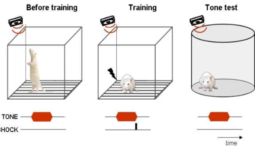
LeDoux, 2000; Maren, 2001) (Fig. 1). Because these responses are
not elicited by the CS before the CS-US association, they are referred
to as learned or conditioned responses.
Figure 1 – Schematic representation of auditory fear conditioning.
Before training, the tone (conditioned stimulus, CS) is a neutral stimulus, thus
eliciting only exploratory behaviors. After the tone being pared with a mild
footshock (unconditioned stimulus, US), when fear of the tone alone is tested,
In what concerns neuronal substrates of fear learning, extensive
evidence, from genetic and pharmacological manipulations to
electrophysiological recordings, points to the amygdala as a key
structure for learning and recall of the CS-US association (Maren,
2001; Han et al., 2009; Koo et al., 2004; Rumpel et al., 2005; LeDoux,
2000; Fendt and Fanselow, 1999; Davis and Whalen, 2001). The
amygdala is composed of several subnuclei (Sah et al., 2003; LeDoux,
2007), the most relevant for fear conditioning being the lateral (LA),
basal (B) and accessory basal (AB) nuclei (many times referred to as
“basolateral amygdala”, BLA), which connects to the central nucleus
(CE) (LeDoux, 2000; Pitkänen et al., 1995; Wilensky et al., 2006).
Neurons in CE then signal to hypothalamic and brainstem regions that
control the defensive and autonomic emotional responses to fear
(LeDoux et al., 1988; Shi and Davis, 1999; Wilensky et al., 2006).
The amygdala receives sensory inputs from several brain areas,
including the thalamus, the hippocampus and cerebral cortex
(McDonald, 1998; Linke et al., 2000; Bordi and LeDoux, 1994a; Li et
al., 1996; Romanski and LeDoux, 1993a; Iwata et al., 1986; Doron and
Ledoux, 1999; Shin et al., 2006; Siguròsson et al., 2010), and it is
widely believed that unimodal inputs enter the amygdala mainly
through its lateral nucleus, as shown by anatomical, behavioral and
physiological studies (LeDoux et al., 1990a, 1990b; Bordi and LeDoux,
1992; Romanski and LeDoux, 1992; Campeau and Davis, 1995a). In
fact, cells in LA respond to both tones and footshock (Romanski et al.,
1993), positioning the LA as a suitable locus for CS-US convergence
in auditory fear conditioning.
Fear conditioning has been shown to enhance auditory responses
underlying learning and memory formation, can also be induced in LA
neurons (Quirk et al., 1995, 1997; Rogan et al., 1997; Rogan and
LeDoux, 1995; Jung et al., 2010; Siguròsson et al., 2010; Ploski et al.,
2010; Fourcaudot et al., 2009; Pan et al., 2009; Blair et al., 2001; Sah
et al., 2008). Moreover, damage to LA has been shown to interfere
with both the acquisition and expression of conditioned fear responses
to auditory CSs (LeDoux et al., 1990a; Campeau and Davis, 1995a,
1995b; Nader et al., 2001; Amorapanth et al., 2000; Goosens and
Maren, 2001; Maren et al., 1996; Maren, 1998, 1999), while blocking
the AMPA receptor GluR1 trafficking in LA impairs LTP as well as fear
conditioning (Rumpel et al., 2005). More importantly, the selective
deletion of LA neurons recruited during learning blocks the expression
of fear memory (Han et al., 2009). Altogether these data strongly
suggest that activity in LA is necessary for formation of CS-US
association.
However, even though the LA as been usually viewed as the
locus for CS-US convergence, focus has been growing on the role of
CE in auditory fear conditioning because this nucleus has been found
to have the same characteristics that originally implicated the LA as a
critical site for fear learning. The CE also receives afferent projections
from the auditory cortex (McDonald, 1998) and the auditory thalamus
(LeDoux et al., 1985b, 1985a; Turner and Herkenham, 1991), and it
has been shown that these projections terminate in the lateral division
of the central nucleus (CEl) (Linke et al., 2000; Turner and
Herkenham, 1991; Pitkänen et al., 1995; McDonald, 1998; Jasmin et
al., 1997; Wilensky et al., 2006), along with nociceptive information
(Bernard et al., 1990; Jasmin et al., 1997; McDonald, 1998). In
center of the CE, also receives projections from the auditory thalamus,
namely the PIN (Turner and Herkenham, 1991; McDonald, 1998;
Linke et al., 2000). Moreover, as for LA, high-frequency stimulation of
thalamic inputs induces NMDA receptor-dependent LTP in the CE
(Samson et al., 2005), the CE-lesions also impair the acquisition of
fear conditioning (Goosens and Maren, 2001; Nader et al., 2001;
Campeau and Davis, 1995b), and the inhibition of protein synthesis
impairs the consolidation of fear memory (Wilensky et al., 2006).
Altogether, these findings make the CE also well suited to integrate
CS and US information during fear conditioning. Therefore, even
though traditionally viewed as the major output structure, the central
nucleus is also a potential critical site for fear learning.
During associative learning, nociceptive information about the
US, ascending from the spinal chord, thus reaches the amygdala via
LA or CE nuclei (Fig. 2-3). On the other hand, considering the
traditionally accepted model, information about auditory CSs may
reach the LA either directly from the auditory thalamus or indirectly via
auditory cortex (Romanski and LeDoux, 1993a; Armony et al., 1995;
McDonald, 1998; Linke et al., 2000; Li et al., 1996; Iwata et al., 1986;
Doron and Ledoux, 1999; Shin et al., 2006; Siguròsson et al., 2010)
(Fig. 2-3). The medial division of medial geniculate nucleus (MGm)
and posterior intralaminar nucleus (PIN), which have multisensory
neurons showing non-tuned auditory responses (except for high
frequencies relating to social vocalizations) (Bordi and LeDoux, 1994b,
1994a), convey the main direct auditory input to the amygdala
(LeDoux et al., 1985b, 1985a; Doron and Ledoux, 1999; Linke et al.,
2000), although also sending diffuse projections to the auditory cortex
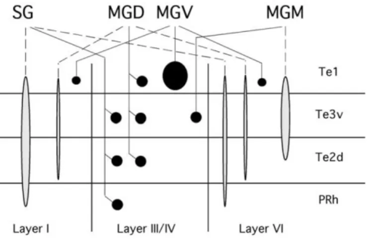
geniculate nucleus (MGd) and the suprageniculate nucleus (SG) also
directly project to the amygdala (Bordi and LeDoux, 1994a; Doron and
Ledoux, 1999; Linke et al., 2000) (Fig. 7).
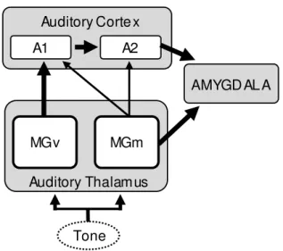
A1 A2
Auditory Corte x
AMYGD AL A
MGv
Auditory Thalamus MGm
Tone
Figure 2 – Schematic showing the main direct (MGv) and indirect (MGm) auditory pathways to the amygdala. Thicker arrows represent major neuronal inputs. A1: primary auditory cortex; A2: secondary auditory cortex;
MGv: ventral division of the medial geniculate nucleus; MGm: medial division
of the medial geniculate nucleus.
Additionally, cortical projections to the amygdala originate in
secondary auditory cortex (A2, also Te2/Te3) and in perirhinal cortex
(PRh) (Doron and Ledoux, 1999; Romanski and LeDoux, 1993a,
ventral portion of A1 (also Te1v), which appears to receive fewer
projections from the MGv when compared to the remainder of A1,
projects substantially to the LA (Doron and Ledoux, 1999; Romanski
and LeDoux, 1993a, 1993b; McDonald, 1998). Considering both
thalamo-amygdala and thlamo-cortical projections, MGv seems to
project to BLA exclusively via cortical relay, while MGm/PIN and MGd
project both directly and indirectly to the amygdala.
This segregation of inputs to the amygdala thus raises the
question of what is the contribution of each pathway for fear learning.
Accumulating evidence has implicated both the direct and indirect
pathways of sound to the amygdala in AFC (Romanski and LeDoux,
1992; McKernan and Shinnick-Gallagher, 1997; Rutkowski and
Weinberger, 2005; Boatman and Kim, 2006). For instance,
glutamatergic transmission in the medial geniculate nucleus (including
both MGm and MGv) is required for the recall of extinction memory
(Orsini and Maren, 2009) and standard AFC has been shown to
produce CS-driven frequency specific receptive field plasticity in the
non-lemniscal MGm/PIN and MGd neurons (Edeline and Weinberger,
1991a, 1992), as well as in the lemniscal MGv neurons (Edeline and
Weinberger, 1991b) and primary AC (Weinberger, 2007a, 2007b,
1998; Suga et al., 2002; Liu et al., 2007; Rutkowski and Weinberger,
2005; Edeline and Weinberger, 1993; Ma and Suga, 2009). It might
thus be that both the AC and the MGm/PIN learn during auditory fear
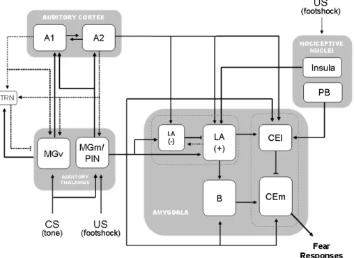
Figure 3 – Schematic showing the neuronal circuit underlying auditory fear learning. For simplicity, only the main thalamic inputs to the amygdala (MGm) and to the auditory cortex (MGv) are represented. For the whole set of
thalamo-amygdala and thalamo-cortical projections see Fig. 6 and Fig. 7 in
the following section of this chapter. Full lines represent feedforward
projections and dashed lines represent feedback projections. Arrow-ended
lines represent excitatory inputs, and dash-ended lines represent inhibitory
inputs. MGv: ventral division of the medial geniculate nucleus; MGm: medial
division of the medial geniculate nucleus; PIN: posterior intralaminar nucleus;
A1: primary auditory cortex; A2: secondary auditory cortex; TRN: thalamic
reticular nucleus; LA: lateral nucleus of the amygdala; (-): inhibitory
interneurons; (+):principal neurons; B: basal nucleus of the amygdala; CEl:
lateral subdivision of the central nucleus of the amygdala; CEm: medial
On the other hand, several studies have also shown that the
amygdala is under tight inhibitory control (Pan et al., 2009; Shaban et
al., 2006; Shin et al., 2006; Bauer and LeDoux, 2004). A significant
feedforward inhibition has been shown to arise from both thalamic and
cortical projections to LA interneurons, even though feedforward
inhibition from thalamic projections is stronger than that coming from
cortex (Shin et al., 2006). Therefore, because local inhibitory networks
have been shown to gate synaptic plasticity in the amygdala (Pan et
al., 2009), cortical and thalamic inputs may also differently impact on
fear learning by inhibitory mechanisms.
Overall, in what concerns the auditory inputs to the amygdala,
each pathway alone seems to be sufficient for the acquisition of fear
memory, as revealed by pre-training lesions of the whole AC (primary,
secondary and perirhinal cortices) or MGm (Romanski and LeDoux,
1992). However, these same lesions, if performed after learning, seem
to affect the recall of the auditory fear memory, with cortical lesions
having the strongest impact on expression of fear responses
(Boatman and Kim, 2006), while temporary inactivation of the auditory
cortex renders animals behaviorally deaf (Talwar et al., 2001).
It has nevertheless been proposed that the indirect cortical
pathway (“high route”) is crucial for discrimination between fearful and
neutral sounds, while the direct thalamic pathway (“low route”)
provides a rapid but less accurate relay of auditory information to the
amygdala (LeDoux, 2000). This would allow a fast response in the
case of danger, but the indirect route would stop the animal from
generalizing its fear to a wide range of sounds. Accordingly, it has
been shown that fear conditioning produces an increase in
consistent with the brief latency in transmission to the LA directly from
the thalamus. In contrast, AC has been reported to develop plasticity
slower than LA neurons (Quirk et al., 1997). It has also been shown
that the earliest tone-evoked responses in MGm/PIN occur 7–9 ms
after tone onset (Bordi and LeDoux, 1994a), while electrical
stimulation of MGm/PIN produces responses in the amygdala about
5ms later (Clugnet and LeDoux, 1990), which is consistent with the
short-latency LA responses induced by fear conditioning. On the other
hand, previous physiological studies have identified a core region of
the auditory cortex, the primary auditory cortex (A1), in which learning
has been shown to expand the representational area of the CS based
on the learned importance (Rutkowski and Weinberger, 2005;
Weinberger, 2007b) (Fig. 11, section below).
Despite being traditionally accepted, little evidence supports the
high/low route hypothesis. Moreover, auditory cortex lesions have in
fact been shown not to affect the generalization across a gradient of
tone frequencies (Armony et al., 1997), and a role for the thalamic
pathway in differential fear responding has been previously suggested
for conditioned bradycardia in rabbits (Jarrell et al., 1986, 1987), while
increasing CREB levels in the MGm/PIN has been shown to result in
broad auditory fear generalization (Han et al., 2008). Although the
neural circuit underlying auditory fear conditioning has been
intensively studied and its key players are now quite well
characterized, further studies are thus still required to clarify the
co-existence of parallel streams of information in the auditory fear circuit
and elucidate individual contributions to associative learning of fear
I.II The auditory system
The auditory system is a complex set of interconnected structures
which allow the transduction of sound waves into neuronal signals so
that ascending auditory information can be relayed into the central
nervous system. The ability to acquire and process acoustic
information about the environment, to localize and identify sound
sources, communicate with conspecifics and heed auditory warnings,
provides a selective advantage in an ever changing and challenging
world. Hearing is a fundamental adaptation for survival and
reproduction, whether it concerns communication within the species,
warning against predators or locating preys, and it assumes particular
significance in animals, namely in rodents, for which auditory signals
are an important part of social interaction (Portfors, 2007).
The system underlying auditory processing is a multi-level
assembly of interconnected structures. Two parallel pathways exist in
the auditory system, an ascending auditory pathway, conveying
auditory information from the organ of Corti to the auditory cortex (Fig.
4), and a descending stepwise projection from the cortex and all the
way down to the cochlea (Paxinos, 2004) (Fig. 10), overall forming a
plastic system with multiple loops.
The neural representation of sensory information undergoes a
series of fundamental transformations as it ascends the central
nervous system (Ehret, 1997; King et al., 2001; Pollak et al., 2003;
Shamma and Micheyl, 2010). At the early stages of auditory
processing, neurons of the auditory nerve and cochlear nucleus
encode elemental properties of sound, such as amplitude, while at
neural receptive fields which are tuned to discrete acoustic features
(Ehret, 1997; King et al., 2001; Pollak et al., 2003; Shamma and
Micheyl, 2010).
A fundamental organizing principle of the auditory system is its
tonotopic organization, preserved from the cochlea to the auditory
cortex (Fig. 5), and resulting from different sensitivity of auditory
neurons to frequency ranges (Ehret, 1997). The spectral sensitivity
profile of a neuron has commonly been characterized as a frequency
tuning curve, representing thresholds along the frequency domain
(Ehret, 1997) (Fig. 5 and Fig. 8). Regarding frequency processing,
numerous parallel and serial pathways converge in a common
destination in the auditory system, the inferior colliculus (IC). From the
IC and upward, the auditory pathway can be divided into a “core
pathway” with tonotopic organization and very selective responding
neurons (the lemniscal system), and a non-tonotopic and multisensory
“belt pathway” (the non-lemniscal system) (Ehret, 1997; Paxinos,
2004). This functional segregation sets the basis for the differential
flow of auditory information, and motivated the questions raised under
the present work.
a) The ascending flow of auditory information
As sensory information ascends from the cochlea to the auditory
cortex, it passes through several nuclei, each representing a neuronal
substrate of auditory information. Neurons at each level in the auditory
pathway exhibit complex and specific response properties which
al., 2001; Pollak et al., 2003; Shamma and Micheyl, 2010). Altogether,
the auditory nuclei process the different components of auditory
objects, like frequency, amplitude, distance or sound localization, thus
representing the complexity of the auditory world (Ehret, 1997; King et
al., 2001; Pollak et al., 2003; Shamma and Micheyl, 2010).
The peripheral sensory organ of the auditory system is the organ
of Corti. Sound waves are transmitted mechanically through the outer
and middle ear to the sensory hair cells of the organ of Corti, in the
cochlear partition of the inner ear (Ehret, 1997; Paxinos, 2004). The
mechanical energy underlying acoustic information is then transduced
in the cochlea into bioelectrical energy in the receptor potentials of hair
cells, and relayed to the cochlear nucleus via the auditory nerve fibers
(Ehret, 1997). The frequency component of sounds is mapped along
the cochlea basilar membrane and its overlying organ of Corti
(Paxinos, 2004). The cochlear frequency map sets the basis for the
tonotopic organization ultimately represented in the auditory cortex
(Fig. 5 and Fig. 8). Signals of the cochlear nerve are then separated
into a number of parallel ascending tracts, each with particular
conduction velocities and relays. The cochlear nerve fibers, encoding
stimulus frequency and intensity, terminate within the cochlear
nucleus, which is the first nucleus of the central auditory pathway, and
the first relay center in the ascending auditory pathway (Paxinos,
2004). The cochlear nucleus is particularly engaged in localization of
sound sources. Cells in the ventral cochlear nucleus provide accurate
information about the timing of acoustical stimuli, which is valuable for
locating sound sources in the horizontal axis, while the dorsal nucleus
is thought to participate in locating sound sources along the vertical
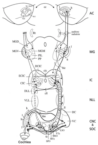
Figure 4- Ascending pathways of auditory information (adapted from Paxinos, 2004). Main auditory nuclei represented are: Cochlea (cochlea);
Cochlear nuclear complex (CNC); Superior olivary complex (SOC); Nuclei of
the lateral lemniscus (NLL); Inferior colliculus (IC); Medial geniculate body
(MG) and Auditory cortex (AC).
Cochlear nerve Inferior colliculus Auditory cortex Trapezoid body Medial geniculate Frequency, kHz S o u n d in te n s it y , d B b a
0.2 1 5 20 0.2 1 5 20
0.2 1 5 20 0 -40 -80 0 -40 -80
Figure 5 – Frequency selectivity in the auditory system of the mammalian brain. (a) Auditory frequency maps in auditory nuclei (adapted from Kandler et al., 2009). AN, auditory nerve; CN, cochlear nucleus; MSO:
medial superior olive; LSO: lateral superior olive; MNTB: medial nucleus of
the trapezoid body; HF, high frequency; LF, low frequency. (b) Tuning curves
of neurons in different auditory nuclei (adapted from Mann, 2002). Tuning
curves plot the sound intensity needed to increase the response of a neuron
above spontaneous firing, and it represents the response area of the neuron.
The frequency at which responses are elicited at the lowest intensity (tip of
There are three main output pathways of the cochlear nucleus: the
superior olivary complex, the ventral acoustic stria and the lateral
lemniscus (Ehret, 1997; Paxinos, 2004). The superior olivary complex
is the first stage were robust convergence of auditory information from
both ears takes place (Ehret, 1997). It consists of three nuclei, which
are tonotopically organized: lateral superior olive (LSO), medial
superior olive (MSO) and medial nucleus of the trapezoid body (MTZ)
(Paxinos, 2004). In rodents, there is also a fourth nucleus, the superior
paraolivary nucleus (SPO) (Paxinos, 1998, 2004; Saldaña and
Berrebi, 2000). The MSO responds mainly to low frequencies while the
LSO responds to all tonal frequencies (Paxinos, 2004). Therefore,
species characterized by high upper frequency limits of their hearing
range (e.g. rat), tend to have large LSO and small MSO. Furthermore,
the MSO is engaged in localization of sound sources in the azimuthal
axis through processing information about auditory delays of sound
reaching each ear (interaural delays) and it contains a map of
sound-source localization in the azimuth plane (Kandel et al., 2000). The
LSO is also involved in the localization of sound sources, but through
intensity cues, based on loudness of sound reaching each ear (Kandel
et al., 2000).
The lateral lemniscus (LL), which is tonotopically organized
(Merchán and Berbel, 1996; Paxinos, 2004), is a tract of axons in the
brainstem that carries information about sound from the cochlear
nucleus to various brainstem nuclei and ultimately to the contralateral
inferior colliculus (Paxinos, 2004).
The ascending auditory tracts from cochlear nucleus, superior
olivary complex and lateral lemniscus converge towards the auditory
obligatory relay centre for most ascending auditory tracts (Fig. 4).
Furthermore, it processes and integrates almost all ascending acoustic
information from lower centres and determines the form in which
information is conveyed to higher regions in the forebrain (Pollak et al.,
2003). Neurons in the IC are involved in the integration of multi-modal
sensory perception, processing of frequency- and
amplitude-modulated sounds and sound localization, based on detection and
representation of interaural timing and intensity differences (Kandel et
al., 2000; Pollak et al., 2003; Paxinos, 2004).
The IC consists of a central nucleus (CIC), an external cortex
(ECIC) and a dorsal cortex (DCIC), each with distinctive functional and
connective properties (Faye-Lund and Osen, 1985). The CIC is
characterized by a laminar structure which is the basis for its tonotopic
organization (Faye-Lund and Osen, 1985; Malmierca et al., 1993,
1995). A narrow range of best frequencies is represented within each
isofrequency lamina of the CIC, with neurons presenting V-shaped
tuning curves, while single units in the DIC and ECIC have a clearly
poorer tonal selectivity, characterized by broad and irregular tuning
curves (Ehret, 1997). The CIC has ascending projections to the medial
geniculate body (Peruzzi et al., 1997; Oliver et al., 1999) and
well-developed commissural fiber systems (Malmierca et al., 2003).
Moreover, the CIC projects in a strictly tonotopic manner to the ventral
division of the medial geniculate body (Linke, 1999a; Peruzzi et al.,
1997; Oliver et al., 1999).
The ECIC receives input from the cerebral cortex as well as from
many non-auditory structures, and its neurons have been shown to
respond not only to auditory but also to somatosensory input (for a
medial divisions of the medial geniculate body, while the DCIC
projects only to the dorsal division of the medial geniculate body
(Linke, 1999a; Peruzzi et al., 1997; Oliver et al., 1999).
Even though the majority of IC projecting neurons is glutamatergic,
recent studies have found GABA-positive projection neurons in the
CIC and a lower proportion in the DCIC and ECIC (Peruzzi et al.,
1997). Because few GABAergic cells are present in the rat medial
geniculate body (Winer and Larue, 1988), inhibitory inputs from the
CIC may be important for bottom-up regulation of firing patterns in
thalamic neurons.
From the inferior colliculus and upward, the auditory pathway can
be divided into a tonotopic “core pathway” (the lemniscal system), and
a non-tonotopic and multisensory “belt pathway” (the non-lemniscal
system) (Ehret, 1997; Paxinos, 2004). The medial geniculate body
(MGB), the main target for ascending projections from the inferior
colliculus (Linke, 1999a; Peruzzi et al., 1997; Oliver et al., 1999), is the
auditory centre of the thalamus. The MGB contains several divisions
defined on the basis of cytoarchitecture and fiber connections (Linke
et al., 2000; Linke and Schwegler, 2000; LeDoux et al., 1985b, 1985a;
Ledoux et al., 1987; Winer et al., 1999, 1999). The ventral division
(MGv) and the dorsal division (MGd) constitute its main core, while the
thalamic nuclei surrounding the MGB in its adjacent posterior, medial,
and rostral parts (the medial division of the MGB [MGm], the posterior
intralaminar nucleus [PIN], the suprageniculate nucleus [SG] and the
peripeduncular nucleus [PP]), integrate the caudal paralaminar nuclei
(Linke and Schwegler, 2000; Linke, 1999a, 1999b). Due to response
1994b, 1994a), these nuclei are generally considered a single module
(Armony et al., 1995).
The lemniscal core of the MGB is the tonotopically organized
ventral division (MGv), which has narrowly tuned neurons (Bordi and
LeDoux, 1994b, 1994a). In rat, MGv neurons show characteristic
frequencies (CFs) along the whole tested spectrum (1-30kHz), while
MGm and PIN neurons tend to show higher CFs (generally above
10Khz) (Bordi and LeDoux, 1994b). Another aspect of the MGV is the
laminar arrangement of afferent fibers and principal neurons (Winer et
al., 1999, 1999). In the rat, three subdivisions with different laminar
patterns occur, namely the ventral nucleus, the ovoid nucleus, and the
marginal zone (Winer et al., 1999). Furthermore, MGv neurons are
organized in a gradient of frequencies which extends along the
dorso-ventral axis, with lower frequencies being represented mainly in the
dorsal part and higher frequencies mainly in the ventral part of the
MGv (Bordi and LeDoux, 1994b). Studies in cats show that while the
LV has the dorso-ventral gradient going from low to high frequencies,
the OV is tonotopically organized but with a gradient from low to high
frequencies along an axis going from its dorsomedial to its
ventrolateral part (Ehret, 1997). However, no clear ventro-lateral
gradient has been identified in the rat (Bordi and LeDoux, 1994b).
The main input to the MGV comes from the ipsilateral CIC
(González-Hernández et al., 1991; Ledoux et al., 1987; Peruzzi et al.,
1997), although a small projection from the contralateral CIC is also
present (Paxinos, 2004). The ipsilateral input has excitatory and
inhibitory components (Peruzzi et al., 1997), and many neurons in the
MGV receive convergent excitatory and inhibitory input from the IC,
and a few only inhibitory (Bartlett and Smith, 1999). MGV is also
reciprocally connected with the tonotopically organized primary
auditory cortex (Kimura et al., 2003, 2005; Hazama et al., 2004; Winer
et al., 1999, 1999; Winer and Larue, 1987; Shi and Cassell, 1997),
sending selective projections to layers III and IV of Te1 (Kimura et al.,
2003) (Fig. 6).
The dorsal division of the MGB, part of the nonlemniscal system, is
morphologically and anatomically complex, with five subnuclei whose
function is poorly understood (Bordi and LeDoux, 1994b; Ehret, 1997;
Paxinos, 2004). Like neurons in the MGv, it also receives excitatory
and inhibitory inputs from the IC, excitatory inputs from the cortex, and
inhibitory inputs from the reticular thalamic nucleus (Bartlett and Smith,
1999, 2002). However, unlike MGv, the MGD is not tonotopically
organized (Winer et al., 1999) and is characterized by a large
proportion of neurons which do not respond to acoustic stimuli or
which are broadly tuned, with delayed responses that habituate faster
(Bordi and LeDoux, 1994b). Furthermore, multimodal
auditory-somatosensory responses are also found in this nucleus, mostly in its
rostral part (Ledoux et al., 1987; Bordi and LeDoux, 1994a).
The major source of inputs to the MGD are the nonlemniscal parts
of the inferior colliculus (Paxinos, 2004), though it also receives input
from the spinal chord (Ledoux et al., 1987). On the other hand, MGd
projects to all layers of primary auditory cortex, and to layers III and IV
of the secondary auditory cortical areas (Kimura et al., 2003), to the
insular cortex (Winer et al., 1999), and to the lateral nucleus of the
amygdala (Doron and Ledoux, 1999; Linke et al., 2000) (Fig. 6 and
Figure 6 – Schematic representation of thalamocortical ascending projections (adapted from Kimura et al., 2003). Circles represent selective projections and extended terminations represent diffuse projections.
The medial division of the MGB (MGm) is also part of the
non-lemniscal system of ascending auditory information. It has no
tonotopic organization, though cells with higher CFs tend to be located
more ventrally in the MGm (Bordi and LeDoux, 1994b). Contrary to the
MGv, neurons in the MGm show broadly responding to a wider range
of frequencies, though slightly narrower for higher frequencies, and
tend to show higher characteristic frequencies, generally above 10Khz
(Bordi and LeDoux, 1994b, 1994a). This nucleus sends diffuse
projections to layer VI of all areas in the auditory cortex, though mainly
targeting the secondary auditory cortex (areas Te2 and Te3) (Kimura
et al., 2003) (Fig. 6). Moreover, MGm also projects to non-auditory
regions and represents the main auditory input to the amygdala
(LeDoux et al., 1985b, 1985a; Doron and Ledoux, 1999; Linke et al.,
2000) (Fig. 7).
The PIN is part of the intralaminar and midline thalamic nuclei. Like
in the MGm, PIN neurons tend to have higher CFs (generally above
16Khz), though generally more broadly tuned (Bordi and LeDoux,
1994b). Furthermore, despite its major input being auditory, and
similarly to MGm, PIN neurons also respond to tactile, thermal,
nociceptive, vestibular and visceral stimulation (Bordi and LeDoux,
1994a; Weinberger, 2010). In these nuclei, three types of neurons are
found: those responding only to auditory stimuli, those responding to
both auditory and somatosensory stimuli, and those responding only
to somatosensory stimuli. Moreover, even unimodal somatosensory
cells show increased responses with simultaneous presentation of
somatosensory and auditory stimuli (Bordi and LeDoux, 1994a).This
nucleus receives inputs from the IC (Ledoux et al., 1987; Linke,
Linke and Schwegler, 2000) (Fig. 6), also constituting one of the direct
sensory inputs to the amygdala (Linke et al., 2000; Doron and Ledoux,
1999) (Fig. 7).
Additionally to the MGm and PIN neurons, the suprageniculate
(SG) is also a site of auditory-somatonsensory convergence (Ledoux
et al., 1987; Bordi and LeDoux, 1994a). Like in the MGm/PIN, neurons
in the SG tend to have high CFs (generally above 16Khz), and are
generally broadly tuned (Bordi and LeDoux, 1994a). This nucleus also
represents one of direct sensory inputs to the amygdala (Bordi and
LeDoux, 1994a; Linke et al., 2000) (Fig. 7).
Finally, the peripeduncular nucleus is a polymodal nucleus situated
ventrally to the MGv which was also shown to project to the lateral and
basal amygdala (Bordi and LeDoux, 1994a; Linke et al., 2000) (Fig. 7).
The auditory cortex constitutes the ending point in the ascending
auditory pathways, particularly for the thalamic efferents (Kimura et al.,
2003; Winer et al., 1999; Winer and Larue, 1987; Shi and Cassell,
1997). In the rat, the cortical map is generally categorized into three
temporal areas: Te1 (core), Te2 and Te3 (belt areas), defined on the
basis of the “gray level index” measured in Nissl staining (Zilles et al.,
1980; Paxinos, 1998). Temporal area Te1 is considered to be the
primary auditory cortex (A1) (Romanski and LeDoux, 1993a, 1993b),
and areas Te2 and Te3 are considered the secondary cortices
(Arnault and Roger, 1990; Paxinos, 2004). Furthermore, physiological
studies have identified a core auditory field which has frequency
selective neurons, surrounded by belt areas with less sharp frequency
representation (Doron et al., 2002; Rutkowski et al., 2003) (Fig. 8a). In
the rat, two tonotopically organized core fields, namely the primary
non-tonotopically organized belt fields, namely the posterodorsal (PDB),
dorsal (DB) and anterodorsal (ADB) belt fields have been identified
(Doron et al., 2002; Rutkowski et al., 2003). Both A1 and AAF are
located within Te1 (Doron et al., 2002; Rutkowski et al., 2003) (Fig.
8a).
Regarding frequency tuning, rat A1 neurons show a mean BW10dB
of approximately 1 octave (Kilgard et al., 2001; Rutkowski et al., 2003)
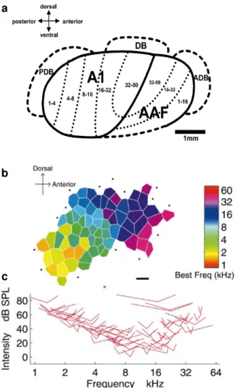
(Fig. 8). Furthermore, mapping studies in A1 have found a low to high
CF gradient that runs in the posterior to anterior direction (ranging
from about 1 kHz to 50 KHz, according to the range of frequencies
tested), with isofrequency lines oriented along the dorsoventral contour
of the cortex, while AAF shows a reversal of frequency organization
relative to A1 (Doron et al., 2002; Rutkowski et al., 2003) (Fig. 8a-b).
When compared to neurons in A1, AAF neurons exhibit broader
frequency tuning, as well as shorter first spike latencies and
significantly higher thresholds (Doron et al., 2002; Rutkowski et al.,
2003). Neurons in PDB, DB and ADB are characterized by strong
responses to white noise and show either poor or no responses to
pure tones (Doron et al., 2002; Rutkowski et al., 2003). The
differences in response properties found between the core and belt
fields may reflect a functional specificity in processing different
features of auditory stimuli.
The auditory cortex is reciprocally connected with the MGB,
although in the rat the reciprocity is not absolute (Winer and Larue,
1987). A1 receives selective ascending projections from the MGV
(Romanski and LeDoux, 1993a; Winer et al., 1999; Kimura et al.,
2003), though it also receives diffuse projections from the caudal parts
cortex receives projections from the MGd, MGM and SG (Romanski
and LeDoux, 1993a; Winer et al., 1999; Kimura et al., 2003) (Fig. 6). In
addition, the callosal fibers interconnect homotopic and heterotopic
areas of the left and right auditory cortex (Rüttgers et al., 1990).
At the endpoint of the ascending flow of auditory information, the
auditory cortex, namely its posterior and ventral regions (Te1v, Te3v
and Te2c) and the interconnected perirhinal cortex (Romanski and
LeDoux, 1993a), in conjunction with the subcortical projections from
the auditory thalamic nuclei (MGm, PIN, MGd and SG) (Bordi and
LeDoux, 1994b, 1994a; Linke et al., 2000), are further on route to
convey processed auditory information to the amygdala, thereby
c b a
Figure 8 – Frequency selectivity in auditory cortex (adapted from Kilgard et al., 2001).(a) tonotopic map of core (primary auditory cortex [A1]) and belt
regions (anterior [AAF], posterodorsal [PDB], dorsal [DB] and anterodorsal
[ADB] auditory fields) of the auditory cortex. (b-c) example of a tonotopic map
(b) and tuning curves (c) of primary AC from naïve rat. Color polygons
represent characteristic frequency (CF), and the tip of V-shaped curves
a) The descending flow of auditory information
Overall, the corticofugal auditory system forms multiple feedback
loops along the descending auditory pathway. The corticothalamic
projection forms the shortest auditory feedback loop, whereas the
projection to cochlear hair cells through olivocochlear fibres forms the
longest auditory feedback loop (Suga and Ma, 2003). Altogether, the
corticofugal modulation loops seem to be important for the
improvement and reorganization of subcortical auditory signal
processing (Suga et al., 2000; Suga and Ma, 2003; Suga, 2008).
Regarding topdown projections in the auditory system, the auditory
cortex sends feedback projections to the MGB (Kimura et al., 2005;
Hazama et al., 2004; Winer and Larue, 1987; Arnault and Roger,
1990; Shi and Cassell, 1997), the thalamic reticular nucleus (RTN)
(Kimura et al., 2005; Zhang et al., 2008), the IC (Druga et al., 1997) as
well as the cochlear nuclear complex (Weedman and Ryugo, 1996). In
what concerns descending corticothalamic projections, it has been
shown that the MGv receives the strongest cortical input, MGd
receives moderate projections and MGm is the thalamic nucleus
receiving the least cortical feedback (Winer and Larue, 1987). In
particular, Te1 has been shown to project to MGv and MGd; Te2
projects to MGd, posterior paralaminar thalamic nuclei and sparsely to
MGm; and Te3 projects to MGv, MGd, posterior paralaminar thalamic
nuclei and sparsely to MGm (Winer and Larue, 1987; Arnault and
Roger, 1990; Shi and Cassell, 1997; Kimura et al., 2005). The
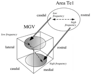
high-to-low frequency gradient on the primary auditory cortex has also been
shown to give rise to corticothalamic projections targeting the MGV in
a tridimensional arrangement, so that higher frequencies are mainly
represented in the ventral-medial-rostral plane of the nucleus, and the
lower frequencies are mainly represented in its dorso-latero-posterior
Figure 10 – Descending pathways of auditory information (adapted from Paxinos, 2004). Main auditory nuclei here represented are: Cochlea
(cochlea); Cochlear nuclear complex (CNC); Superior olivary complex (SOC);
Nuclei of the lateral lemniscus (NLL); Inferior colliculus (IC); Medial geniculate
Corticofugal inhibition of MGB neurons acts likely via feedback
projections from the RTN. Placed between the reciprocal
thalamo-cortical projections (Kimura et al., 2005; Zhang et al., 2008), the RTN
consists on a sheet of GABAergic cells situated along the rostral and
lateral surface of the dorsal thalamus (Guillery et al., 1998), and it
receives ascending projections from all the thalamic nuclei (Paxinos,
2004; Kimura et al., 2005). This inhibitory pathway has been shown to
play a role in the tonotopic control of frequency tuning in thalamic
neurons (Cotillon-Williams et al., 2008).
Further descending in the auditory pathway, the dorsal nucleus of
the IC has been shown to receive its inputs largely from the auditory
cortex (Saldaña et al., 1996). The neocortical terminals make a
tonotopic banded pattern like that of the ascending projections to the
CIC (Saldaña et al., 1996; Druga et al., 1997). On the other hand, the
external cortex receives inputs from the cerebral cortex, the medial
geniculate body as well as from many non-auditory structures
(Paxinos, 2004). Finally, descending projections from the IC target the
superior olivary complex and the cochlear nuclear complex (Paxinos,
2004), thus bringing back processed auditory signals to the receiving
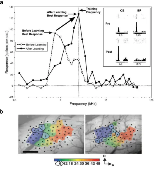
Figure 11 – Receptive field plasticity in the AC. (a) CS-specific tuning shift of a single cell in AC of guinea pig, resulting from auditory fear conditioning
(adapted from Weinberger, 2007a). After conditioning, responses to the CS
frequency increase and become the new characteristic frequency (CF). (b)
tonotopic map of A1 in a naïve rat (left), and in a rat trained with 6KHz CS
(right) (adapted from Rutkowski and Weinberger, 2005). Colored polygons
indicate the estimated A1 area representing the CF according to the color bar
shown below the maps.