Rev Bras Med Esporte _ Vol. 13, Nº 1 – Jan/Fev, 2007
43e
1. Faculdade de Educação Física, Universidade de Brasília (Brasília / DF).2. Programa de Pós-Graduação em Educação Física da Universidade Cató-lica de Brasília (Brasília / DF). Grupo de Estudos Avançados em Saúde e Exercícios (Brasília / DF).
3. Faculdade de Ciências da Saúde, Universidade de Brasília (Brasília / DF). Grupo de Estudos Avançados em Saúde e Exercícios (Brasília / DF). Received in 17/ 10/ 05. Final version received in 21/3/06. Approved in 19/7/ 06.
Correspondence to: Paulo Gentil, CLS 203, bl.A, lj.05 – 70233-510 – Brasília, DF. Tel.: (61) 8118-4732, fax: (61) 3322-7972. E-mail: paulogentil @hotmail. com
Comparison among the EMG activity of the pectoralis
major, anterior deltoidis and triceps brachii during the
bench press and peck deck exercises
Valdinar de Araújo Rocha Júnior1, Paulo Gentil2, Elke Oliveira3 and Jake do Carmo1
O
RIGINALA
RTICLEKeywords: Resistance training. Maximum repetitions. Electromyography. Muscle recruitment.
ENGLISH VERSION
ABSTRACT
The identification of the characteristics of each movement and its adjustment to the training goals are tasks that demand the in-teraction of many knowledge areas. These tasks are essential to the success in sports activities and training programs designed with athletic, aesthetic or healthy purposes. The objective of the present study was to compare the electromyographic (EMG) activ-ity of the pectoralis major (PM), anterior deltoids (DA) and triceps brachii (TB) muscles during the barbell bench press (SP) and the peck deck (PD) exercises. EMG activity of TB, PM and DA were assessed during 10 maximum repetitions performed in SP and PD in 13 trained men. The results did not show any differences be-tween exercises for PM and DA activity; however, TB activity was higher for SP than PD exercise. During SP, the PM muscle activity was higher than TB. There were no differences between PM and DA, or between DA and TB. During the PD exercise, the PM and DA muscle activities were higher than TB. There were no differ-ences between PM and DA. It was concluded that the prime mov-ers of both exercise are DA and PM, and there are no differences between them. Therefore, both PD and SP could be performed with the purpose to stimulate DA and PM muscles, depending on the availability of the equipments and/or the specificity of the mo-tor tasks.
INTRODUCTION
The identification of each movement’s peculiarities and its suit-ability to the training objectives is a task which demands the inter-action of many fields of knowledge. Such task is crucial for the success in the several sports modalities and training programs with rehabilitation and/or aesthetic purposes. A number of exercises can be adopted for the development of a given muscular group; however, an exercise is usually more indicated for each specific situation. Therefore, the biomechanical study becomes important for exercises selection in each training session in order to optimize the stimuli in each body segment.
Among the used exercises for the development of the muscula-ture of the anterior part of the chest are the barbell bench press and the machine peck deck. Both exercises involve horizontal ad-duction of the scapulo humeral joint; moreover, the bench press also involves the extension of the elbow joint(1). Therefore, the main
difference between the bench press and the peck deck would be the fact that the former is bi-articular while the latter involves only one articulation. Within this context, it is believed that multi-articu-lar exercises require greater neural coordination among the mus-cles(2), therefore, such movements could present a differentiated
pattern in the demand of the primary and accessory motor mus-cles. Conversely, many coaches and enthusiasts claim that the uni-articular exercises, also known as isolation exercises, promote greater musculature activation, what is confirmed by recent stud-ies(3).
The use of machines or free weights may also interfere in the muscular recruitment, once free weight exercises require the con-trol of the implement in three dimensions, which can generate greater activation of the stabilizer muscles(4-5). On the other hand,
it is believed that machine exercises require more overload in the primary motor muscle due to the reduction of the stabilizers ac-tion(6). McCaw and Friday(5) compared the free weight and machine
bench press with 60% and 80% of the workload equivalent to one maximal repetition (1RM) and observed greater muscular activa-tion of the anterior and medium deltoids during the free weight bench press. Nevertheless, no significant differences were report-ed between the exercises in the activity of the pectoralis major and triceps brachii muscles.
The literature is scarce concerning the comparison of the mus-cular activity between the bench press and peck deck exercises. Welsch et al.(7) compare the activity of the pectoralis major and
anterior deltoids muscles in three exercises: the barbell bench press; free weight bench press and free weight peck deck. Ac-cording to the results, there were no differences in the EMG activ-ity of the pectoralis major and anterior deltoids in the exercises. One of the broadest exercises is mentioned by Bompa and Cor-nacchia(8), in this study 56 exercises were compared with the
pur-pose to classify them concerning the integrated EMG signal nor-malized by the maximal voluntary isometric contraction (MVIC). This analysis, limited in its generalization for picking the signal of a single muscle, mention the free weight bench press as the move-ment which generates the heaviest overload over the pectoralis major (93%), followed by the barbell bench press (89%) and by the push-ups between benches (88%). It was not possible to find any study which has compared the EMG activity between the two most popular variations of the two exercises: the barbell bench press and the peck deck.
Another issue which needs further explanation is the difference between the EMG activities of the muscles in the same exercise. In the study by Welsch et al.(7), the authors did not report
report-44e
Rev Bras Med Esporte _ Vol. 13, Nº 1 – Jan/Fev, 2007 ed that the EMG signal of the triceps brachii normalized by themaximal MVIC, was higher comparing to the EMG signal of the pectoralis major during the bench press. The results did not reveal differences between the pectoralis major and anterior deltoids ac-tivities or anterior deltoids and triceps brachii acac-tivities. Nonethe-less, a flaw in the normalization procedure of the EMG signal may have interfered in the comparisons performed by Clemons and Aaron(9).
Several methodological variations have been applied with the purpose to improve the knowledge concerning the bench press and its modifications; however, the literature is scarce concerning comparison parameters with other exercises which are also wide-ly used in strength training. The aim of the present study was to compare the EMG activity of the pectoralis major (PM), anterior deltoids (AD) and triceps brachii (TB) muscles during the barbell bench press (BP) and machine peck deck (PD).
METHODS
Sample
The sample consisted of 13 male individuals, mean age 25.08 (± 2.58) years, weight 75.35 (± 8.49) kg and mean height 175.41 (± 5.10) cm. The mean strength training time of the subjects was 7,.8 (± 4.43) years. All subjects were experienced in the proposed ex-ercises performance and were able to perform a 1RM of the exer-cises with a workload heavier than their weight. The participants signed a free and clarified consent form prior to the experiment. The study was approved by the Ethics Committee of the Universi-ty of Brasília.
Experimental procedures
The EMG of the PM, TB and AD were measured during the per-formance of a maximal series with workload equivalent to 10 RM in both machines in order to evaluate the differences in the mus-cular activation in the exercises BP and PD. The 10 RM test was applied instead of the 1RM percentages with the aim to get the experiment closer to the real training situation as well as to mini-mize variations between exercises and individuals which can oc-cur in the application of the maximal workload percentages(10-11).
Pre-test
In the week prior to the data collection, the individuals performed 10 RM tests in the two exercises according to the procedures pre-viously used by Simão et al.(12). The aim of the tests was to
deter-mine the maximal workload which would be used in order to per-form 10 complete and consecutive movements within 2 seconds for the eccentric phase and 2 seconds for the concentric phase. Would the workload not been precisely measured in the first try, the weight was adjusted in 4 to 10 kg and the individual was sub-mitted to a new test. The minimum interval set between each try was 5 minutes. Only three tries were allowed in each session. The tests were performed in two different occasions separated by at least 48 hours. The results of the two tests were analyzed by the Pearson correlation and the values obtained were 0.99 for the PD and 0.98 for the BP. The workload obtained in the last test per-formed was used in the experiment. Besides the workload estab-lishment, the pre-test was useful for the adaptation of the sub-jects to the experimental protocol.
Test
At the test day, the subjects performed a maximal series of each exercise with the workload equivalent to 10 RM. The exercises were randomly performed among the individuals. The exercises were performed with a minimum interval of 20 minutes.
The exercises were performed in High On® machines by
Righet-to Fitness Equipment (São Paulo-Brazil). In the BP, the subjects were told to perform the eccentric phase placing the bar in a line
close to the center of the sternum, not touching the chest though, to avoid movement of the electrodes. The bench height in the PD machine was adjusted so that the arm of the subject would as-sume a position slightly lower in relation to an imaginary line paral-lel to the ground.
The rhythm of the movements was the same adopted in the pre-test. A metronome with a rhythm of 60 beats per minute was used in order to aid in the maintenance of the movement velocity. The subjects were told to synchronize the beep with the begin-ning and the end of each phase (concentric and eccentric).
Electromyography
Electromyographies brandname Delsys-Bagnoli 2 (DelSys Incor-porated, Boston, MA, USA) with bipolar active surface electrodes of Ag/AgCl were used for the EMG data collection. The rejection capacity of the usual mode of the electromyographies used in the experiment was of 90 dB. The electrodes were placed on the right side (dominant) of the subjects with the aid of specific double-faced adhesive patches after hair removal and cleaning of the site with alcohol.
The electrodes were placed parallel to the muscular fibers. The positioning in the AD and TB muscles followed anatomic recom-mendations by Zipp(13). Differently, for the PM the procedures
adopt-ed by Clemons and Aaron(9) were observed. The identification of
the anatomic points and placement of the electrodes were per-formed by the same researcher.
The EMG was obtained with a 1.000 gain, a sample frequency of 2.000 Hz and the signal was submitted to a passband filter of 20 Hz to 500 Hz. The mean of five repetitions was calculated in order to guarantee that the analyses would be performed with repeti-tions involving correct rhythm and techniques. The first try was always excluded from the calculation since there was a possibility of the bar removal movement as well as the machine breadth be-ing picked by the electromyography. The second try was eliminat-ed since normally the rhythm was not adequate yet in this repeti-tion. The rhythm violation also occurred when the individuals were close to fatigue, which led to the exclusion of the last tries. There-fore, the third to the seventh repetitions were used. After the rat-ification of the signal the normalization was performed by the max-imal peak of contraction of the mean try(14-15) and the RMS (Root
Mean Square) energy was calculated.
Statistical analysis
The data were submitted to descriptive statistics procedures (mean and Standard deviation). The workloads used in the BP and PD exercises were compared through a t-Student test. A 2 x 3 factorial ANOVA (exercises x muscle) was used in order to verify the interaction between the exercises and muscular groups. Would significant differences occur, multiple comparisons procedures with the reliability interval correction by the Bonferroni method were applied. The significance level was of p < 0.05.
RESULTS
The sample characteristics are presented in table 1. Although the 10 RM workload mean for the PD was slightly higher
compar-TABLE 1
Characteristics of the sample
Characteristics Mean ± stand. deviation
Age (years) 25.08 ± 02.58 Weight (kg) 75.35 ± 08.49 Height (cm) 175.41 ± 05.100
Rev Bras Med Esporte _ Vol. 13, Nº 1 – Jan/Fev, 2007
45e
ing to the BP, the values did not reach significant difference (p >0.05). The results of the EMG activity are illustrated in figure 1. The factorial ANOVA revealed a significant interaction between muscles and exercises (p < 0.05).
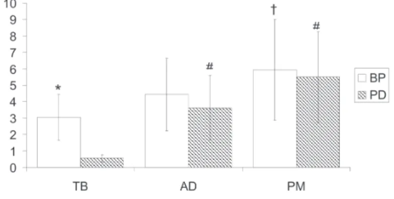
During the BP, the mean values of RMS were of 5.942 (± 3.058) for the PM muscle; 4.444 (± 2.21) for the AD and 3.053 (± 1.403) for the TB. The post-hoc analyses revealed that during the BP there was greater muscular activation of the PM in relation to the TB (p < 0.01). There were no differences between the PM and AD mus-cles and between the TB and AD (figure 1).
For the PD, the mean values of RMS were 5.501 (± 2.771) for the PM muscle; 3.626(± 1.977) for the AD and 0.552 (± 0.227) for the TB. In the PD, higher activation of the AD and PM muscles was verified in relation to the TB (p < 0.01). There were no differ-ences between the EMG activity of the AD and PM muscles (fig-ure 1).
The comparison between exercises indicated higher muscular activation of the TB for the BP in comparison to the PD (p < 0.01). There were no differences between exercises for the activity of the PM and AD muscles (figure 1).
DISCUSSION
The methodological differences make the comparison and prac-tical application of studies which through EMG try to evaluate the efficiency of exercises in the demand of specific muscles. One example is the utilization of protocols which make use of 1RM percentages for the establishment of tests intensity. Hoeger et al.(10) conducted a test in order to verify the number of repetitions
possible to be performed with steady maximal workload for differ-ent exercises. The authors reported that a given percdiffer-entage of 1RM allows an exacerbated number of repetitions for some exer-cises and a reduced number for others. Thus, studies with this methodology, such as the ones by Barnett et al.(16), Glass and
Arm-strong(17) and Bompa and Cornacchia(8), should be carefully
ana-lyzed, once the procedure itself may lead to differences in the ex-ercises due to the overestimation or underestimation of the muscular capacity in different movements.
The results of the present study reveal that both exercises sim-ilarly recruit the PM and AD muscles. Therefore, it would be a mis-take to affirm that only the PM muscles is primary motor in these movements, as commonly suggested in some books(18). These
find-ings are according to previous studies performed in the free weight bench press and peck deck(7,9) and should be considered at the
moment of trainings prescription, since it would be unnecessary and perhaps counter producing, that training involving these exer-cises are complemented with exerexer-cises directed to the AD mus-cle.
In the BP, the RMS values registered for the TB muscle were statistically lower than the ones for the PM and not different in comparison with the AD. These findings are opposite to the re-ports by Clemons and Aaron(9), who found greater muscular
activ-ity of the TB in relation to the PM during the BP. Despite this in-compatibility of results, the present study is more consistent for analysis of the TB in the BP, once in the study by Clemons and Aaron(9) the values of the signal in the concentric phase of the
movement exceeded the value of the MVIC as well as generated percentages above 100% for signal energy, suggesting hence, flaw of the normalization process.
When analyzing the muscular activity in the seating knees ex-tension (extensor table) and in the legs pressure through magnetic resonance, Enocson et al.(3) verified that the muscular activity of
the quadriceps during the extensor table was higher than the quad-riceps activity during the legs pressure. Although such study sug-gests a higher muscular recruitment in uni-articular exercises, the obtained results in the present study do not confirm this hypothe-sis, once no statistically significant difference was found in the activity of the PM and AD muscles between the PD and BP, which suggests that such muscles are equally recruited in the two exer-cises.
Although several authors have reported a differentiated recruit-ment pattern of the stabilizer muscles in machine exercises and free weight exercises(4-6), such disparity was not confirmed in the
present study, since the RMS values of the PM and AD muscles were similar between both exercises, corroborating recent find-ings by Welsch et al.(7). Thus, it is possible to infer that both
exer-cises are equally efficient in the recruitment of these muscles. Welsch et al.(7) recommend the free weight peck deck as a
supple-mentary exercise, since this movement presents shorter activa-tion time of the PM and AD muscles comparing to the BP. Howev-er, an extrapolation of this recommendation for the PD performed in machine should be cautiously seen due to the observed interac-tions in the present study as well as the lack of other reports in the literature about this movement.
The obtained results in the present study refer to a sample con-sisting of trained individuals; thereby, further studies are needed in order to evaluate the responsiveness in individuals with no ex-perience with the tested exercises. Moreover, it is important men-tioning that the calculation of the EMG signal breadth allows the quantitative analysis of the recruitment of motor units, while the results obtained with a resisted exercises program depend on the control of several variables. Therefore, one should be careful when using such results for qualification of the exercises, since it is not possible to predict the adaptations to a training program uniquely based on these data.
CONCLUSION
The PM and AD muscles were equally recruited in the BP and PD exercises, which clashes with the Idea that uni-articular exer-cises promote greater activity of the primary motors due to isola-tion. Therefore, would the aim be to promote stimuli for these muscles, both exercises may be used, depending on the availabil-ity of materials and/or specificavailabil-ity of motor activavailabil-ity in which perfor-mance improvement is searched. During the PD and the BP there was no difference between the RMS activity of the PM and AD muscles, which leads one to conclude that both muscles are equally recruited in the exercises. Such fact can make athletes and resist-ed – training activity practitioners save time when not including exercises specific for the AD muscle in the training sessions. Con-versely, the TB muscle is not relevant in the PD performance. Moreover, it seems to have reduced recruitment in the BP, which justifies the use of these exercises mainly for the development of the chest muscles.
Figure 1 – RMS values for the different muscles during the machine bench press and peck deck exercises (PD – machine peck deck; BP –barbell bench press; TB – triceps brachii, AD – anterior deltoids, PM – pectoralis major) * significant difference between BP and PD (p < 0.01). † significant difference concerning the TB
46e
Rev Bras Med Esporte _ Vol. 13, Nº 1 – Jan/Fev, 2007ACKNOWLEDGMENTS
To Mr. Mauro Siqueira from the Righetto Fitness Equipment for supply-ing the equipment used in the experiment.
All the authors declared there is not any potential conflict of inter-ests regarding this article.
REFERENCES
1. Hay JG, Reid JG. As bases biomecânicas do movimento humano. Rio de Janei-ro: Prentice-Hall do Brasil, 1985.
2. Fleck SJ, Kraemer WJ. Designing resistance trainig programs. 3rd ed. Cham-paing, IL: Human Kinetics, 2004.
3. Enocson AG, Berg HE, Vargas R, Jenner G, Tesch PA. Signal intensity of MR-images of thigh muscles following acute open- and closed chain kinetic knee extensor exercise-index of muscle use. Eur J Appl Physiol. 2005;94:357-63. 4. Hatfield FC. Hardcore bodybuilding: a scientific approach. Chicago:
Contempo-rary Books, 1993.
5. McCaw ST, Friday JJ. A comparison of muscle activity between a free weight and machine bench press. J Strength Cond Res. 1994;8:259-64.
6. Lander JE, Bates BT, Sawhill JA, Hamill J. A comparison between free-weight and isokinetic bench pressing. Med Sci Sports Exerc. 1985;17(3):344-53. 7. Welsch EA, Bird M, Mayhew JL. Electromyographic activity of the pectoralis
major and anterior deltoid muscles during 3 upper-body lifts. J Strength Cond Res. 2005; 9:449-52.
8. Bompa TO, Cornacchia L. Serious strength training. Champaing, IL: Human Ki-netics, 1998.
9. Clemons JM, Aaron. Effect of grip width on the myoelectric activity of the prime movers in the bench press. J Strength Cond Res. 1997;1:82-7.
10. Hoeger WWK, Hopkins DR, Barette SL, Hale DF. Relationship between repeti-tions and selected percentages of one repetition maximum: a comparison be-tween untrained and trained males and females. J Strength Cond Res. 1990;4: 47-54.
11. Tan B. Manipulating resistance training program variables to optimize maximum strength in men: a review. J Strength Cond Res. 1999;13:289-304.
12. Simão R, Farinatti PTV, Polito MD, Maior AS, Fleck SJ. Influence of exercise order on the number of repetitions performed and perceived exertion during resistance exercises. J Strength Cond Res. 2005;19:152-6.
13. Zipp P. Recommendations for the standardization of lead positions in surface electromyography. Eur J Appl Physiol. 1982;50:41-6.
14. Burden AM, Trew M, Baltzopoulos V. Normalization of gait EMGs: a re-examina-tion. J Electromyogr Kinesiol. 2003;13:519-32.
15. Yang JF, Winter DA. Electromyographic amplitude normalization methods: im-proving their sensitivity as diagnostic tools in gait analysis. Arch Phys Med Re-habil. 1984;65:517-21.
16. Barnett C, Kippers V, Turner P. Effects of variations of the bench press exercise on the EMG activity of five shoulder muscles. J Strength Cond Res. 1995;9:222-7.
17. Glass SC, Armstrong T. Electromyographical activity of the pectoralis muscle during incline and decline bench presses. J Strength Cond Res. 1997;11:163-7. 18. Baechle TR, Groves BR. Treinamento de força: passos para o sucesso. 2ª ed.