ABSTRACT
BACKGROUND AND OBJECTIVES: Temporomandibular disorder is a collective term encompassing a wide range of clini-cal orofacial joint and muscle problems. he stomatognatic sys-tem is part of the postural syssys-tem, so changes in one syssys-tem may interfere with the other. his study aimed at observing whether there is change in jaw and head position before and after tem-poromandibular disorder treatment.
METHODS: Participated in the study 16 volunteers, aged above 18 years, of both genders, who looked for assistance in the dentistry course clinic of a Public University, with diagnosis of temporomandibular disorder according to Diagnostic Crite-ria for Temporomandibular Disorder Research. Volunteers were submitted to X-rays (teleradiography with analysis of cephalo-metric points); posture in physiologic centric relation was evalu-ated by computerized photogrammetry and pain intensity was evaluated by the visual analog scale from zero to 10. Patients were evaluated before and after 8 weeks of treatment.
RESULTS:Pain has decreased from 6.43±2.84 to 2.17±2.39, before and after treatment, respectively (p<0.05). Vertical head alignment, in initial angle, has changed from 21.84⁰±17.49⁰ to 11.38⁰±14.61⁰ (p<0.05). Jaw position has changed from A-NB (angle indicating mandible-jaw relationship in the anterior pos-terior direction): 4.95±2.52 millimeters to A-NB: 4.64±2.52 mm (p<0.05).
CONCLUSION:Muscle temporomandibular disorder changes vertical head alignment and interferes with jaw position.
Keywords:Cephalometry, Photogrammetry, Posture, Temporo-mandibular joint.
Clinical study on head and jaw position of patients with muscle
temporomandibular disorder
Estudo clínico da posição da cabeça e mandíbula em pacientes com disfunção
temporomandibular muscular
Daisilene Baena Castillo1, Flaviane Keiko Azato1, Tulio Kalife Coelho1, Paulo Zarate Pereira1, Marcela Galdina Silva2
1. Universidade Federal de Mato Grosso do Sul, Faculdade de Odontologia, Campo Grande MS, Brasil.
2. Centro Universitário Anhanguera de Campo Grande, Graduanda do Curso de Fisiotera-pia, Campo Grande, MS, Brasil.
Submitted in October 20, 2015. Accepted for publication in April 18, 2016. Conlict of interests: none – Sponsoring sources: none.
Correspondence to:
Av. Costa e Silva, s/n - Bairro Universitário Faculdade de Odontologia
79070-900 Campo Grande, MS, Brasil. E-mail: lavianeazato@bol.com.br
© Sociedade Brasileira para o Estudo da Dor
RESUMO
JUSTIFICATIVA E OBJETIVOS: A disfunção temporoman-dibular é um termo coletivo que abrange um largo espectro de problemas clínicos da articulação e dos músculos na área orofa-cial. O sistema estomatognático integra o sistema postural, assim sendo, alterações que ocorrem em um sistema podem interferir no funcionamento do outro. O objetivo deste estudo foi veriicar se há alteração da posição da mandíbula e da cabeça antes e após o tratamento da disfunção temporomandibular.
MÉTODOS: Foram selecionados 16 voluntários, com idade aci-ma de 18 anos, de ambos os gêneros, que buscaram tratamento na clínica do curso de odontologia de Instituição Pública de En-sino Superior, com diagnóstico de disfunção temporomandibu-lar de acordo com os Critérios de Diagnóstico para Pesquisa das Desordens Temporomandibulares. Realizaram-se tomadas radio-gráicas (telerradiograia com análise de pontos cefalométricos); a avaliação postural, em relação cêntrica isiológica, foi veriicada por meio da fotogrametria computadorizada e a intensidade da dor foi avaliada pela escala analógica visual com pontuação de zero a 10. Os pacientes foram avaliados antes e após 8 semanas de tratamento.
RESULTADOS: A percepção à dor diminuiu de 6,43±2,84 para 2,17±2,39, antes e após tratamento, respectivamente (p<0,05). O alinhamento vertical da cabeça, no ângulo inicial, mudou de 21,84⁰±17,49⁰ para 11,38⁰±14,61⁰ (p<0,05). A posição da man-díbula mudou de A-NB (ângulo que indica a relação maxila--mandíbula no sentido anteroposterior): 4,95±2,52 milímetros para A-NB: 4,64±2,52 mm (p<0,05).
CONCLUSÃO: A disfunção temporomandibular muscular pro-move alteração do alinhamento vertical da cabeça e interfere na posição da mandíbula.
Descritores: Articulação temporomandibular, Cefalometria, Fo-togrametria, Postura.
INTRODUCTION
Temporomandibular disorder (TMD) is a collective term involving a large spectrum of clinical orofacial joint and muscles problems. These disorders are primarily character-ized by pain, joint noises and irregular or limited mandible function1.
Orofacial pain is multifactorial since factors triggering such disorders may be physical, psychological, traumatic, patho-logic or functional, such as parafunctions, bruxism and
clenching (act of maintaining unnecessary occluded teeth). Change in head posture is also pointed as a possible causal factor2,3. Forward head posture has been suggested as a
fac-tor interrelated with TMD4,5, however it is not clear whether
it is cause or effect.
Myofascial TMD pain is classified as a regional painful condition characterized by firm and hypersensitive muscle tissue bands, known as trigger points6. Although this
dis-order has not been totally understood, some factors, such as continuous deep painful stimulation source, increased emotional stress levels, sleep disorders, parafunctional hab-its, abnormal posture and muscle tension may be related to myofascial pain.
By means of lateral face radiographies and A-NB angle analysis (angle indicating mandible-jaw relationship in the anterior posterior direction), it was observed that the posi-tion of the mandible in relaposi-tion to the jaw may be altered by increased muscle activity7.
Mandible is stabilized in the skull especially by muscles which also command opening, closing and laterality move-ments. When positioning it to posterior, it is possible that with contracture there is change in biomechanical balance favoring the development of myofascial trigger point. Still, depending on time, intensity and frequency, there is change in intra-articular structures with decreased intra-articular space, which may lead to disc displacement8.
This study aimed at evaluating whether muscle TMD may promote forward head posture and take the mandible to a more posterior position.
METHODS
Sample was made up of 16 volunteers with major complaint of masticatory muscles pain.
Muscle pain was diagnosed during functional tests in pa-tients with at least 20 teeth and was classified as muscle TMD by Research Diagnostic Criteria for Temporoman-dibular Disorders (RDC/TMD)9. Exclusion criteria were
individuals with systemic diseases which could be mistaken for TMD (arthritis, fibromyalgia, sclerosis, inflammatory myopathy), those using or having used anti-inflammatory, anticonvulsant, antidepressant and psycothropic analgesic drugs in the six month previous to the study, those with history of facial or cervical trauma and those with Angle’s occlusal relation Class III.
Evaluation of signs and symptoms
Clinical evaluation was performed according to RDC cri-teria9, by a single researcher. In addition to palpation
rec-ommended by RDC for general evaluation of patients and their pain perception10,11, measurements were taken with
the visual analog scale (VAS)12, when patients were asked to
mark in a zero to 10 scale the point corresponding to pain intensity at that moment.
Posture analysis
After signs and symptoms evaluation and classification of patients in the muscle TMD group, posture was evaluated by photogrametry. Images were recorded by a 10 megapix-els Nikon® digital camera, on a tripod, three meters away
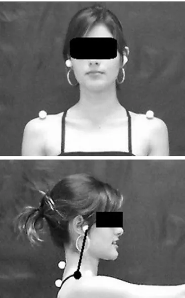
from volunteers in a previously assembled studio for this purpose. Evaluations were always carried out by the same evaluators. Ear tragus and acromion received polystyrene markers with 15mm diameter, for photographic recording, to evaluate vertical head angle (VHA) (Figure 1). After be-ing transferred to a computer, images were analyzed by the posture evaluation software – SAPO®.
Figure 1. Records with markers using Styrofoam balls (polystyre-ne)
Cephalometric analysis
Cephalometric tracings aimed at evaluating mandible po-sition changes with regard to jaw, before and after muscle TMD treatment. For such, we decided to check the distance between point A and line NB, for being bone structures with lower risk of changes, during treatment (Figure 2).
Point N
Point B 4 degrees
Measurement Variable between point A and line of points NB Point A
129
Figure 2. Illustration of measurement from point A to line NB
Digitized images were viewed in a 1366 x 768 pixels moni-tor, with real size gauging, in millimeters. Measurement was the distance between two ruler points of each teleradiogra-phy. For better interpreting anatomic points, lateral telera-diography real size was increased in up to three times.
Treatment
Realizada a avaliação postural, foi feita a análise do RDC no lux-ograma e, foram selecionados os pacientes com DTM muscular. After posture evaluation RDC was analyzed in the flowchart and muscle TMD patients were selected.
Proposed treatment was limited to reversible measures13
with efficacy proven by improved TMD signs and symp-toms. Protocol consisted of: 1) behavioral guidance to avoid hard food, not chewing gum, be aware of posture at sleep; 2) thermotherapy on the affected muscle with wet warm com-press, three times a day, for 20 minutes; 3) local massage af-ter compress with diclofenac diethylammonium, in circular and rake movements; 4) occlusal splint to be used at night. After therapy explanation14, volunteers have periodically
re-turned for follow-up. In the eighth visit, patients were sub-mitted to new posture and radiographic evaluation.
Statistical analysis
Student’s t test was used for statistical analysis considering
significant 5%, both for pain improvement and forward
head posture evaluation, where VHA (vertical head angle) was reported in degrees (⁰), and the distance between line NB and point A was given in mm.
This study was approved by the Research Ethics Committee, UFMS (Opinion 161957).
RESULTS
After eight weeks of cognitive-behavioral therapy associated to anterior occlusal splint, pain intensity evaluated by VAS went from 6.43±2.84 to 2.17±2.39 (p<0.001) (Figure 3).
10,0
9,0
8,0
7,0
6,0
5,0
4,0
3,0
2,0
1,0
0,0
VAS (baseline) VAS (inal)
VA
S
Time
Figure 3. Visual analog scale at treatment beginning and com-pletion
VAS = visual analog scale.
Possible change in forward head posture was evaluated by vertical alignment (forward posture) before and after therapy. Results with regard to baseline and final VHA, re-spectively, were 21.84⁰±17.49 and 11.38⁰±14.61 (p<0.05) (Figure 4).
2,5
2,0
1,5
1,0
0,5
0,0
VHA (treatment, baseline) VHA (treatment, inal)
VHA (degr
ees)
Groups
Figure 4. Measurement, in degrees, of forward head posture be-fore and after temporomandibular disorder treatment
VHA = vertical head alignment
This result shows that there are significant differences be-tween vertical head alignment in the treatment group before and after therapeutic intervention.
cephalo-metric point A and line resulting from the union of points N and B was calculated, before and after treatment. We de-cided to get also the image with occlusal splint since occlu-sal relationship could interfere with mandible positioning (Table 1).
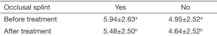
Table 1. Mean ± standard deviation of mandible displacement in mm, of patients with temporomandibular disorder, with and without ante-rior occlusal splint at radiography time (n=27)
Occlusal splint Yes No
Before treatment 5.94±2.63a 4.95±2.52a
After treatment 5.48±2.50b 4.64±2.52b
Different letters in lines, intervariables (before and after treatment), indicate signiicant difference (p<0.05). Kolgomorov-Smirnov sample distribution test. Student’t t test.
DISCUSSION
Most skull weight rests on anterior cervical spine region and on temporomandibular joints (TMJ), so, orthostatic posi-tion of the skull-cervical spine relaposi-tionship is maintained by a complex muscle mechanism involving head, neck and shoulder muscles3,16,17. It is to be expected that masticatory
muscles hyperactivity, in addition to generating pain and mouth opening limitation, promotes changes both in head and mandible position.
In our study, conservative therapies, that is, healthy behav-ioral habits, warm and wet compress, massages and ante-rior occlusal splint have significantly improved pain. The efficiency of self-management therapies, counseling, mas-sage with diclofenac, use of occlusal splints associated to heat and compresses is found in the literature14,18-20. Manual
therapy associated to self-care exercises at home has excel-lent short-term results21,22.
A study23 evaluating the result of treatment applied to
pa-tients with masticatory muscles pain has observed that cog-nitive-behavioral therapy is effective, however, when associ-ated to total or anterior occlusal splint, pain relief is faster, in addition to being a conservative and low cost treatment20.
Hard occlusal splints inhibit mandible neuromuscular activ-ity24, decreasing, during sleep, the number of muscle
con-tracture events.
Our results have shown a significant change with regard to anterior head tilting. Vertical angle, at beginning of treat-ment was 21.84±17.49⁰; at treattreat-ment completion it was 11.38±14.61⁰. So, one may state that TMD actively par-ticipates in forward head posture. Other authors have also confirmed that the more severe is the TMD, the more pro-nounced is forward head posture25,26.
When evaluating the postural tonic system27, it is observed
that mandible and tongue are connected to anterior head and neck muscle chain, and the hyoid bone plays an impor-tant role in the inter-relation of such structures; jaw, for be-ing fixed in the skull, is related to posterior chains. So, mas-ticatory muscles hyperactivity may lead to mandible changes since this is the only mobile bone of the skull, maintained in
position by occlusal teeth relationship.
With results obtained before and after treatment, with sig-nificant pain improvement and increased tolerance to pres-sure, one may infer that there has been mandible position change. A previous study28 has observed correlation between
sagital mandible position and body posture at thoracic and cervical regions.
Pain may increase muscle tone resulting in forward head posture and mandible retraction. A study29 has reported that
in people with forward head posture, the condyle goes to a more posterior position with regard to natural head position. In our study, we tried to observe how much this movement represented in the anterior skull portion (distance between point A and line of points NB), and has observed difference in mandible position before and after treatment, respective-ly, of 4.95±2.52 mm and 4.64±2.52 mm. It is interesting to evaluate occlusal relationship, the observe whether there is need for occlusal adjustment (removing premature and de-flective contacts), aiming at providing mandible orthopedic balance.
In light of these data, one may infer that a possible muscle tension due to TMD may change the whole rehabilitation planning, be it orthodontic or prosthetic, in adult patients. A planned rehabilitation with the mandible displaced from the ideal position may promote constant tension in crani-al-cervical posture and may cause a postural problem with painful discomfort along the years if there is not a natural body reconditioning.
Our results are in line with other studies3,30 which state that
patient’s posture should be balanced, that is, in the natural position when obtaining mandible-jaw relationship. This is a fact which should not be neglected when planning a reha-bilitation program, due to inter-relations existing between cranial-cervical posture and the development and function of dental-facial structures.
Face to this close relationship between posture system and masticatory system, it was observed that multidisciplinary assistance, especially between dentists and physiotherapists, is first line indication for the success both of TMD treat-ment and of rehabilitator, because when starting such proce-dure, it is indicated that patients have their muscle tonicity balanced, so that records are real and treatment is effective and efficient.
CONCLUSION
TMD treatment promotes vertical head alignment change and interferes with mandible position, normalizing it.
REFERENCES
1. Axelsson R, Tullberg M, Ernberg M, Hedenberg-Magnusson B. Symptoms and signs of temporomandibular disorders in patients with sudden sensorineural hearing loss. Swed Dent J. 2009;33(3):115-23.
2. Pedroni CR, De Oliveira AS, Guaratini MI. Prevalence study of signs and symptoms of temporomandibular disorders in university students. J Oral Rehabil. 2003;30(3):283-9. 3. Shiraishi CF, Salgado AS, Kerppers II, Furmann M, Oliveira TB, Ribeiro LG, et al.
Journal. 2014;12:83-6.
4. Lee WY, Okeson JP, Lindroth J. he relathionship between forward head posture and temporomandibular disorders. J Orafac Pain. 1995;9(2):161-7.
5. Nicolakis P, Nicolakis M, Piehslinger E, Ebenbichler G, Vachuda M, Kirtley C, et al. Relationship between craniomandibular disorders and poor posture. Cranio. 2000;18(2):106-12.
6. Shah JP, Gilliams EA. Uncovering the biomechanical milieu of myofascial trigger points using in vivo microdialysis: an application of muscle pain concepts to myofas-cial pain syndrome. J Bodyw Mov her. 2008;12(4):371-84.
7. Tecco S, Crincoli V, Di Bisceglie B, Caputi S, Festa F. Relation between facial mor-phology on lateral skull radiographs and sEMG activity of head, neck, and trunk muscles in Caucasian adult females. J Electromyogr Kinesiol. 2011;21(2):298-310. 8. Fricton JR, Schifman EL. Reliability of a craniomandibular index. J Dent Res.
1986;65(11):1359-64.
9. Dworkin SF, LeResche L. Research diagnostic criteria for temporomandibular disor-ders: review, criteria, examinations and speciications, critique. J Craniomandib Di-sord. 1992;6(4):301-55.
10. Gomes MB, Guimarães FC, Guimarães SM, Claro-Neves AC. Limiar de dor à pressão em pacientes com cefaléia tensional e disfunção temporomandibular. Cienc Odontol Bras. 2006;9(4):84-91.
11. Jensen R, Rasmussen BK, Pedersen B, Lous I, Olesen J. Cephalic muscle tenderness and pressure pain threshold in a general population. Pain. 1992;48(2):197-203. 12. Bailey B, Gravel J, Daoust R. Reliability of the visual analog scale in children with
acute pain in the emergency department. Pain. 2012;153(4):839-42.
13. Cordeiro PC, Bonato LL, Dias IM, Guimarães JP. Evaluation of stabilizing plate the-rapeutic efects on diferent types of temporomandibular disorders - painful evolution of patients treated in a reference center. Braz Dent Sci. 2014;17(4):17-26. 14. Azato FK, Castillo DB, Coelho TMK, Taciro C, Pereira PZ, Zomerfeld V, et al.
In-luence of temporomandibular disorders management on pain and global posture Inluência do tratamento das desordens temporomandibulares na dor e na postura global. Rev Dor. 2013;14(4):280-3.
15. Software para Avaliação Postural – SAPO. Versão 0.68 – Julho/2007. Disponível em http://code.google.com/p/sapo-desktop/source.
16. Cauás M, Alves IF, Tenório K, Brasiliense FJ, HC Filho JB, Guerra CM. Incidências de hábitos parafuncionais e posturais em pacientes portadores de disfunção da articu-lação craniomandibular. Rev Cir Traumatol Buco-Maxilo-Fac. 2004;4(2):121-9. 17. Deljo E, Filipovic M, Babacic R, Grabus J. Correlation analysis of the hyoid bone
position in relation to the cranial base, mandible and cervical part of vertebra with particular reference to bimaxillary relations / teleroentgenogram analysis. Acta Inform Med. 2012;20(1):25-31.
18. Ay S, Dogan ṢK, Evcik D, Baṣer OC. Comparison the eicacy of phonophoresis and ultrasound therapy in myofascial pain syndrome. Rheumatol Int. 2011;31(9):1203-8. 19. Gomes CA, El Hage Y, Amaral AP, Politti F, Biasotto-Gonzalez DA. Efects of massage therapy and occlusal splint therapy on electromyographic activity and the intensity of signs and symptoms in individual with temporomandibular disorder and sleep bru-xism: a randomized clinical trial. Chiropr Man herap. 2014;22(1):43.
20. de Freitas RF, Ferreira MA, Barbosa GA, Calderon PS. Counselling and self-manage-ment therapies for temporomandibular disorders: a systematic review. J Oral Rehabil. 2013;40(11):864-74.
21. Tuncer AB, Ergun N, Tuncer AH, Karahan S. Efectiveness of manual therapy and home physical therapy in patients with temporomandibular disorders: a randomized controlled trial. J Bodyw Mov her. 2013;17(3):302-8.
22. Chan YC, Wang TJ, Chang CC, Chen LC, Chu HY, Lin SP, et al. Short-term efects of self-massage combined with home exercise on pain daily activity, and autonomic function in patients with myofascial pain dysfunction syndrome. J. Phys her Sci. 2015;27(1):217-21.
23. Conti PC, de Alencar EM, da Mota Corrêa AS, Lauris JR, Porporatti AL, Costa YM. Behavioural changes and occlusal splints are efective in the management of mastica-tory myofascial pain: a short-term evaluation. J Oral Rehabil. 2012;39(10):754-60. 24. Arima T, Takeuchi T, Tomonaga A, Yachida W, Ohata N, Svensson P. Choice of
bio-materials – Do soft occlusal splints inluence jaw-muscle activity during sleep? A pre-liminary report. Appl Surf Sci. 2012;262:159-62.
25. Grade R, Caramês J, Pragosa A, Carvalhão J, Sousa S. Postura e disfunção temporoman-dibular: controvérsias actuais. Rev Port Estomatol Cir Maxilofac. 2008; 49(2):111-7. 26. Biasotto-Gonzalez DA, Andrade DV, Gonzalez TO, Martins MD, Fernandes KP,
Cor-rêa JC, et al. Correlação entre disfunção temporomandibular, postura e qualidade de vida. Rev Bras Cresc Desenvolv Hum. 2008;18(1):79-86.
27. Bricot B. Posturologia. São Paulo. Ed. Ícone, 2001. 49-75p.
28. Lippold C, Segatto E, Végh A, Drerup B, Moiseenko T, Danesh G. Sagital back con-tour and craniofacial morphology in preadolescents. Eur Spine J. 2010;19(3):427-34. 29. Ohmure H, Miyawaki S, Nagata J, Ikeda K, Yamasaki K, Al-Kalaly A. Inluence of
forward head posture on condylar position. J Oral Rehabil. 2008;35(11):795-800. 30. Solow B, Sandham A. Cranio-cervical posture: a factor in the development and