SURFACE ELECTROMYOGRAPHY: PROPOSAL
OF A PROTOCOL FOR CERVICAL MUSCLES
Eletromiograia de superfície:
proposta de um protocolo para músculos cervicais
Klyvia Juliana Rocha de Moraes (1), Daniele Andrade da Cunha(2),
Luciana Ângelo Bezerra (3), Renata Andrade da Cunha(4), Hilton Justino da Silva(5)
ABSTRACT
Purpose: to present a proposal of a surface electromyography evaluation method for cervical muscles
speciic and detailed protocol, with a standardized collection method of electrical signal in these
muscles. Method: the researchers took as reference the existing publications about this subject which
evidenced a need for standardization, clarity, better reproducibility and greater speciicity for the surface electromyography evaluation of the upper trapezium and sternocleidomastoid muscles ibers. The proposal preparation process for the current protocol included the cleaning of the target area, placing the electrodes, required tasks in order to collect and register the electrical signal and interpretation of the electromyography signal parameters. This evaluation method was carried out in 24 healthy volunteers of both genders, with an average age of 26 years. We used the electromyography Miotool 400 with 4 channels. Result: an evaluation surface electromyography method for upper trapezium
and sternocleidomastoid muscles ibers was developed and tested in order to determine the best form
of electrical signal data collection for these muscles. Conclusion: we submitted a protocol proposal
to evaluate the cervical muscles by a surface electromyography, allowing the healthy professionals
and researchers to get more information about this electrical potential evaluation method for the
sternocleidomastoid and the upper trapezium muscles ibers. This knowledge will be an adjuvant in a more speciic therapy.
KEYWORDS: Electromyography; Electrodes; Site Selection; Electric Impedance
(1) Physiotherapist; Preceptor Internship in Physical Therapy
from the University Salgado de Oliveira, Federal University of Pernambuco, UFPE, Recife- PE, Brazil; Master of Patho -logy from Federal University of Pernambuco -UFPE.
(2) Speech Therapist; Coordinator Course of Speech in Facul -dade Integrada of Recife, Facul-dade Integrada of Recife, FIR, Recife- PE, Brazil; PhD in Nutrition from Federal Uni -versity of Pernambuco -UFPE.
(3) Physiotherapist; Physiotherapist Clinic Pepita Duran, Recife-PE, Brazil; Specialist in Cardiorespiratory Physical Therapy from the Gama Filho University - UGF.
(4) Physiotherapist; Physiotherapist Neurofunctional home, Recife-PE, Brazil; Specialist in Neurofunctional Physical Therapy from Faculdade Integrada of Recife -FIR.
(5) SpeechTherapist; Vice-Coordinator of the post-graduate studies in Pathology, Master level, Professor of Speech
Pathology course at the Federal University of
Pernam-buco - UFPE, Federal University of PernamPernam-buco – UFPE, Recife-PE, Brazil; PhD in Nutrition from Federal University
of Pernambuco -UFPE.
Conlict of interest: non-existent
INTRODUCTION
The Stomatognathic System is described in
the literature as a functional unit of human body
comprising components of the body skeletal, dental arch, muscles and other structures, bringing a close relationship with cervical muscles1.
The literature shows the myofascial tissue as an inseparable functional entity. The occurrence of muscular synergism justiies the interdependence of muscle tissue as well as a continuity of solution1 that
reveals the occurrence of disturbances which follow
muscular chains1,2.
Campignion3 suggested that the voluntary action
of the cervical muscles, represented by the upper trapezius ibers and sternocleidomastoid muscles
reproducibility17,19, clarity and speciicity to evaluate
the sternocleydomastoid22 and upper trapezius
ibers.
Thus, the objective of this paper is to present a proposal of protocol for sEMG evaluation for cervical muscles speciic and detailed in an attempt to standardize these results, contributing to the standardization of tests by different professionals, collaborating with the academic background of
students in the health area and favoring the
compar-ison of indings from different study centers.
METHOD
During the 2009 and 2010 years, a multidisci
-plinary group of Brazilian researchers in electromy
-ography with extensive experience in research and clinical area, met periodically to discuss the needs of a speciic protocol for sEMG analysis of cervical muscles, more precisely, to the Sternocleidomas
-toid muscle (SCM), and upper ibers of trapezius
muscle.
The process of elaboration the current proposal of protocol included the skin cleaning, the electrodes placement position, tasks required to collect the
electrical signal and parameters to be collect from the electromyographic signal.
The data of muscle electrical potential were collected by the electromyography equipment (Miotool 400) connected to the LG notebook with Windows® Vista Premium operating system with
110GB HD, Intel® Dual-Core Processor 1.60GHz, 2
GB of RAM, 32 BITs, with the Miograph 2.0 software. The electromyography showed up with a gain of 1000; 4 sensors SDS500 with grab type terminals; 1 ground cable all MIOTEC® brand and 1 USB commu
-nication cable to capture the electrical potential of
SCM ibers and upper ibers of trapezius muscles, bilaterally. Bipolar surface EMG electrodes were used (Ag/AgCl electrodes - Tyco Helthcare, Medi
-trace 100-Kendall) with conductive gel and properly secured with 3M™ Micropore™ Tape.
The researchers applied this proposal of protocol
in the laboratory of electromyography of the Patho-physiology of the Stomatognathic System research group on the Campus of the Federal University of Pernambuco (UFPE) in 24 volunteers of both
genders with the mean age of 26 years, to verify its reproducibility as well as investigating the best maneuver that would generate the best electromyo -graphic signal to the evaluated muscle.
All volunteers were students of Federal Univer
-sity of Pernambuco (UFPE), chosen randomly, with no complaints of pain in the cervical region or shoulders, and they were not under treatment for postural changes or claw. The cervical region muscle group, resulting in greater coordination of
movement, proximal stabilization and functional gain in the same region. The evaluation of skeletal cervical muscle, has important value because the inluence of posture and stress on the musculo -skeletal changes is a primary overload commonly observed4.
Researchers have shown that the disorders of the cervical muscles are a serious and frequent problem that interest in developing standards for adequate muscular evaluation, in multidisciplinary teams, in order to minimize or prevent such problem5.
There are published reports that the postural
control of head and neck depends on the
integra-tion of the sternocleidomastoid and upper trapezius ibers muscles, controlling the gravitational torque, inluencing the functionality of the cervical region6.
A practical way to clinically investigate the musculoskeletal conditions, particularly in the
cervical muscles7 and verify their physiological
conditions8, with regard to action, is the Surface
Electromyography (sEMG)8-11.
This monitoring can be used as a safe, easy
and noninvasive method9,10 to measure the
elec-trical muscle activity during function9, in addition to
being used as an auxiliary to the study of muscle kinesiology12. A study by Ericson and Fernstrom13
examined how the electrical activity of upper ibers of trapezius muscle changed during the modiication
of the activity.
Therefore, there is a great importance for the knowledge on in vivo muscle physiology, in the
differential diagnosis and monitoring of possible
disorders. Knowing what is normal, can help the diagnosis with conditions that they are abnormal14
and may provide a more thorough and reliable of patient outcomes and the effectiveness of therapy applied15.
Some researches indicated the need of a
stan-dardization of the collection of EMG data in order
to have more reliable recordings9,16. There are
some reports in the literature regarding the
method-ological dificulties of sEMG recordings, describing some limitations such as laws in research protocols by the dificulty in reproducing the positioning of the electrodes, as well as a lack of more speciic
protocols 16.
The regulatory standards for EMG decision, analysis, interpretation and the records of signs
have been a concern to the implementers of this
technique. They present as a solution, the prepara
-tion of a practical guide that aims to standardize the
procedures in the electromyographic studies17,18.
The researchers took as reference the existing
publications about the proposed subject7,9,17,18,22,24,27
amplitude of electrical signal, however, strengthen
the possibility of the effect crosstalk from other muscles19,26. Thus, the choice of ideal electrode,
location and interelectrode distance will be mecha
-nisms to minimize this effect7,11.
In the current protocol, the choice of small elec
-trode size was standardized to it well to the studied muscles, self-adhesive and interelectrode distance
of 1.5cm according to guidelines published by International Society of electrophysiology and
Kine-siology-ISEK/ Surface Electromyography for the Non-Invasive Assessment of Muscles-SENIAM11,19,27.
Researchers report the electrode placement should be in the motor point of the muscle belly
where it will include the Innervation Zones (IZ) located along the muscle iber near the midpoint of the muscle belly to optimize the accuracy of the
electrical signal and to portray a character of greater
reproducibility of the sEMG technique7. Merletti et
al.28 report that the differences in sEMG signals may
arise from changes in the IZ location of the muscles, emphasizing the need to respect them.
For the upper ibers of each trapezius muscle, the electrodes were placed at half the distance between
the acromion line and seventh cervical vertebra of the spine (C7)27, longitudinal ibers (1 channel
for each upper ibers of trapezius muscle)27,29.
The ground electrode was placed over the right
humerus lateral epicondyle to reduce interpositions of external electrical noise.
The actions that represented the best collec
-tion of sEMG record of the muscles studied were
based on muscle function testing15 to know: the
cervical lexion–rotation to the right (manually resisted) and cervical lexion–rotation to the left (manually resisted), for the increased the activation of SCM muscle. For the increased the activation of upper ibers of trapezius muscle, elevation of both shoulders, simultaneously (manually resisted). These actions were taken as a basis for normal
-ization of sEMG signal and to proposed activities, being named Maximum Resistive Voluntary Activity (MAVR). Thus, for the SCM muscle, is determined the MAVR to the right SCM (M
AVR of the SCMR); to th left
SCM (M
AVR of the SCML); and to the upper ibers of
right and left trapezius (M
AVR of the UTFR and MAVR
of the UTFL).
The normalization process of sEMG signal was
based on ISEK19. However, the Maximum Activity
Voluntary Resistive (M
AVR) with manual loading was
availed instead of considering the Maximum Volun
-tary Isometric Contraction (MVIC) with a known load
and mensurable.
For manual resistance imposed, there is no the possibility of measuring the same, however, to be
attributed to M
AVR for the normalization sEMG the
was assessed by photographic recording (digital photogrammetry), in anterior view, right lateral view, left lateral view and posterior view, in order
to position the cervical region for electromyographic
evaluation. All these evaluations were performed in the morning, according to the circadian cycle. All
volunteers signed an informed consent term about
the research, keeping a copy of it with them. During the electromyography evaluation, envi
-ronmental noise was controlled closing doors and windows and turning off the air conditioning to make the room more appropriate for the collection. This research was approved by the Ethics Committee and Research of the Cancer Hospital of Pernam
-buco (CEP/HCP), under protocol nº 41/2009.
RESULTS
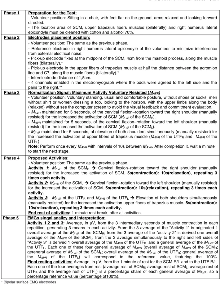
Figure 1 shows the phases of protocol of the electromyographic evaluation for the SCM and upper ibers of trapezius muscles.
DISCUSSION
The protocol presented in this paper was
prepared to have the largest contingent of possible information at the time of electromyographic
evalu-ation, facilitating its reproduction. It is simple and easily applied which encourages future studies.
The skin of the muscles evaluated was cleaned with cotton and alcohol 70%, as well as the lateral epicondyle region of the right humerus, according
to the recommendations of Surface
Electromyog-raphy for the Non-Invasive Assessment of Muscles-SENIAM19. This region was slightly scorched in
the direction of SCM and upper ibers of trapezius muscles. The literature reports that the skin cleaning
aims to reduce the impedance and eliminates any interference from the electrical signal20,21.
To ind the muscle belly, the concentric contrac
-tion for each studied muscle was requested, as reference, based on Falla et al.7 and from this,
followed by placing the electrodes. For each SCM muscle, the electrodes were ixed at the midpoint of the muscle belly along its ibers7,18,19, 4cm below
the insertion on the mastoid process (one channel
for each SCM muscle) with the patient at rest22.
This distance of 4cm is suggested to avoid inter
-ference in the electrical signal by the ibers of the
platysma muscle22. These interferences has been
described by some researchers as the crosstalk
phenomenon 7,9,11,23-25, which captures the electrical
signal from adjacent muscles and could interfere in the evaluated signal7,11.
Researches also indicate the size of the elec
* Bipolar surface EMG electrodes
** Channel 1 and 2 for the upper portion left and right trapezius muscle, respectively. Channel 3 and 4 for of left and right SCM muscle,
respectively.
Figure 1 – Phases of protocol of the electromyographic evaluation for the SCM and upper trapezius
ibers muscles
Phase 1 Preparation for the Test:
- Volunteer position: Sitting in a chair, with feet flat on the ground, arms relaxed and looking forward directed.
- The location area of SCM, upper trapezius fibers muscles (bilaterally) and right humerus lateral epicondyle must be cleaned with cotton and alcohol 70%.
Phase 2 Electrodes placement position:
- Volunteer position: The same as the previous phase.
- Reference electrode in right humerus lateral epicondyle of the volunteer to minimize interference from external electrical noise.
- Pick-up electrode fixed at the midpoint of the SCM, 4cm from the mastoid process, along the muscle fibers (bilaterally).*
- Pick-up electrode in the upper fibers of trapezius muscle at half the distance between the acromion line and C7, along the muscle fibers (bilaterally).*
- Interelectrode distance of 1,5cm.
- 4 channels enabled in the electromyograph where the odds were agreed to the left side and the pairs to the right.**
Phase 3 Normalization Signal: Maximum Activity Voluntary Resisted (MAVR)
- Volunteer position: Voluntary standing, usual and comfortable posture, without shoes or socks, men without shirt or women dressing a top, looking to the horizon, with the upper limbs along the body (relaxed) without see the computer screen to avoid the visual feedback and commitment evaluation.
- MAVR maintained for 5 seconds, of the cervical flexion–rotation toward the right shoulder (manually
resisted) for the increased the activation of SCM (MAVR of the SCMR).
- MAVR maintained for 5 seconds, of the cervical flexion–rotation toward the left shoulder (manually
resisted) for the increased the activation of SCM (MAVR of the SCML).
- MAVR maintained for 5 seconds, of elevation of both shoulders simultaneously (manually resisted) for
the increased the activation of upper fibers of trapezius muscle (MAVR of the UTFR and MAVR of the
UTFL).
Note: Perform once every MAVR with intervals of 10s between MAVR. After completion it, wait a minute
to start the next stage.
Phase 4 Proposed Activities:
- Volunteer position: The same as the previous phase.
Activity 1: MAVR of the SCMR Cervical flexion–rotation toward the right shoulder (manually
resisted) for the increased the activation of SCM. 5s(contraction): 10s(relaxation), repeating 3 times each activity.
Activity 2: MAVR of the SCMLCervical flexion–rotation toward the left shoulder (manually resisted)
for the increased the activation of SCM. 5s(contraction): 10s(relaxation), repeating 3 times each activity.
Activity 3: MAVR of the UTFR and MAVR of the UTFL Elevation of both shoulders simultaneously
(manually resisted) for the increased the activation upper fibers of trapezius muscle. 5s(contraction): 10s(relaxation), repeating 3 times each activity.
End rest of activities: 1 minute rest break, after all activities.
Phase 5 EMGs singal analizy and interpretation:
Activity 1,2 and 3: Average, in µV, from the 3 intermediary seconds of muscle contraction in each
repetition, generating 3 means in each activity. From the 3 average of the "Activity 1" is originated 1 overall average of the MAVR of the SCMR; from the 3 average of the "activity 2" is derived one overall
average of the MAVR of the SCML; from the 3 average simultaneously to the right and left sides of
"Activity 3" is derived 1 overall average of the MAVR of the UTFR and a general average of the MAVR of
the UTFL. Each one of these four general average of MAVR (overall average of MAVR of the SCMR;
gereneral average of MAVR of the SCML; overall average of the MAVR of the UTFR; general average of
the MAVR of the UTFL) will correspond to the reference value, featuring the 100%.
Final resting activities: Average, in µV, from the 1 minute of rest for the SCM R/L and to the UTF R/L.
Each one of the four averages from rest (average rest of SCMR; average rest of SCML; average rest of UTFR and the average rest of UTFL) is a percentage share of each general average of MAVR, so a percentage reference value (percentage of100%).
average of the M
AVR of the SCML; overall average of
the M
AVR of the UTFR and general average of the MAVR
of the UTFL) represent this stabilization moment of EMGs signal, which serves as the benchmark, so the 100% of activities.
The electric potential related to the inal resting activities (end rest of SCMR; end rest of SCML; end
rest of UFTR and end rest of UFTL), start to match a percentage share of the overall averages of MAVR,
so a percentage of the reference value34,35. Being
this the reference value the most stabilish moment
of EMGs signal36,37.
CONCLUSION
This paper presented a proposal of protocol for sEMG evaluation for cervical muscles allowing
more information about the evaluation of the
elec-trical potential of SCM and upper ibers of trapezius muscles to physiotherapists, other health profes
-sionals and specialists. This knowledge will serve in speciic adjuvant therapies.
It is suggested that more publications of
proto-cols for other speciic muscles happen to strengthen the technique as well as its reliable and universal reproduction in an attempt to minimize the possible biases and differences between evaluators.
ACKNOWLEDGMENT
The authors thank the National Council of Tech
-nological and Scientiic Development (CNPq), which had a inancial support with Edictal Universal MCT/ CNPq 14/2009 - Faixa B - Process: 476412/2009-9. examiners were aware of the signal spectral graph
on the computer screen, imposed referring to
maximum activity. Each of the 3 M
AVR for normaliza
-tion lasted 5 seconds, interspersed by 10 seconds of rest between executions with only one repetition
for each test.
Although there is a large contingency about the
normalization of EMG signal from the MVIC19,25,30-33,
the literature does not address the normalization form of sEMG signal with known and measurable loads, proposed by MVIC for the muscles evaluated in this protocol, nor not to the facial muscles, thus
justifying the use of M AVR.
After normalization, the implementation of activi
-ties proposed for the SCM (M
AVR of the SCMR; MAVR
of the SCML; characterizing the 1 and 2 activities of the igure 1) and the upper trapezius ibers muscles
(M
AVR of the UTFR and MAVR of the UTFL;
character-izing the 3 activity of the igure 1) were done. Each activity, differently the normalization, was repeated 3 times with 5 seconds of contraction for 10 seconds of relaxation (ratio 1:2), adapted from Barbosa and Gonçalves34 and Kakihara, Sens and Ferreira35, with
1 minute rest break at the end rest of the activities.
In EMGs signal interpretation the literature shows that the moment of major stabilization of the electromyographic signal is one who understands the middle region of this signal, so the intermediary seconds of each muscle contraction, represented in the signal spectrum EMGs36,37. Thus, the average
in microvolts (µV) of 3 intermediate seconds of muscle contraction, in each of the repetitions in the proposed activities, producing a single average for each activity (activities 1, 2 and 3, referring to the
general average of the M
research electromyography applications. Clin
Neurophysiol. 2002,113:57–63.
8.Malta J, Campolongo GD, Barros TEP, Oliveira RP. Eletromiograia aplicada aos músculos da mastigação. Acta Ortop Bras. 2006;14(2):106-7. 9. Moraes KJR, Cunha RA, Lins OG, Cunha DA, Silva HJ. Eletromiograia de superfície: padronização da técnica. Neurobiol. 2010; 73(3):151-8.
10. Bassani E, Candotti CT, Pasini M, Melo M, La Torre M. Avaliação da ativação neuromuscular em indivíduos com escoliose através da eletromiograia de superfície. Rev Bras de Fis. 2008;12(1):13-9. 11.Tank FF, Silva GT, Oliveira CG, Garcia MAC. Inluência da distância intereletrodos e da cadência de movimento no domínio da frequência do sinal de EMG de superfície. Rev Bras Med Esporte. 2009; 15(4):272-6.
12. Amadio AC, Duarte M. Fundamentos biomecânicos para análise do
movimento. São Paulo: Laboratório de Biomecânica EEFUSP; 1996.
162p.
13. Ericson MO, Fernstrom EA. Upper-Arm Elevation During Ofice Work. Ergonom. 1996;39(10):1221-30. 14. Veiga PHA. Análise eletromiográica como base para o tratamento das luxações recidivas da patela. Fisioter em Mov. 2007;20(1):11-6.
15. Pereira TB, Bergmann A, Ribeiro ACP, Silva JG, Dias R, Ribeiro MJP, et al. Padrão da atividade mioelétrica dos músculos da cintura escapular após
REFERENCES
1. Vázquez-Nava F, Vázquez REM, Saldivar GAH, Beltrán GFJ, Almeida AVM, Vázquez RCF. Allergic Rhinitis, Feeding and Oral Habits, Toothbrushing
and Socioeconomic Status. Caries Res. 2008; 42:141-7.
2. Rydeard R, Leger A, Smith D. Pilates-based Therapeutic Exercise: Effect on Subjects with Nonspeciic Chronic Low-back Pain and Functional Disability: a randomized controlled trial. J Orthop Sports Phys Ther. 2006;36(7):472-84.
3.Campignion P. Aspectos Biomecânicos, Cadeias Musculares e Articulares – método GDS. São Paulo: Summus, 2003.
4.Menoncin LCM, Jurkiewicz AL, Silvério KCA, Camargo PM, Wolff NMM. Alterações musculares e esqueléticas cervicais em mulheres disfônicas. Arq. Int. Otorrinolaringol. 2010; 14(4):461-6.
5. Mercer JA, Bezodis N, Delion D, Zachry T, Rubley MD. EMG sensor location: Does it inluence the ability to detect differences in muscle contraction conditions? J Electromyogr Kinesiol. 2006;16:198-204.
6.Falla D, Farina D. Neuromuscular adaptation in experimental and clinical neck pain. J Electromyogr Kinesiol. 2008; 18:255-61.
7. Falla D, Dall’Alba P, Rainoldi A, Merletti R, Jull G. Location of innervation zones of sternocleidomastoid and scalene muscles– a basis for clinical and
RESUMO
Objetivo: apresentar uma proposta de avaliação da eletromiograia de superfície em músculos cer
-vicais de forma especíica e detalhada, com a inalidade de padronizar o método de coleta do sinal
elétrico nesta musculatura.Método: os pesquisadores tomaram como referência as publicações já
existentes, acerca do tema proposto, na qual foi evidenciada a necessidade de padronização, maior reprodutibilidade, clareza e maior especiicidade para a avaliação eletromiográica de superfície dos músculos esternocleidomastóideo e das ibras superiores do trapézio. O processo de elaboração da proposta do protocolo abrangeu a limpeza da região avaliada, a colocação e posicionamento dos eletrodos, as tarefas realizadas para a coleta do sinal elétrico, e os parâmetros a serem registrados e interpretados do sinal eletromiográico. Este método de avaliação aplicou-se em 24 voluntários saudáveis de ambos os sexos, com média de idade em 26 anos, sendo utilizado o eletromiógrafo da marca Miotool 400 com 4 canais. Resultado: um método de avaliação eletromiográico de superfície
nos músculos esternocleidomastídeo e ibras superiores do trapézio foi elaborado, e testado para demonstrar a melhor forma de coleta do sinal elétrico para estes músculos. Conclusão:
apresen-tamos uma proposta de protocolo para a avaliação da eletomiograia de superfície nos músculos cervicais, permitindo aos proissionais da saúde e estudiosos do tema, maiores informações sobre o método de avaliação do potencial elétrico dos músculos esternocleidomastóideo e das ibras supe
-riores do trapézio. Estes conhecimentos servirão como coadjuvantes numa terapia mais especíica.
26. Delsys (2010). Neuromuscular research center. Boston University [Acesso em 13 fev 2010]; Disponívem em: http:// www.delsys.com/library/
papers.
27. SENIAM (2010). “SENIAM: European
Recommendations for Surface
Electromyography.” [Acesso em: 13 fev 2010]; Disponível em: http:// www.seniam.org .
28.Merletti R, Rainoldi A, Farina D. Surface
electromyography for non-invasive
characterization of muscle. Exerc Sport Sci Rev.
2001;29:20-5.
29.Kamom E, Gormley J. Muscular activity pattern
for skilled performanced and during learning of a
horizontal exercise. Ergonom. 1968;11:345- 57. 30.Bueno RC, Fortes JBP, Camacho SP. Eletromiograia do músculo quadríceps femural: inluência do treinamento especíico no disparo neuromotor periférico. Mov e Percep. 2007;
8(11):55-70.
31. Linnamo V, Strojnik V, and Komi P V. Maximal
force during eccentric and isometric actions at
different elbow angles. Eur J Appl Physiol. 2006; 96(6): 672-8.
32. Arruda ARC, Rosa RC, Freitas FS, Léo JA, Shimano AC, Bertoncello D. Elaboração de equipamentos para mensuração de força isométrica de punho e antebraço. ConS Saúde. 2008;7(1):61-7. 33. Vera-Garcia FJ, Moreside JM, McGill SM. MVC techniques to normalize trunk muscle EMG in healthy women. J of Electromyogr and Kinesiol. 2010;20:10-6.
34. Barbosa FSS, Gonçalves M. Protocolo para identiicação da fadiga dos músculos eretores
da espinha por meio da dinamometria e da
eletromiograia. Fis em Mov. 2005; 18(4):77-87. 35. Kakihara CT, Sens YAS, Ferreira U . Effect of functional training for the pelvic loor muscles with or without electrical stimulation in cases of urinary incontinence following radical prostatectomy. Rev Bras Fisioter. 2007;11(6):481-6.
36. Ferrario VF, Tartaglia GM, Galletta A, Grassi GP, Sforza C. The inluence of occlusion on jaw and neck muscle activity: a surface EMG study in healthy young adults. J of Oral Rehabil. 2006; 33:
341-8.
37. Ferrario VF, Tartaglia GM, Luraghi FE, Sforza C. The use of surface electromyography as a tool
in differentiating temporomandibular disorders from
neck disorders. Manual Therapy. 2007; 12: 372-9. linfadenectomia axilar no câncer de mama. Rev
Bras Ginecol Obstet. 2009;31(5):224-9.
16.Santiago JR. Análise do estudo eletromiográico dos músculos estomatognáticos [dissertação]. Santos (Mestrado em Patoisiologia de Órgãos e Sistemas): Universidade Metropolitana de Santos; 2000, 94p.
17. Basmajlan JV, DeLuca CJ. Muscle alive: their
function revealed by electromyography. 5th ed. 1985.
18. DeLuca CJ. The use of electromyography in biomechanics. J Biomech. 1997; 13:135-63.
19. Hermens JH, Freriks B, Klug CD, Rau G. Development of recommendations for SEMG sensors and sensor placement procedures. J Electromyogr Kinesiol. 2000; 14:361-74.
20. Barros, RML; Russomanno TG; Brenzikover R, Figueroa PJ. A method to synchronise video cameras using the audio band. J Biomech. 2006; 39:776-80.
21. Santos GM, Say KG, Pulzato F, Oliveira AS, Bevilaqua-Grossi D, Monteiro-Pedro V. Relação eletromiográica integrada dos músculos vasto medial oblíquo e vasto lateral longo na marcha em sujeitos com e sem síndrome de dor femoropatelar. Rev Bras Med Esporte. 2007; 13(1):17-21.
22. Costa D, Vitti M, Tosello DO. Electromyographic
study of the sternocleidomastoid muscle in head movements. Electromyogr and clin neurophysiol. 1990;30(7):429-34.
23. Silva GT, Tank FF, Alves RB, Barbier LK, Oliveira CG, Garcia MAC. Inluência da distância intereletrodos no domínio do tempo do sinal de EMG de superfície em contrações isotônicas do músculo bíceps braquial. Arq em Mov. 2008; 4(2):16-33. 24. Malek MH, Housh TJ, Coburn JW, Weir JP, Schmidt RJ, Beck TW. The effects of interelectrode
distance on electromyographic amplitude and
mean power frequency during incremental cycle ergometry. J Neurosci Methods. 2006;151:139-47. 25.Schwartz FP, Soares FA, Salomoni S, Rocha AF, Nascimento FAO, Romariz ARS. Análise de Filtros Espaciais em Sinais EMG de Superfície nas Condições do Máximo Volume de Contração. IFMBE Proceed. 2007;18:95-8.
http://dx.doi.org/10.1590/S1516-18462011005000133
RECEIVED ON: 01/27/2011
ACCEPTED ON: 05/08/2011
Mailing Address:
Klyvia Juliana Rocha de Moraes Rua Hélio Falcão, 176, Apt 803 Boa Viagem – Recife/PE
CEP: 51021-070