Swallowing rehabilitation of dysphagic
tracheostomized patients under mechanical
ventilation in intensive care units: a feasibility study
INTRODUCTION
In the intensive care unit (ICU), pulmonary protection mechanisms are usually abnormal,(1) and dysphagia is a common inding.(2) Some researchers have investigated an association between oropharyngeal dysphagia and the presence of an endotracheal tube and cufed tracheostomy(3-6) because these patients may present silent tracheal aspiration.(5-7) Additionally, during the tracheostomy weaning process, patients may experience diiculty in swallowing saliva, and the likelihood of developing aspiration pneumonia is considerably high.(6-9)
It has been clearly demonstrated that swallowing can be rehabilitated using some therapeutic strategies.(10) Compensatory maneuvers are designed to minimize the signs and symptoms of dysphagia and include changes in posture, enhancement of oral sensitivity(11) and changes in food characteristics such as Katia Alonso Rodrigues1, Flávia Ribeiro
Machado2, Brasília Maria Chiari1, Heloísa
Baccaro Rosseti2, Paula Lorenzon3, Maria Inês
Rebelo Gonçalves1
1. Departament of Fonoaudiology, Universidade Federal de São Paulo - São Paulo (SP), Brazil. 2. Discipline of Anesthesiology, Pain and Intensive Care, Universidade Federal de São Paulo - São Paulo (SP), Brazil. 3. Departament of Otorrinolaryngology, Universidade Federal de São Paulo - São Paulo (SP), Brazil.
Objective: he aim of the present study was to assess the feasibility of the early implementation of a swallowing rehabilitation program in tracheostomized patients under mechanical ventilation with dysphagia.
Methods: his prospective study
was conducted in the intensive care units of a university hospital. We included hemodynamically stable patients under mechanical ventilation for at least 48 hours following 48 hours of tracheostomy and with an appropriate level of consciousness. he exclusion criteria were previous surgery in the oral cavity, pharynx, larynx and/or esophagus, the presence of degenerative diseases or a past history of oropharyngeal dysphagia. All patients were submitted to a swallowing rehabilitation program.
Conflicts of interest: None.
Submitted on July 1, 2014 Accepted on February 22, 2015
Corresponding author:
Katia Alonso Rodrigues
Rua Passo da Pátria, 1.407, apto. 62, bloco A3 Zip code: 05085-000 - São Paulo (SP), Brazil E-mail: [email protected]
Responsible editor: Gilberto Friedman
Reabilitação da deglutição em pacientes traqueostomizados
disfágicos sob ventilação mecânica em unidades de terapia
intensiva: um estudo de factibilidade
ABSTRACT
Keywords: Tracheostomy; Respiration, artiicial; Deglutition disorders/rehabilitation; Dysphagia; Intensive care units
An oropharyngeal structural score, a swallowing functional score and an otorhinolaryngological structural and functional score were determined before and after swallowing therapy.
Results: We included 14 patients. he mean duration of the rehabilitation program was 12.4 ± 9.4 days, with 5.0 ± 5.2 days under mechanical ventilation. Eleven patients could receive oral feeding while still in the intensive care unit after 4 (2 - 13) days of therapy. All scores signiicantly improved after therapy.
Conclusions: In this small group of patients, we demonstrated that the early implementation of a swallowing rehabilitation program is feasible even in patients under mechanical ventilation.
volume, viscosity, temperature and taste.(12,13) he aim of these therapeutic strategies is to restore physiological swallowing; these strategies include mobility exercises, sensory motor integration and swallowing maneuvers.(14,15)
hus, there is a reasonable rationale for the early diagnosis and treatment of oropharyngeal dysphagia in critically ill patients. A rehabilitation program can contribute to minimize the negative aspects of food restriction, including patient discomfort, muscular atrophy, decreased oropharyngeal structure sensitivity and nutritional deiciencies. It can also contribute to reducing the risks related to the presence of a feeding tube and bronchial aspiration. It is very likely that the early return of swallowing ability in the setting of mechanical ventilation, even in a small volume, may contribute to better recovery of the health and general well-being of inpatients in the ICU setting.
Early speech therapy in the ICU has received growing attention by researchers and clinicians.(16,17) A recent study showed that among the 222 patients submitted to a rehabilitation program in an Italian acute care hospital, 14% were referred from the intensive care unit.(18) However, rehabilitation interventions remain uncommon in the management of tracheostomized patients under mechanical ventilation, and studies in this subject are scarce. An Australian retrospective observational study analyzed 140 critically ill patients and reported a 78% incidence of speech-language pathology. he irst assessment was performed on average only 14 days after tracheostomy insertion, and the median time to oral intake was 15 days.(19) Some commentaries regarding the relevance of this intervention(20-32) and some case reports have been published.(33-36) However, there is a lack of information on swallowing rehabilitation outcomes in prospective studies that address the efectiveness of swallowing function or the feasibility of reintroducing an oral diet during mechanical ventilation.
hus, the aim of this study was to assess, in a small number of patients, the feasibility of implementing an early swallowing rehabilitation program in tracheostomized patients under mechanical ventilation with dysphagia.
METHODS
Patients
his is a prospective, non-controlled, intervention
of a university public hospital, each one with a particular population. hey comprised a general (24 beds), a medical (6 beds), a respiratory (8 beds), a cardiology (10 beds), an emergency department (10 beds) and a nephrology (9 beds) intensive care unit. We included patients under mechanical ventilation with a tracheostomy for at least 48 hours and a diagnosis of dysphagia. hey needed to have an appropriate level of consciousness, deined by spontaneous eye opening and the ability to obey commands, hemodynamic stability without a need for vasoactive drugs, and minimum mechanical ventilation parameters, characterized as follows: pressure support ventilation ≤ 20cmH2O, positive end-expiratory pressure (PEEP) ≤ 8cmH2O, fraction of inspired oxygen (FiO2) ≤ 50 and respiratory rate ≤ 30 inspirations per minute he exclusion criteria included recent surgery involving the resection of oral cavity, pharyngeal, laryngeal and/or esophageal structures, the presence of a nasal or skull base fracture preventing otorhinolaryngological examination, the presence of degenerative diseases characterized by outbreaks and remissions, the lack of upper airway patency, grade III dysphagia, otorhinolaryngological exam intolerance, low survival expectancy or the absence of dysphagia (Table S1 in the electronic supplementary materials). After inclusion, we excluded patients in whom the assessment could not be adequately made as described below.
his study was approved by the Research Ethics Committee of the Universidade Federal de São Paulo
(UNIFESP) under the protocol number 1802/06, and all participants or legal representatives signed an informed consent form.
Procedure
he study comprised three phases: (1) initial assessment for patient selection, with evaluations by a speech pathologist and an otorhinolaryngologist; (2) swallowing rehabilitation program; and (3) post-treatment reassessments.
he initial assessment included the evaluation of upper airway patency using a Passy-Muir®
included intolerance to remain with a delated cuf and impossibility of inserting and adjusting a speaking valve due to intolerance or a lack of upper airway patency.
Subsequently, patients underwent initial otorhinolaryngological and speech therapy assessments. In this phase, the exclusion criteria included intolerance to the iberoptic endoscopic evaluation of swallowing, grade III oropharyngeal dysphagia, deined as massive tracheal aspiration of food at the video nasal endoscopic examination, a tracheostomy tube size that would not allowed the passage of expired air, the presence of bilateral vocal fold paralysis in the adduction position, severe laryngeal and tracheal stenosis, severe laryngotracheomalacia, granuloma or tumor and the occurrence of death before the end of evaluations. he speech pathologist’s assessment of oropharyngeal structures took into account tonus of the lips and tongue as well as mobility of the lips, tongue, jaw and larynx. he functional evaluation of swallowing was based on lip sealing, food and/or saliva stasis in the oral cavity, the swallowing trigger time, laryngeal elevation and synchronism between swallowing and breathing. At this time point, patients also underwent the modiied blue dye test as a complementary test for the evaluation of dysphagia.(33)
he otorhinolaryngological assessment was carried out using a bedside video nasal endoscopic examination of swallowing and included evaluation of the following aspects: mobility of vocal folds, swallowing trigger time, food stasis in pharyngeal recesses, laryngeal penetration, tracheal aspiration (according to the Rosenbek scale),(37) pharyngeal clearance after swallowing, laryngeal sensitivity and cough relex. Laryngeal sensitivity was tested by lightly touching the epiglottis with the tip of the scope. his assessment was performed both by the otorhinolaryngologist and the speech-language pathologist in patients using the speaking valve. A value between zero and three was assigned by the examiner, where zero corresponded to an absence of alteration (normal), one corresponded to mild alteration, two corresponded to moderate alteration and three indicated severe alteration. We used these variables to assess the degree of dysphagia from 0 to III, with grade 0 corresponding to a score from 0 - 2 (absence of dysphagia), grade I (mild) corresponding to a score from 3 to 6, grade II (moderate) corresponding to a score from 7 to 18 and grade III (severe) corresponding to a score from 19 to 29.
Based on these results, the research team also developed an oropharyngeal structural score (OSS), a swallowing functional score (SFS) and an otorhinolaryngological structural and functionalscore (OSFS), which are described in detail in the electronic supplementary materials (Tables S2 to S4). We graded each item of these scores according to the severity from 0 to 3, and a pre-established weight was given according to its functional relevance in swallowing. hus, a higher score denoted a greater compromise of the swallowing functions, with the OSS score varying from 0 to 27, the SFS score varying from 0 to 17, and the OSFS score varying from 0 to 29.
After the baseline assessment, the rehabilitation program was initiated. A single oral-motor technique was selected for each observed deicit, aiming to standardize the intervention and to reduce muscular fatigue. Every day, each technique was initially performed 10 times in a series intercalated with rest, and the amount of work was reevaluated in each session. Swallowing training techniques comprised indirect therapy (swallowing of saliva) and direct therapy (swallowing of food). he techniques used were as follows: strengthening and motility exercises of the lips, tongue and cheeks; thermal-tactile stimulation; chin down posture; sustained /i/ vowel and melodic curves maneuvers; vocal fold adduction exercises; and coughing and efortful swallowing exercises. We used paste consistency and thin liquids. If disorders of oropharyngeal structures, swallowing delay or reduced laryngeal elevation were detected in the baseline assessment, we began with paste consistency. We used the Passy-Muir®
speaking valve in each of the rehabilitation sessions.
We deined the treatment duration as the period between the irst and the last day of the efective therapy; thus, we included days of interruptions secondary to changes in ventilation parameters or in the level of consciousness. At the end of the treatment period or at the time of ICU discharge, whichever came irst, the assessments by the speech pathologist and otorhinolaryngologist were repeated to enable the evaluation of treatment outcomes.
Statistical analysis
median and minimum/maximum values. Paired Student’s t test was applied to data with a normal distribution. When normality was rejected, the Wilcoxon test was used. Categorical variables were expressed as the number and percentage and analyzed by the McNemar test. All p-values were two-sided, and a p-value < 0.05 was considered statistically signiicant. Statistical analysis was conducted using Statistical Package for the Social Science (SPSS) software, version 15.0 for Windows.
RESULTS
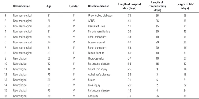
A total of 97 tracheostomized patients receiving mechanical ventilation who were admitted to ICUs from October 2006 to October 2007 were screened. Of those, 81 patients did not participate in the study for various reasons. Sixteen dysphagic patients matched all of the inclusion criteria; 8 were neurological patients, and 8 were non-neurological patients. the mean age was 56.6 ± 25.4, and a total of 11 males and 5 females were included. he mean hospital stay was 46.9 ± 17.0 days, the mean duration of mechanical ventilation was 32.4 ± 11.6 days, and the mean duration of tracheostomy was 16.5 ± 11.2 days. he demographic and individual characteristics of these patients are described in table 1.
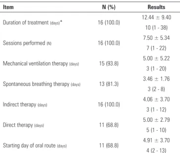
All patients underwent the rehabilitation program; two died during the course of therapy. here was 100% agreement between the assessments conducted by the speech-language pathologist and the otorhinolaryngologist in the diagnosis of oropharyngeal dysphagia. he rehabilitation program characteristics are described in table 2.
Data collected pre and post swallowing therapy for the 14 surviving patients were compared. Analysis of the scores showed a signiicant improvement on all scales (OSS: pre - 9.0 (3.0 - 15.0), post - 2.5 (0.0 - 8.0), p = 0.0007; SFS: pre - 4.5 (3.0 - 6.0), post - 1.0 (0.0 - 3.0), p = 0.001 and OSFS: pre - 8.0 (6.0 - 10.0), post - 3.0 (0.0 - 6.0), p = 0.0004), as shown in igure 1.
Before speech therapy, four patients presented grade 1 dysphagia, and 10 patients presented grade 2 dysphagia. In the group with grade 1 dysphagia, two patients (50.0%) achieved full improvement; however, in those patients with grade 2 dysphagia, four (40.0%) achieved full improvement, and two (20.0%) patients achieved partial improvement characterized by grade 1 dysphagia. All 14 patients were receiving tube feeding before the onset of therapy. After the rehabilitation program, it was possible for 10 of these patients to receive oral intake associated with enteral feeding; one patient could receive
Table 1 - Demographic data and characterization of individuals in the sample
Classification Age Gender Baseline disease Length of hospital stay (days)
Length of tracheostomy
(days)
Length of MV (days)
1 Non neurological 21 F Uncontrolled diabetes 75 38 59
2 Non neurological 26 M ARDS 41 26 35
3 Non neurological 86 M Pleural effusion 41 15 35
4 Non neurological 81 M Chronic renal failure 55 30 43
5 Non neurological 78 M Renal transplant 63 19 35
6 Non neurological 34 M Firearm wound 47 29 36
7 Non neurological 51 F Renal transplant 88 20 48
8 Non neurological 81 F Femur fracture 49 10 31
9 Neurological 62 M Hydrocephalus 37 18 27
10 Neurological 82 F Parkinson’s disease 55 16 32
11 Neurological 14 M Spinal cord injury 25 3 14
12 Neurological 75 F Alzheimer’s disease 36 3 18
13 Neurological 60 M Stroke 31 6 21
14 Neurological 21 M Brain injury 26 2 22
15 Neurological 75 M Parkinson’s disease 42 4 24
16 Neurological 59 M Botulism 39 25 38
Table 2 - Overall features of the speech rehabilitation program
Item N (%) Results
Duration of treatment (days)* 16 (100.0) 12.44 ± 9.40
10 (1 - 38)
Sessions performed (N) 16 (100.0) 7.50 ± 5.34
7 (1 - 22)
Mechanical ventilation therapy (days) 15 (93.8) 5.00 ± 5.22
3 (1 - 20)
Spontaneous breathing therapy (days) 13 (81.3) 3.46 ± 1.76
3 (2 - 8)
Indirect therapy (days) 16 (100.0) 4.06 ± 3.70
3 (1 - 12)
Direct therapy (days) 11 (68.8) 5.00 ± 2.79
5 (1 - 10)
Starting day of oral route (days) 11 (68.8) 4.91 ± 3.70
4 (2 - 13)
* Includes the days of treatment interruption. The results are expressed as the mean ± standard deviation, median (minimum - maximum).
Figure 1 - Comparison between the oropharyngeal structural score, swallowing functional score and otorhinolaryngological structural and functional score pre and post treatment. OSS - oropharyngeal structural score; SFS - swallowing functional score; OSFS - otorhinolaryngological structural and functional score. Results show the significant reduction of all scores; OSS, p = 0.007, SFS, p = 0.001 and OSFS, p = 0.004. (OSS and OSFS - paired t test, SFS - Wilcoxon paired test).
an exclusively oral diet, and 3 patients could not receive an oral diet. he average amount of oral intake was 180ml (11 patients) for the paste consistency diet and 87.5ml (4 patients) for the thick liquid diet.
DISCUSSION
his study showed that a speech therapy intervention on ICU patients under mechanical ventilation is feasible and might help to improve the swallowing function and oropharyngeal dysphagia severity. We did not ind any studies in the literature analyzing the impact of swallowing
rehabilitation on dysphagia in patients on mechanical ventilation except for case reports(33-36) and the opinions of specialists in the ield.(20-32) hus,this feasibility study in a small number of patients suggests that a clinical trial with an adequate sample size and clinical outcomes should be conducted.
hrough the assessment of oropharyngeal structures, we observed that the most evident pre-program abnormalities, i.e., lip and tongue tonus and larynx mobility, showed improvement, although without statistical signiicance, after the treatment. Even the less frequent abnormalities (lip, tongue and jaw mobility) also showed non-signiicant improvement. his inding suggests that the proposed isolated exercises were able to improve the range of movement and the tonus of each oropharyngeal structure, which may have led to better swallowing.(38) We found similar results in the functional parameters (swallowing trigger time and laryngeal elevation). Although none of these parameters signiicantly improved after treatment, likely because of our small sample size, this inding is in accordance with the signiicant improvement found in the scores. As for the structural and functional analysis of swallowing based on the OSFS score, a signiicant improvement was observed in the laryngeal sensitivity and the cough relex.(5) Tracheal penetration, food stasis in pharyngeal recesses and the swallowing trigger time showed non-signiicant improvements.
breathing and swallowing are essential mechanisms to prevent tracheal aspiration. For this reason, a weight of 2 was assigned to these variables. In the OSFS, a weight of 2 was assigned to the cough relex, laryngeal penetration and tracheal aspiration.
According to Cowley et al. and Moraes et al., daily assistance is necessary, especially during the transition from tube feeding to oral feeding.(23,41) In our study, although we predeined a daily follow-up, this was not possible due to limitations inherent to the patients themselves such as changes in ventilation parameters and in the level of consciousness. In this study, the mean number of sessions per patient was 7.5, with a mean treatment duration of 12 days, including both the swallowing sessions and the days of interruption. It is possible that our results would have been more relevant if the daily assistance approach had been feasible.
Our swallowing therapy was based on data from the baseline swallowing assessment. All patients in this study initially received indirect therapy over a mean period of 4 days. he indication of indirect therapy as the initial approach was based on the presence of a signiicant number of swallowing deicits related to the range of movement and tonus of the lips, tongue, mandible and larynx. he objective of indirect therapy was to prepare the oropharyngeal muscles using swallowing and voice maneuvers and techniques, with the inal goal of reintroducing oral feeding. Additionally, a period of one to two days was necessary to adjust the speaking valve. Direct therapy was implemented according to the patient’s progression. Speciic swallowing training was initiated with 3 to 5ml of paste consistency diet, administered orally. Although several authors have advocated that direct therapy could be given to these patients,(25,27,35,42-44) the safety of this procedure has never been studied. In this study, the initial use of indirect therapy followed by direct therapy may have contributed to our positive outcomes. hree patients were unable to manage food in the oral cavity and were not able to progress to direct therapy during their stay at the ICU. Although oral feeding could not be attained in these patients, the general improvement in the aspects related to the oropharyngeal structures has allowed for better communication regarding speech - voice and articulation, at least in a subjective analysis.(45) In addition, because the rehabilitation treatment continued after discharge from the ICU, indirect therapy may still
have contributed to improving the swallowing function throughout the patient’s stay at the hospital ward.
his study has both strengths and limitations. For the strengths, irst, we compared the results of clinical and otorhinolaryngological assessments before and after swallowing therapy. Additionally, the evaluation of swallowing was complete and included clinical and objective aspects using detailed scores. However, there were some limitations. First, the scores used had not been previously validated; thus, it is not possible to assure that they truly express the severity of dysphagia. However, the negative impact of the lack of validation was minimized by the fact that the same scores were used both before and after the intervention. Second, the number of patients evaluated was small, which certainly compromises the generalizability of the results. As mentioned previously, this is a case series and should be thus considered as a feasibility study. hird, we did not have a randomized control group. he lack of a control group hindered the analysis of potential spontaneous improvement during the swallowing rehabilitation program. Fourth, we analyzed a speciic subset of critically ill patients, all of whom had a tracheostomy, were ventilated with minimal parameters, and were clinically stable, awake and cooperative at the time of the procedure. Consequently, other populations with diferent features should be further investigated. As this was the irst feasibility study, as a safety measure, we decided not to include hemodynamically unstable patients or patients with high levels of PEEP or pressure support. However, based on our promising initial results, a future study could certainly include those patients. Finally, in most of the ICUs, a speech therapist is not available; this might hamper the applicability of our scores as well as the use of the rehabilitation program.
CONCLUSION
In this study, we demonstrated that an early
rehabilitation program is feasible in a small group of patients still under mechanical ventilation. Our results should help to design a clinical trial with an adequate sample size and clinically applicable outcomes.
ACKNOWLEDGMENTS
Objetivo: Avaliar a factibilidade da implantação precoce de um programa de reabilitação da deglutição em pacientes traqueostomizados com disfagia e sob ventilação mecânica.
Métodos: Estudo prospectivo realizado em unidades de te-rapia intensiva de um hospital universitário. Incluímos pacien-tes hemodinamicamente estáveis e submetidos à ventilação me-cânica por pelo menos 48 horas e há no mínimo 48 horas com traqueostomia e nível adequado de consciência. Os critérios de exclusão foram cirurgia prévia na cavidade oral, faringe, laringe e/ou esôfago, presença de doenças degenerativas ou história pre-gressa de disfagia orofaríngea. Todos os pacientes foram subme-tidos a um programa de reabilitação da deglutição. Antes e após o tratamento de reabilitação da deglutição, foram determinados
um escore estrutural orofaríngeo, um escore funcional de deglu-tição, e um escore otorrinolaringológico estrutural e funcional.
Resultados: Foram incluídos 14 pacientes. A duração média do programa de reabilitação foi de 12,4 ± 9,4 dias, com média de 5,0 ± 5,2 dias sob ventilação mecânica. Onze pacientes pu-deram receber alimentação por via oral enquanto ainda perma-neciam na unidade de terapia intensiva após 4 (2 - 13) dias de tratamento. Todos os escores apresentaram melhora signiicante após o tratamento.
Conclusões: Neste pequeno grupo de pacientes, a implan-tação de um programa precoce de reabiliimplan-tação da deglutição foi factível, mesmo em pacientes sob ventilação mecânica.
RESUMO
Descritores: Traqueostomia; Respiração artiicial; Transtornos de deglutição/reabilitação; Disfagia; Unidades de terapia intensiva
REFERENCES
1. Simonian MA, Goldberg NA. Swallowing disorders in the critical care patient. In: Carrau RL, Murry T. Comprehensive management of swallowing disorders. San Diego: Singular; 1999. p. 363-8
2. Zielske J, Bohne S, Brunkhorst FM, Axer H, Guntinas-Lichius O. Acute and long-term dysphagia in critically ill patients with severe sepsis: results of a prospective controlled observational study. Eur Arch Otorhinolaryngol. 2014;271(11):3085-93.
3. Elpern EH, Jacobs ER, Bone RC. Incidence of aspiration in tracheally intubated adults. Heart Lung. 1987;16(5):527-31.
4. DeVita MA, Spierer-Rundback L. Swallowing disorders in patients with prolonged orotracheal intubation or tracheostomy tubes. Crit Care Med. 1990;18(12):1328-30.
5. Elpern EH, Scott MG, Petro L, Ries MH. Pulmonary aspiration in mechanically ventilated patients with tracheostomies. Chest. 1994;105(2):563-6. 6. Tolep K, Getch CL, Criner GJ. Swallowing dysfunction in patients receiving
prolonged mechanical ventilation. Chest. 1996;109(1):167-72.
7. Pannunzio TG. Aspiration of oral feedings in patients with tracheostomies. AACN Clin Issues. 1996;7(4):560-9.
8. Lima CA, Siqueira TB, Travassos EF, Macedo CM, Bezerra AL, Paiva Júnior MD, et al. Influence of peripheral muscle strength on the decannulation success rate. Rev Bras Ter Intensiva. 2011;23(1):56-61.
9. Hernandez G, Pedrosa A, Ortiz R, Cruz Accuaroni Mdel M, Cuena R, Vaquero Collado C, et al. The effects of increasing effective airway diameter on weaning from mechanical ventilation in tracheostomized patients: a randomized controlled trial. Intensive Care Med. 2013;39(6):1063-70. 10. Macht M, Wimbish T, Bodine C, Moss M. ICU-acquired swallowing
disorders. Crit Care Med. 2013;41(10):2396-405.
11. Rosenbek JC, Roecker EB, Wood JL, Robbins J. Thermal application reduces the duration of stage transition in dysphagia after stroke. Dysphagia. 1996;11(4):225-33.
12. Lazarus CL, Logemann JA, Rademarker AW, Kahrilas PJ, Pajak T, Lazar R, et al. Effects of bolus volume, viscosity, and repeated swallows in nonstroke subjects and stroke patients. Arch Phys Med Rehabil. 1993;74(10):1066-70. 13. Logemann JA, Pauloski BR, Colangelo L, Lazarus C, Fujiu M, Kahrilas PJ.
Effects of a sour bolus on oropharyngeal swallowing measures in patients with neurogenic dysphagia. J Speech Hear Res.1995;38(3):556-63. 14. Logemann JA. Rehabilitation of oropharyngeal swallowing disorders. Acta
Otorhinolaryngol Belg. 1994;48(2):207-15.
15. Baumgartner CA, Bewyer E, Bruner D. Management of communication and swallowing in intensive care: the role of the speech pathologist. AACN Adv Crit Care. 2008;19(4):433-43.
16. Padovani AR, Andrade CR. Perfil funcional da deglutição em unidade de terapia intensiva clínica. einstein (São Paulo). 2007;5(4):358-62. 17. Padovani AR, Moraes DP, Sassi FC, Andrade CR. Avaliação clínica da
deglutição em Unidade de Terapia Intensiva. CoDAS. 2013;25(1):1-7. 18. Schindler A, Vincon E, Grosso E, Miletto AM, Di Rosa R, Schindler O.
Rehabilitative management of oropharyngeal dysphagia in acute care settings: data from a large Italian teaching hospital. Dysphagia. 2008;23(3):230-6. 19. Freeman-Sanderson A, Togher L, Phipps P, Elkins M. A clinical audit of
the management of patients with a tracheostomy in an Australian tertiary hospital intensive care unit: Focus on speech-language pathology. Int J Speech Lang Pathol. 2011;13(6):518-25.
20. Weber B. Eating with a trach. Am J Nurs. 1974;74(8):1439.
21. Weisinger W, Goldsmith T. Artificial ventilation: its impact on communica-tion and swallowing. Probl Respir Care. 1988;1:204-16.
22. Godwin JE, Heffner JE. Special critical care considerations in tracheostomy management. Clin Chest Med. 1991;12(3):573-83. Review.
23. Cowley RS, Swanson B, Chapman P, Kitik BA, Mackay LE. The role of rehabilitation in the intensive care unit. J Head Trauma Rehabil. 1994;9:32-42. 24. Dikeman KJ, Kazandjian MS. Oral communication options. In: Dikeman
KJ, Kazandjian MS. Communication and swallowing management of tracheostomized and ventilator-dependent adults. San Diego: Singular; 1995. p. 141-95.
25. Langmore SE. Dysphagia in neurologic patients in the intensive care unit. Semin Neurol. 1996;16(4):329-40.
26. Logemann JA. Evaluation and treatment of swallowing disorders. Texas: Pro-ed; 1998.
27. Murray KA, Brzozowski LA. Swallowing in patients with tracheotomies. AACN Clin Issues. 1998;9(3):416-26; quiz 456-8.
28. Hauck KA. Communication and swallowing issues in tracheostomized/ ventilator-dependent geriatric patients. Top Geriatr Rehabil. 1999;15:56-70. 29. Goldsmith T. Evaluation and treatment of swallowing disorders following
endotracheal intubation and tracheostomy. Int Anesthesiol Clin. 2000;38(3):219-42.
30. Britton D, Jones-Redmond J, Kasper C. The use of speaking valves with ventilator-dependent and tracheostomy patients. Curr Opin Otolaryngol Head Neck Surg. 2001;9:147-52.
31. Conway D, Parker C. Should we allow ventilated patients with a tracheostomy to eat and drink? Hosp Med. 2004;65(12):764.
32. Ward E, Jones C, Solley M, Cornwell P. Clinical consistency in tracheostomy management. J Med Speech Lang Pathol. 2007;15(1):7-26.
34. Siebens AA, Tippett DC, Kirby N, French J. Dysphagia and expiratory air flow. Dysphagia. 1993;8(3):266-9.
35. Phelan BA, Cooper DA, Sangkachand P. Prolonged mechanical ventilation and tracheostomy in the elderly. AACN Clin Issues. 2002;13(1):84-93. 36. Antunes MF, Santos AM, Santos JS, Vecina AL, Coronatto AG, Ferreira
OB, et al. Uso da válvula de fonação em paciente traqueostomizado dependente de ventilação mecânica na UTI de um hospital privado: relato de caso. Rev Bras Ter Intensiva. 2006;18 Supl:76.
37. Rosenbek JC, Robbins JA, Roecker EB, Coyle JL, Wood JL. A penetration-aspiration scale. Dysphagia. 1996;11(2):93-8.
38. Steele CM, Bailey GL, Polacco RE, Hori SF, Molfenter SM, Oshalla M, et al. Outcomes of tongue-pressure strength and accuracy training for dysphagia following acquired brain injury. Int J Speech Lang Pathol. 2013;15(5):492-502. 39. Crary MA, Mann GD, Groher ME. Initial psychometric assessment of a
functional oral intake scale for dysphagia in stroke patients. Arch Phys Med Rehabil. 2005;86(8):1516-20.
40. Martino R, Silver F, Teasell R, Bayley M, Nicholson G, Streiner DL, et al. The Toronto Bedside Swallowing Screening Test (TOR-BSST): development and validation of a dysphagia screening tool for patients with stroke. Stroke. 2009;40(2):555-61.
41. Moraes AM, Coelho WJ, Castro G, Nemr K. Incidência de disfagia em Unidade de Terapia Intensiva de adultos. Rev CEFAC. 2006;8(2):171-7. 42. Belafsky PC, Blumenfeld L, LePage A, Nahrstedt K. The accuracy of the
modified Evan´s blue dye test in predicting aspiration. Laryngoscope. 2003;113(11):1969-72.
43. Davis LA, Thompson Stanton S. Characteristics of dysphagia in elderly patients requiring mechanical ventilation. Dysphagia. 2004;19(1):7-14. 44. Freeman-Sanderson A, Togher L, Phipps P, Elkins M. A clinical audit of
the management of patients with a tracheostomy in an Australian tertiary hospital intensive care unit: Focus on speech-language pathology. Int J Speech Lang Pathol. 2011;13(6):518-25.