PET and the multitracer concept in the
study of neurodegenerative diseases
Henry Engler1, Andres Damian2, Cecilia Bentancourt2
ABSTRACT. The complexity of the pathological reactions of the brain to an aggression caused by an internal or external noxa represents a challenge for molecular imaging. Positron emission tomography (PET) can indicate
in vivo, anatomopathological changes involved in the development of different clinical symptoms in patients with neurodegenerative disorders. PET and the multitracer concept can provide information from different systems in the brain tissue building an image of the whole disease. We present here the combination of 18F-flourodeoxyglucose (FDG)
and N-[11C-methyl]-L-deuterodeprenyl (DED), FDG and N-[11C-methyl] 2-(4’-methylaminophenyl)-6-hydroxybenzothiazole
(PIB), PIB and L-[11C]-3’4-Dihydrophenylalanine (DOPA) and finally PIB and [15O]H2O.
Key words: PET, neurodegeneration, F-flourodeoxyglucose, PIB, Alzheimer’s disease, Creutzfeldt-Jakob disease. PET E O CONCEITO DE MULTITRAÇADOR NO ESTUDO DE DOENÇAS DEGENERATIVAS
RESUMO. A complexidade das reações patológicas do cérebro à agressões causadas por noxa interna ou externa representa um desafio para a imagem molecular. Tomografia por emissão de positron (PET) pode indicar, in vivo, alterações anatomopatológicas envolvidas no desenvolvimento de diferentes sintomas clínicos em pacientes com desordens neurodegenerativas. PET e o conceito de multitraçador pode fornecer informações de diferentes sistemas no tecido cerebral, construindo assim uma imagem da doença como um todo. Nós apresentamos neste artigo a combinação de
18F-flourodeoxyglucose (FDG) e N-[11C-methyl]-L-deuterodeprenyl (DED), FDG e N-[11C-methyl]
2-(4’-methylaminophenyl)-6-hydroxybenzothiazole (PIB), PIB e L-[11C]-3’4-Dihydrophenylalanine (DOPA) e finalmente, PIB e [15O]H2O .
Palavras-chave: PET, neurodegenerativas, F-flourodeoxyglucose, PIB, Doença de Alzheimer, Doença de Creutzfeldt-Jakob.
INTRODUCTION
T
he study of neurodegenerative diseaseshas experienced a qualitative leap since the introduction of molecular imaging of pathological processes in vivo. PET tracers
have the potential to reveal changes in the diferent stages of many diseases in the brain. hey improve the diagnoses increasing our understanding of the pathological processes in the central nervous system.
Diferent tracer combinations can be the way to characterize brain diseases with a higher level of accuracy. he multitracer con-cept can help us to achieve a more accurate classiication of brain diseases and open the way for better therapeutic strategies.
In this paper we highlight the use of the multitracer concept for the characterization
of neurodegenerative diseases and its impli-cations in patient management. he Table 1 describes the diferent tracers we are going to cover in this review.
FDG AND DED
18F-lourodeoxyglucose (FDG) is actively
transported into the cell by a group of glucose transport proteins (GLUT) and phosphory-lated. FDG is trapped in the cells as FDG-6-Phosphate avoiding the glycolysis and giving an indirect measure of the glucose uptake in the tissues.
N-[11C-methyl]-L -deuterodeprenyl (DED) binds irreversibly to the enzyme Mono-Amino-oxydase -B (MAO-B), which is expressed by reactive astrocytes and can be used as a marker for reactive astrocytosis.1-8
This study was conducted at the Uruguayan Centre of Molecular Imaging (CUDIM), Montevideo, Uruguay.
1MD. PhD and 2MD - Uruguayan Centre of Molecular Imaging (CUDIM), Montevideo, Uruguay.
Andres Damian. Uruguayan Centre of Molecular Imaging (CUDIM) – Av. Ricaldoni 2010 – Montevideo – Uruguay – E-mail: [email protected]
Disclosure: The authors report no conflits of interest.
Received September 02, 2015. Accepted in final form November 05, 2015.
he combination of DED, indicating reactive astrocy-tosis and FDG, showing changes in the neuronal func-tion, allows us to diferentiate between diseases with simultaneous astrocytosis and neuronal death from diseases with astrocytosis and concomitant increased glucose uptake indicating inlammation.
We have used this combination of tracers to dif-ferentiate Creutzfeldt-Jakob disease (CJD) from other degenerative processes. CJD is a prion disease clinically characterized by sudden dementia, ataxia and myoclo-nia. he anatomopathological hallmarks in the brain include: spongiform changes, astrocytosis and neuronal death.
Today there is no cure for CJD, but some other dis-eases can be treated and therefore it is crucial to achieve an early diagnosis. Inlammation can be produced by an exogenous (bacteria, virus, etc.) or an endogenous agent (autoimmune diseases or paraneoplastic limbic enceph-alitis)9 that is caused by antineuronal antibodies pro-duced in reaction to body neoplasms. In these patients
the clinical symptomatollogy can appear as dementia of rapid onset. Sometimes the laboratory tests can help in the diagnosis, but in other cases it is not possible to diferentiate between these clinical entities with this information.
he diferent patterns obtained with PET and appro-priated tracer combinations may allow a diferential diag-nosis modifying the treatment. It is important not only to exclude CJD but also to suggest other diagnoses.
In one patient with dementia of rapid onset and symptoms similar to those found in CJD, high FDG uptake was observed in the limbic system (Figure 1). he medial temporal lobes including the amygdala, the hippocampus and the parahippocampus were the most afected regions.2
his pattern of uptake was very diferent to the pat-tern described with FDG in patients with conirmed CJD.2 Paraneoplastic limbic encephalitis was suspected, a whole body FDG PET was performed and a tumour was found in the left lung.
Figure 1. Patient with suspect CJD. The patient, however, had paraneoplasic limbic encephalitis. Left, coronal view: high FDG uptake bilaterally in the medial temporalcortex. Center and right: transaxial view. The FDG pattern is differ-ent to that seen in confirmed cases of CJD.
Table 1. PET molecular probes and their clinical use.
Radiotracer Uptake mechanism Clinical use
18F-flourodeoxyglucose (FDG) Uptake by glucose transporters and trapped into the
cell after phosphorylation by the enzyme hexoquinase
Alzheimer’s disease, frontotemporal degeneration, Creutzfeldt Jakob disease, lymbic encephalitis, etc.
[11C-methyl] 2-(4’-methylaminophenyl)-
6-hydroxybenzothiazole (PIB)
Binding to amyloid-β peptide Alzheimer’s disease, amylodosis
L-[11C]-3’4-Dihydrophenylalanine (DOPA) Measure of the DOPA- decarboxylase activity at
the level of presynaptic terminals. Evaluation of presynaptic integrity
Parkinson’s disease, atypical parkinsonisms, endocrine tumours
N-[11C-methyl]-L-deuterodeprenyl (DED) Monoaminooxidase-B (MAO-B) inhibitor. Uptake by
reactive astrocytes.
Creutzfeldt Jakob disease, Alzheimer’s disease
[15O]H2O Gold standard for non invasive measurement of
cerebral blood flow (CBF)
An autopsy of the patient revealed an adenocarci-noma of the lung and tumoural antibodies against the brain were demonstrated. he PET examination could quickly distinguish this patient from the other patients with CJD.
In this patient, the quantitative FDG examination proved to be suicient to make the diferentiation between these clinical entities.
Other patient with suspected CJD had a glucose uptake pattern similar to that found in Alzheimer’s dis-ease (AD). he metabolism was bilaterally decrdis-eased in the temporoparietal regions, but it was conserved in the central parts of the brain and the occipital cortex.
he FDG examination indicating hypometabolism in the parietotemporal areas with conserved metabolism in basal ganglia, cerebellum and sensory motor cortex suggested the possibility that the patient had AD instead of CJD, but further investigations (biopsy of a salivary gland) revealed
Sjögren’s disease. Treatment with corticoids had a dramatic efect in the patient’s symptoms.
Parieto-occipital hypometabolism is a conspicuous inding observed mainly in MRI-negative neuropsychi-atric systemic lupus erythematosus (SLE). As this cere-bral region is located at the boundary of the blood supply territories of all three major arteries, it could be the most vulnerable zone of the cerebrum and may be afected at early stages of the cerebrovascular disease.10
An autoimmune disease, producing inlammatory meningo-encephalitis or a perivascular inlammation that afect this region might create a pattern of glucose uptake which is not possible to diferentiate with FDG from AD. Patients with Sjörgen’s disease can present with symptoms of AD.11
It is important to diferentiate between these dis-eases because a dementia with an onset corresponding to CJD or AD may be caused by immunological mecha-nisms: (System Lupus Erithematosus (SLE), Sjögren disease or a paraneoplasic phenomenon) and therefore can be treated with speciic drugs. he result, particularly in the case of Sjögren’s disease or SLE, can be dramatic, reversing a dementia condition in hours by administra-tion of corticoids and cyclophosphamide.11 Even demen-tias produced by antineuronal tumour antibodies can be reversed by the extirpation of the tumour.12 In a patient with CJD we demonstrate the congruence between pathological indings and PET results, as expressed by the combination DED/FDG.13
In the case of rapid onset dementias, a brain investi-gation with FDG could be followed by a scan of the whole body to quickly discard the possibility of an unknown
primary tumour producing antineuronal antibodies. If a brain scan shows hypermetabolism and the body scan is negative, autoimmune diseases or infectious diseases may be suspected. If the brain scan indicates asymmetric hypometabolism, the examination with DED can reveal high ratio DED/FDG suggesting CJD. Hypometabolism similar to that found in AD, but negative to PIB reten-tion, may be caused by a disease afecting the vascular system.
FDG AND PIB
he combination of FDG and PIB has helped us to under-stand better the dynamic process in the in vivo amyloid
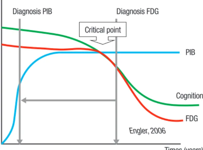
formation. he deterioration in cognition seems related to the decrease in the brain’s glucose metabolism, but not directly to the increase in amyloid depositions.
he accumulation of amyloid in the brain appears to be an early process in the development of AD that increases to a certain level (possibly many years before the debut of the symptoms) and then reaching an equi-librium between aggregation and degradation.14
he glucose uptake decreases slightly in the begin-ning of the disease, reaching a critical point in which it becomes pronounced enough to be detected by PET FDG.
Because the metabolic changes occur later in the development of the disease, FDG can be used to con-irm a diagnosis when the symptoms indicate dementia. he tracer however, is not able to discriminate between healthy persons and AD patients in the early stage of the disease in asymptomatic individuals, whereas PIB ofers the possibility to detect amyloid depositions when the symptoms are not evident.
FDG in combination with PIB increases the possibil-ity to perform diferential diagnoses. If the PIB examina-tion is negative and FDG reveals a pattern similar to that found in AD, it is possible that a disease with vascular anatomopathological substratum (autoimmune disease?) underlies the symptoms.
Several questions arise from the evidences presented above.
1) Is the presence of amyloid in the brain a process that can be related to aging without consequences for normal
func-tions? It have been suggested a common origin in the
plaques were absent in some of the patient brains up to 88 years of age.
Other nondemented patients presented with widely distributed neuritic as well as difuse plaques through-out the neocortex and limbic structures. It has been pro-posed that they represent “preclinical” AD.16
We suggest that the presence of amyloid is always a sign of degeneration and the depositions will disturb and injure the brain, causing dementia.
Like a city in which the recollection of refuse has been stopped and the garbage is blocking the streets preventing cars from circulating, the amyloid deposi-tions and neuroibrillary tangles block interneuronal communication.
he brain has a highly developed capacity to compen-sate damage produced by the presence of strange sub-stances accumulating in cytoplasm and interstitium and perturbing functions.
his capacity to compensate is not exclusive of the brain. All the organs in the human body have a reserve that allows normal functions when the tissue is afected by a disease.
We have seen in PET clinical routine extensive tumours invading a whole brain hemisphere before they cause symptoms.
Symptoms of Parkinson’s disease (PD) appear when the degeneration of the presynaptic pathway has reached 30-50% of the striatonigral component. he diagnosis with PET can often be made because the damage is extended enough. We suggest that the brain compen-sates the slow and progressive biochemical and mechanic damage originated by the increasing presence of amyloid depositions and neuroibrillary tangles until a critical point when the collapse of the function is a fact without the possibility of return.
Although some studies have conirmed that asymp-tomatic patients may present positive PIB scans, there is a general agreement in the idea that PIB is very sensitive and has a high negative predictive value in the detection of AD. A negative PIB scan would be very important to discard AD and suggesting other possible diagnoses.17-19
If a person having amyloid depositions could live long enough, she or he should develop symptoms of AD. his hypothesis is supported by recent metaanalysis indicat-ing that with longer follow-up periods, the accuracy of PIB to predict AD conversion in Mild Cognitive Impair-ment (MCI) patients improves signiicatively.20
Education, and aspects of occupational experience have been indicated as factors that may delay the clini-cal manifestations of AD.21 Other researchers suggest that the efect of education is modest.22 he mechanism
underlying such a delay could be the presence in educated or trained people of more “activated” neurons from the “reserve pool”, which could supply the efects of neuronal death.
2) Not all patients fulfilling international criteria for AD
show the presence of amyloid depositions. A discussion
concerning new classiications of dementia diseases seems necessary. Frontotemporal dementia (FTD) is a syndrome incuding many entities with diferent anato-mopathological background.
We suggest that the AD diagnosis must be changed to “Alzheimer’s syndrome” or “Cognitive Deicit of Alzheimer’s type” (CDAT) because there are many dis-eases expressing the same symptoms and producing similar changes in glucose uptake.
he diagnosis of AD must be established in patients with “Alzheimer’s syndrome” who have positive PET examinations with PIB or other well-proved amyloid markers. he absence of amyloid depositions is the most questioning result to the diagnosis of AD.
We have examined patients with “Alzheimer’s syn-drome” without amyloid depositions and it is possible for these patients to have, for example, a disease with a diferent pathological basis (vasculitis?), which is neces-sary to investigate.
Since physicians do not treat only symptoms, we need to clarify the underlying pathology to ind the proper treatment for patients expressing similar clinical symptoms.
MCI is considered a transitional stage between nor-mal aging and dementia, especially in early AD. It is known that MCI may have multiple causes, including AD and other forms of dementia, as well as depression and various psychiatric disorders. Because MCI is a frequent syndrome, there is a need to establish new methods for predicting the progression to AD.
In a previous study23 we demonstrate that PIB reten-tion in MCI patients is an intermediate step between healthy controls (HCs) and AD patients. here are MCI patients who can have either high or low PIB retention. Eleven of twenty-one MCI patients showed high PIB retention in the frontal, parietal and temporal cortices.
detect in this study decreased glucose metabolism in cor-tical brain areas as earlier reported in MCI patients.24,25 Nevertheless, in some individual MCI patients, decreased regional cerebral metabolic rate of glucose consumption (rCMRglc) was found in such areas as the parietal and temporal cortices. he non-converters in the MCI group had signiicant higher glucose uptake than AD patients whereas for the converters the diference was less.
In clinical routine we have observed increased glu-cose uptake in some of the patients with the diagnosis
MCI.26,27 he irst reaction of neurons against an
unspe-ciic injury is a hypermetabolic response. Injured neurons increase the glucose uptake by an enhanced glycogen-esis, possibly as a form to economize energy necessary to repair the damages.
Hypermetabolism in patients with MCI may indicate some inlammatory process afecting the brain. hese patients, however, might have a negative PIB scan.
Diseases such as autoimmune reactions may produce a perivascular disease or a vasculitis, which, in the acute period, may course with slight hypermetabolism, but in the chronic period, produce hypometabolism. Given that the most sensitive part of the brain is the boundary of blood supply between cerebral arteries, the temporopari-etal and the frontotemporal regions will be afected. In a more advanced stadium the patient may have symptoms of AD and the FDG may indicate a pattern similar to the one found in this disease.
he MCI patients classiied as converters had amy-loid depositions, but the FDG uptake was similar to the controls. he possibilities are that they had increased glucose uptake in the beginning of the amyloid deposi-tions that normalized later; however, it is possible that amyloid depositions do not increase the glucose uptake because they occur slowly under years of progress as a silent, devastating chronic deterioration.
More studies are needed to either conirm or ques-tion these interpretaques-tions. Based on our indings we suggest that diseases with cognitive impairment caus-ing hypermetabolism in the brain and without amyloid depositions are of a more acute nature. hese diseases afect the corridors between arteria cerebri anterior media and posterior and are possibly caused by an auto-immune or inlammatory base. he symptoms may be reverted spontaneously or by treatment.
AD probably courses with normal metabolism until the damage, induced by the accumulation of plaques and tangles, reaches the critical point in which the neurons can no longer compensate the dysfunction and the symp-toms appear (Figure 2).
here is possibly a critical point in which the capacity
of the brain to compensate damage is overcome, with the consequence that symptoms appear. Cognitive impair-ment follows the decrease in glucose uptake but not the increase of amyloid deposition. FDG is a useful tracer to conirm the diagnosis of AD, but often when damage of brain tissue is pronounced. PIB may detect the presence of the protein before the neurodegenerative changes cause symptoms because the amyloid deposition occurs early in the course of the disease.
PIB AND DOPA
Amyloid-containing deposits are a neuropathological feature in a wide range of dementias and movement disorders.
In a previous study28 we concluded that the normal PIB results in PD do not exclude the possibility that PIB binds to Lewy bodies, Lewy neurites or Amyloid-beta in PD.
Maetzler et al. examined ten patients with Parkin-son’s disease with dementia (PDD).29 Only two PDD patients displayed increased PIB binding to cortical amyloid comparable to AD patients. he remaining eight patients showed control-like cortical indings, but elevated PIB binding in the pons and mesencephalon. It has been suggested that, in addition to nonspeciic binding, PIB uptake in the brainstem may also relect PDD-related amyloid.29
Another study demonstrated the presence of a high ainity binding site for benzothiazole derivatives, includ-ing [3H]-PIB, on alpha-synuclein (AS) ilaments gener-ated in vitro.30
However, no discernible interaction of [3H]-PIB
Diagnosis PIB Diagnosis FDG
PIB
Cognition
FDG
Times (years)
Engler, 2006
Critical point
was detected with amygdala sections from PD cases containing frequent AS-immunoreactive Lewy bodies. hese indings suggest that the density and/or accessi-bility of AS binding sites in vivo is signiicantly less than
those associated with amyloid-beta peptide lesions. Lewy bodies pathology is therefore unlikely to contrib-ute signiicantly to the retention of PIB in PET imaging studies.30
PIB AND [
15O]H2O CBF
In previous studies we have used FDG images based on a 40-60-minute summation as a template to realign PIB images because they give a good anatomic subtract allowing the deinition of diferent brain regions.2,23,31-33 An automatic procedure has been applied to transfer the set of regions of interest (ROI) from FDG images to PIB images.34
In the case of PIB, it is diicult to deine the brain cor-tex in HCs in a late PIB summation (40-60 min) because the tracer is retained only in the white matter disturbing the automatic realignment of images.
In the irst study performed in Uppsala,33 we found that an early summation of the PIB activity frames (6 min) gave images that could be compared with later images of the FDG summation.
he early PIB summation images were realigned to the late FDG images and the rest of the PIB activity frames (7-60 min) were “co-resliced” using the realigned early summation as template.
In this way the ROIs drawn in the late FDG activity image could be transferred automatically to the later (40-60 min) PIB summation.
he similarity of the FDG images with the images of the early PIB summation suggested a positive relation-ship between the cumulative uptake of PIB in a short time interval following bolus administration and the cerebral blood low (CBF) measured [15O]H2O.
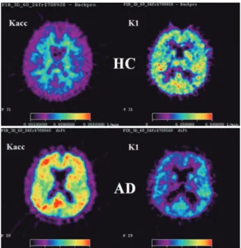
A kinetic model with one reversible and one irrevers-ible tissue compartment and three rate constants was used to investigate the PIB net accumulation (Kacc) and unidirectional inlux (K1) across the blood brain barrier (BBB) in HC and AD-patients.35
his is in contrast to previous studies in which mea-sures of PIB retention were obtained from reversible kinetic models.36,37 he blood input function was deter-mined with arterial sampling in 4 HC-subjects and 4 AD-patients. Parametric maps of Kacc = K1k3/(k2+k3) and K1 were created using a linear algorithm.
Results were compared using the ratio between target uptake and a reference region in a late interval, in HC and AD patients from studies.31,33 he parameter Kacc
Figure 3. Parametric maps of the net accumulation rate constant Kacc (left) and the unidirectional influx rate constant K1 (right). Upper row: HC subject, lower row: AD-patient.
and the late uptake ratio were found to have similar regional distributions.
he rate constant K1 for PIB was found to be com-paratively larger, demonstrating high extraction of PIB into the brain tissue and indicating that this parameter might relect CBF.
he possibility to replace K1 by a simple index of the PIB uptake in an early time interval following tracer administration was explored, but needs further validation.
he regional K1 values were lower in AD patients than in HCs (Figure 3). Most AD patients had higher Kacc values than HCs in cortical areas, but some patients had values similar to HCs. However, these patients had lower K1 than HCs and did not difer from the other patients regarding this feature.
Many groups have supported this hypothesis, show-ing that PIB can be used as a functional and pathological tracer in AD.38-40 Recently Chen et al. compared the early dynamic PIB scans with [15O]H2O CBF studies, showing a high correlation with CBF in patients with AD.41 hese
indings conirm that PIB can also be used as a functional tracer in AD to explore areas of impaired CBF.
Author contributions. All authors contributed equally in
manuscript preparation.
REFERENCES
1. Ekblom J, Jossan SS, Bergström M, Oreland L, Walum E, and Aquilonius SM. Monoamine oxidase-B in astrocytes. Glia 1993;8:122-132. 2. Engler H, Lundberg PO, Ekbom K, et al. Multitracer study with positron
emission tomography in Creutzfeldt-Jakob disease.” Eur J Nucl Med Mol Imaging 2003;30:85-95.
3. Fowler JS, Logan J, Volkow ND, Wang GJ. Translational neuroimaging: positron emission tomography studies of monoamine oxidase. Mol Imaging Biol 2005;7:377-387.
4. Jossan SS, Gillberg PG, Gottfries CG, Karlsson I, Oreland L. Monoamine oxidase B in brains from patients with Alzheimer’s disease: A biochemical and autoradiographical study. Neuroscience 1991;45:1-12.
5. Oreland L, Jossan SS, Hartvig P, Aquilonius SM, Långström B. Turn-over of monoamine oxidase B (MAO-B) in pig brain by positron emis-sion tomography using 11C-L-deprenyl. J Neural Transm 1990; 32(Suppl.):55-59
6. Choo ILH, Carter SF, Schöll ML, Nordberg A. Astrocytosis measured by
11C-deprenyl PET correlates with decrease in gray matter density in the
parahippocampus of prodromal Alzheimer’s patients. Eur J Nucl Med Mol Imaging 2014;41:2120-2126.
7. Carter SF, Scholl M, Almkvist O, et al. Evidence for Astrocytosis in Prodromal Alzheimer Disease Provided by 11C-Deuterium-L-Deprenyl: A Multitracer PET Paradigm Combining 11C-Pittsburgh Compound B and 18F-FDG. J Nucl Med 2012;53:37-46.
8. Carter SF, Nordberg A, Schöll M, et al. Investigating astrocytosis with 11C-deuterium-L-Deprenyl in mild cognitive impairment and mild Alzheimer’s disease: A multi tracer PET paradigm combining 11C-PIB and 18F-FDG. Alzheimers Dement 2011;7: S110.
9. Corsellis JA, Goldberg GJ, Norton AR. Limbic encephalitis’ and its asso-ciation with carcinoma,” Brain 1968;91:481-496.
10. Otte A, Weiner SM, Peter HH, et al. Brain glucose utilization in systemic lupus erythematosus with neuropsychiatric symptoms: a controlled posi-tron emission tomography study,” Eur J Nucl Med 1997;24:787-791. 11. Caselli RJ, Scheithauer BW, O’Duffy JD, Peterson GC, Westmoreland
BF, Davenport PA. Chronic inflammatory meningoencephalitis should not be mistaken for Alzheimer’s disease. Mayo Clin Proc 1993;68:846-853. 12. Provenzale JM, Barboriak DP, Coleman RE. Limbic encephalitis:
comparison of FDG PET and MR imaging findings. AJR Am J Roent-genol 1998;170:1659-1660.
13. Engler H, Nennesmo I, Kumlien E, et al. Imaging astrocytosis with PET in Creutzfeldt-Jakob disease: case report with histopathological findings. Int J Clin Exp Med 2012;5:201-207.
14. Engler H, Forsberg A, Almkvist O, et al.“Two-year follow-up of amyloid deposition in patients with Alzheimer’s disease,” Brain 2006;129: 2856-2866.
15. Arriagada PV, Marzloff K, Hyman BT.Distribution of Alzheimer-type patho-logic changes in nondemented elderly individuals matches the pattern in Alzheimer’s disease. Neurology 1992;42:1681-1688.
16. Price JL and Morris JC. Tangles and plaques in nondemented aging and ‘preclinical’ Alzheimer’s disease. Ann Neurol 1999;45:358-368. 17. Rowe CC and Villemagne VL. Brain amyloid imaging. J Nucl Med
Technol 2013;41:11-18.
18. Rabinovici G, Lehmann M, Rosen H, et al. Diagnostic Accuracy of Amyloid and FDG PET in Pathologically-Confirmed Dementia (S8.005). Neurology 2014;82:(Suppl.):S8.005.
19. Nordberg A, Carter SF, Rinne J, et al. A European multicentre PET study of fibrillar amyloid in Alzheimer’s disease. Eur J Nucl Med Mol Imaging 2013;40:104-114.
20. Ma Y, Zhang S, Li J, et al. Predictive accuracy of amyloid imaging for progression from mild cognitive impairment to Alzheimer disease with different lengths of follow-up: a meta-analysis. Medicine (Baltimore) 2014;93:e150.
21. Stern Y, Alexander GE, Prohovnik I, et al. Relationship between lifetime
occupation and parietal flow: implications for a reserve against Alzheim-er’s disease pathology. Neurology 1995;45:55-60.
22. Fritsch T, McClendon MJ, Smyth KA, Ogrocki PK. Effects of educa-tional attainment and occupaeduca-tional status on cognitive and funceduca-tional decline in persons with Alzheimer-type dementia. Int Psychogeriatr 2002; 14:347-363.
23. Forsberg A, Engler H, Almkvist O, et al. PET imaging of amyloid depo-sition in patients with mild cognitive impairment. Neurobiol. Aging 2008;29:1456-1465.
24. Anchisi D, Borroni B, Franceschi M, et al. Heterogeneity of brain glucose metabolism in mild cognitive impairment and clinical progression to Alzheimer disease. Arch Neurol 2005;62:1728-1733.
25. Chételat G, Desgranges B, De la Sayette V, Viader F, Eustache F, Baron JC. Mild cognitive impairment: Can FDG-PET predict who is to rapidly convert to Alzheimer’s disease? Neurology 2003;60:1374-1377. 26. Kreutzberg GW, Emmert H. Glucose utilization of motor nuclei during
regeneration: A [14C]2-deoxyglucose study. Exp Neurol 1980;70: 712-716.
27. Smith CB, Crane AM, Kadekaro M, Agranoff BW, Sokoloff L. Stimulation of protein synthesis and glucose utilization in the hypoglossal nucleus induced by axotomy. J Neurosci 1984; 4:2489-2496.
28. Johansson A, Savitcheva I, Forsberg A, et al. “[(11)C]-PIB imaging in patients with Parkinson’s disease: preliminary results”. Parkinsonism Relat Disord 2008;14:345-347.
29. Maetzler W, Reimold M, Liepelt I, C. et al. “[11C]PIB binding in Parkin-son’s disease dementia.,” Neuroimage 2008;39:1027-1033.
30. Ye L, Velasco A, Fraser G, et al. In vitro high affinity alpha-synuclein binding sites for the amyloid imaging agent PIB are not matched by binding to Lewy bodies in postmortem human brain. J Neurochem 2008; 105:1428-1437.
31. Engler H, Forsberg A, Almkvist O, et al. Two-year follow-up of amyloid deposition in patients with Alzheimer’s disease. Brain 2006;129: 2856-2866.
32. Engler H, Santillo AF, Wang SX, et al. In vivo amyloid imaging with PET in frontotemporal dementia. Eur J Nucl Med Mol Imaging 2008;35:100-106. 33. Klunk WE, Engler H, Nordberg A, et al. “Imaging brain amyloid in
Alzheimer’s disease with Pittsburgh Compound-B. Ann Neurol 2004;55: 306-319.
34. Andersson JLR, Thurfjell L. Implementation and Validation of a Fully Automatic System for Intra- and Interindividual Registration of PET Brain Scans. J Comput Assist Tomogr 1997;21:136-144.
35. Blomquist G, Engler H, Nordberg A, et al. Unidirectional Influx and Net Accumulation of PIB. Open Neuroimag J 2008;2:14-125.
36. Lopresti BJ, Klunk WE, Mathis CA, et al. Simplified quantification of Pittsburgh Compound B amyloid imaging PET studies: A comparative analysis. J Nucl Med 2005; 46:1959-1972.
37. Price JC, Klunk WE, Lopresti BJ, et al. Kinetic modeling of amyloid binding in humans using PET imaging and Pittsburgh Compound-B. J Cereb Blood Flow Metab 2005;25:1528-1547.
38. Meyer PT, Hellwig S, Amtage F, et al. Dual-biomarker imaging of regional cerebral amyloid load and neuronal activity in dementia with PET and 11C-labeled Pittsburgh compound B. J Nucl Med 2011;52:393-400. 39. Forsberg A, Engler H, Blomquist G, Långström B, Nordberg A. The
use of PIB-PET as a dual pathological and functional biomarker in AD. Biochim Biophys Acta 2012;1822: 380-385.
40. Sojkova J, Goh J, Bilgel M, et al. Voxelwise Relationships Between Distri-bution Volume Ratio and Cerebral Blood Flow: Implications for Analysis of β-Amyloid Images. J Nucl Med 2015;56:1042-1047.