Human islet xenotransplantation in rodents: A literature
review of experimental model trends
Leandro Ryuchi Iuamoto,
I* Andre´ Silva Franco,
IFa´bio Yuji Suguita,
IFelipe Futema Essu,
ILucas Torres
Oliveira,
IJuliana Mika Kato,
IMatheus Belloni Torsani,
IAlberto Meyer,
IIWellington Andraus,
IIEleazar Chaib,
IILuiz Augusto Carneiro D
’
Albuquerque
IIIFaculdade de Medicina, Universidade de Sao Paulo, Sao Paulo, SP, BR.IIDepartamento de Gastroenterologia, Faculdade de Medicina, Universidade de Sao
Paulo, Sao Paulo, SP, BR.
Among the innovations for the treatment of type 1 diabetes, islet transplantation is a less invasive method of
treatment, although it is still in development. One of the greatest barriers to this technique is the low number
of pancreas donors and the low number of pancreases that are available for transplantation. Rodent models
have been chosen in most studies of islet rejection and type 1 diabetes prevention to evaluate the quality and
function of isolated human islets and to identify alternative solutions to the problem of islet scarcity. The
purpose of this study is to conduct a review of islet xenotransplantation experiments from humans to rodents,
to organize and analyze the parameters of these experiments, to describe trends in experimental modeling and
to assess the viability of this procedure. In this study, we reviewed recently published research regarding islet
xenotransplantation from humans to rodents, and we summarized the findings and organized the relevant
data. The included studies were recent reports that involved xenotransplantation using human islets in a rodent
model. We excluded the studies that related to isotransplantation, autotransplantation and
allotransplanta-tion. A total of 34 studies that related to xenotransplantation were selected for review based on their relevance
and current data. Advances in the use of different graft sites may overcome autoimmunity and rejection after
transplantation, which may solve the problem of the scarcity of islet donors in patients with type 1 diabetes.
KEYWORDS:
Islet Transplantation; Allograft; Transplantation; Heterologous; Islets of Langerhans.
Iuamoto LR, Franco AS, Suguita FY, Essu FF, Oliveira LT, Kato JM, et al. Human islet xenotransplantation in rodents: A literature review of experimental model trends. Clinics. 2017;72(4):238-243
Received for publication onAugust 10, 2016;First review completed onNovember 2, 2016;Accepted for publication onDecember 16, 2016 *Corresponding author. E-mail: [email protected]
’
INTRODUCTION
According to the International Diabetes Federation (IDF),
diabetes mellitus currently affects 382 million people, with a
projected increase to 592 million people by 2035 (1).
The etiology of type I diabetes mellitus is unknown;
however, histopathological findings indicate an autoimmune
destruction of
-cells, an association with HLA alleles and
environmental factors, such as exposure to bovine milk.
Diabetes mellitus was historically considered a fatal disease
that resulted in hyperglycemic coma. However, since the
discovery of the therapeutic application of insulin in the
1920s, diabetes mellitus has become a chronic disease that
causes many complications, including retinopathy,
nephro-pathy, vasculopathy and neuropathy.
In 1894, the first case of islet transplantation as a treatment
for diabetes was described by Dr. Watson Williams and
Hareshant. Notably, this case occurred before the insulin
isolation of Banting, Best and Collip in 1921. In the early
twentieth century, Dr. W. Williams attempted to implant
sheep pancreatic fragments in the subcutaneous tissue of a
15-year-old male with ketoacidosis. However, the xenograft
was rejected because of a lack of immunosuppressive
techniques. In 1972, Dr. P. Lacey demonstrated the
reversi-bility of diabetes in rodents by using islet implantation (2).
The first successes in islet allografts in the surgical treatment
of diabetes occurred in 1990 with Scharp et al., who achieved
insulin independence in a patient with type 1 diabetes mellitus
for one month. However, many technical difficulties were
found during the reproduction of this experiment.
One of the greatest barriers to the development of islet
transplantation is the low number of pancreas donors and
the low number of pancreases that can be used for
trans-plantation (3). According to the Network of Organ
Procure-ment and Transplantation, fewer than 20% of the pancreases
that are collected from a total of 8,000 donors are available
for transplantation. In addition, many pancreas donors do
not meet the selection criteria, and many islets are handled
DOI:10.6061/clinics/2017(04)08
Copyright&2017CLINICS–This is an Open Access article distributed under the terms of the Creative Commons License (http://creativecommons.org/licenses/by/ 4.0/) which permits unrestricted use, distribution, and reproduction in any medium or format, provided the original work is properly cited.
incorrectly, negatively affecting the transplant procedure. (4)
Other inconveniences are the high cost of islet isolation, the
poor durability of insulin independence, autoimmunity and
rejection after transplantation (2,3,5,6).
To supply the scarcity of islets, animal donors, such as pigs,
could provide an alternative source of cells for transplantation
(7). However, xenotransplantation is challenged by the
pos-sible risk of infection from pathogens within the donor
ani-mal. Specifically, all pigs contain multiple copies of porcine
endogenous retrovirus and at least three variants of pig
endogenous retrovirus (PERV), which can infect human cells
in vitro. Thus, there is a risk of PERV infection associated with
the xenotransplantation of pig islets to immunosuppressed
human patients (8,9).
In this context, to evaluate the quality and function of
isolated human islets (10), the rodent has been chosen over
other animals in most studies that involve islet rejection and
the prevention of type 1 diabetes (3).
Manikandan et al. (11) studied the antioxidant effect of
black tea on the regeneration of pancreatic
-cells and
obser-ved a positive therapeutic effect in rodent studies. Recently,
Gu et al. (12) described an alternative therapeutic strategy to
treat type 1 diabetes, namely, treatment by nanoparticles,
which sustainably promotes the self-regulation of
glucose-mediated insulin secretion. This effect is observed for a longer
period of time than the insulin injections that are currently used
for treatment.
Although there have been many positive results related to
the xenotransplantation of human islets to rodents, researchers
have rarely achieved a breakthrough in the clinical treatment
of islet transplantation, perhaps because of the differences
between the human immune system and the rodent model.
These differences have stimulated the development of
humanized rodent models, which allow the detailed study
of human immune system cells and transplanted human
islets in vivo (3).
The purpose of this study is to review islet
xenotrans-plantation experimental attempts from humans to rodents, to
organize the parameters of these experiments and to analyze
the viability of these procedures.
’
METHODOLOGY
We reviewed studies regarding islet xenotransplantation
from humans to rodents. The relevant data from recently
published studies from 2006 to 2016 were summarized and
organized.
Eligibility Criteria
Types of Studies
.
The study designs of previous reviews
and experimental studies were included.
Types of Participants
.
Donor participants were humans
from whom islets were isolated and transplanted to rodents
(recipient).
Types of Intervention
.
The interventions were islet
xenotransplantation from humans to rodents. There were
different graft sites and types of islet recipients. In the
present review, only the studies that relate to human to
rodent islet xenotransplantation were selected.
Types of Parameters Analyzed
.
Several parameters were
considered, namely, strain, gender, age and weight of the
recip-ient, xenotransplantation site, graft survival time (follow up),
number of transplanted islets and diabetes induction method.
Exclusion Criteria
Articles discussing transplantation in porcine, tilapia and
nonhuman primates (which are some of the more common
species that are used for transplantation) were excluded from
the review to focus on the articles that relate to islet
xeno-transplantation from humans to rodents. Studies using stem
cells or that had an unclear methodology were excluded
from our review.
Research letters, articles not published in English and
articles for which the full text was unavailable were not
considered in this review.
Following the PubMed search, we reviewed the references
from the retrieved publications and obtained the entire text
of the publications for potential inclusion in the review.
Literature Search
Using the Medline database, the literature was searched
for English-language articles that were published from
January 2006 to January 2016.
We performed a manual search of the references and
con-tacted experts in the field.
Search Strategy
.
We searched for published articles by
using the Medline database with the keywords "rodent islet
transplantation".
We also selected the most recent works that were
pub-lished from January 2006 to January 2016 by using the following
search terms:
‘‘
(((((rodent human islet xenotransplantation)
NOT tilapia) NOT porcine) NOT nonhuman primate)
NOT pig) AND (
‘‘
2006
’’
[Date - Completion]:
‘‘
2016
’’
[Date
-Completion])
’’
.
Articles that were published before 2006 were not included
in the analysis because of a lack of information, relevance and
current data.
Data Extraction
.
The data from each study were
inde-pendently extracted by 3 of the authors. Disagreements were
resolved by consensus. If no consensus was achieved, a
fourth author was consulted.
’
RESULTS
A total of 1,819 articles from 2006 to 2016 were found, but
only 225 articles were related to xenotransplantation and
were thus selected based on their relevance and current
information. We selected 91 articles and analyzed them; 34 of
these articles were had good methodological quality, such as
updated information that is necessary for this review and a
description of all comparative parameters related to islet
xenotransplantation from human donors to rodents.
The results are organized and displayed in Tables 1 and 2.
’
DISCUSSION
Islet transplantation is an innovation for type 1 diabetes
treatment that is less invasive and that has a 20-fold lower
morbidity rate than pancreas transplantation (2,4,6,16).
Some studies have reported an 80% rate of insulin
independence during the first postoperative year in the
patients who were treated with islet transplantation.
How-ever, graft survival rates remain low (2).
The islet transplantation technique has been developed to
provide an adequate supply of insulin, which solves the
prob-lem of donor shortage for diabetic patients (17). From 1991 to
2000, 450 islet transplantation attempts were performed in
patients with type 1 diabetes with only an 8% success rate.
We discuss the analyzed studies in more detail below.
Recipient characteristics
In this study, we reviewed the articles describing xenograft
transplantation in rodents. The majority of the animals were
between 9 and 16 weeks old and were male (32.4% male;
17.6% female; 50% N/A). See Table 1. Although more studies
used C57BL/6 mice in the xenograft experiments (22%),
followed by NOD-SCID and BALB/c mice (14% each), no
significant difference was observed in the results that were
obtained using other strains.
Diabetes induction method
The standard diabetes induction method was the use of
streptozotocin. The median dose was 170 mg/kg (50-250
mg/kg).
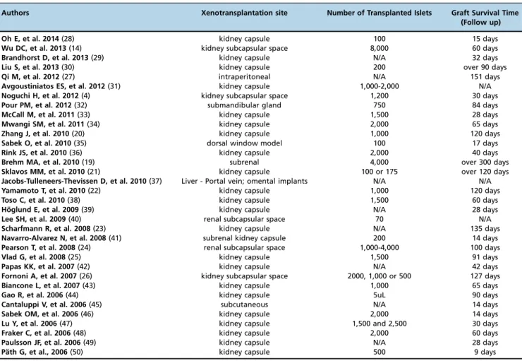
Islet xenotransplantation site
The authors used different sites for the xenografts
(Table 2), but the kidney capsule (91.2% of the studies) was
the most frequently used site for transplantation. Other sites,
such as the intraperitoneal space, liver (portal vein),
subcu-taneous space, submandibular gland and dorsal window
model, were used in a small number of studies.
The highest graft survival time was more than 300 days,
which was obtained by Brehm MA et al. (19). This study
used the subrenal space as the site of xenograft
transplanta-tion. Other studies that used the kidney capsule as the
xeno-transplantation site, such as the studies by Zhang J et al. (20),
Sklavos MM et al. (21), Yamamoto T et al. (22) Scharfmann R
et al. (23), Pearson T et al. (24), Vlad G et al. (25) and Fornoni A
et al. (26), reported more than 100 days of graft survival time.
Although the majority of articles show higher survival rates
Table 1
-
Comparative analysis of the types of rodents used and their clinical characteristics to evaluate the viability of the procedure: Strain, Gender, Age and Diabetes induction method.Authors Recipient Gender Age Diabetes induction method Viability
Yes No
Oh E, et al. 2014 (28) NOD-SCID mice N/A 10-14 weeks Streptozotocin 180 mg/kg X
Wu DC, et al. 2013 (14) BALB/c mice N/A 6-12 weeks Streptozotocin 250 mg/kg X
Brandhorst D, et al. 2013 (29) C57BL/6 mice N/A N/A N/A X
Liu S, et al. 2013 (30) C57BL/6 mice Male 10 weeks Streptozotocin 200 mg/kg X
Qi M, et al. 2012 (27) BALB/c mice N/A N/A N/A X
Avgoustiniatos ES, et al. 2012 (31) N/A N/A N/A Streptozotocin (dose: N/A) X
Noguchi H, et al. 2012 (4) N/A N/A N/A Streptozotocin 220 mg/kg X
Pour PM, et al. 2012 (32) Syrian Golden hamsters Female 8 years Streptozotocin 50 mg/kg X
McCall M, et al. 2011 (33) C57BL/6 mice N/A N/A Streptozotocin (220mg/kg -BALB/c; 180mg/kg - B6-RAG-/-)
X
Mwangi SM, et al. 2011 (34) athymic nude Foxn1-nu mice N/A 6 weeks Streptozotocin 75 mg/kg X
Zhang J, et al. 2010 (20) NOD/LtJ mice Female N/A N/A X
Sabek O, et al. 2010 (35) N/A Female 10-12 weeks N/A X
Rink JS, et al. 2010 (36) N/A N/A N/A Streptozotocin 220 mg/kg x
Brehm MA, et al. 2010 (19) NOD SCID gamma mice N/A 12-16 weeks Spontaneous: 3-5 week-old x
Sklavos MM, et al. 2010 (21) C57BL/6 and BALB/c Male 6-8 weeks Streptozotocin 240 mg/kg x
Jacobs-Tulleneers-Thevissen D, et al. 2010 (37)
Rowett rats Male 7-10 weeks Streptozotocin 60 mg/kg x
Yamamoto T, et al. 2010 (22) N/A N/A N/A Streptozotocin 200 mg/kg x
Toso C, et al. 2010 (38) C57BL/6 mice Female and Male N/A Streptozotocin 175 mg/kg x
Ho¨glund E, et al. 2009 (39) C57BL/6 mice Male N/A N/A x
Lee SH, et al. 2009 (40) SCID-Beige mice N/A 8 weeks Streptozotocin 40 mg/kg x
Scharfmann R, et al. 2008 (23) SCID mice Male N/A N/A x
Navarro-Alvarez N, et al. 2008 (41) SCID mice Male 10-12 weeks Streptozotocin 200 mg/kg x
Pearson T, et al. 2008 (24) NOD-SCID mice N/A N/A Streptozotocin 150 mg/kg x
Vlad G, et al. 2008 (25) NOD-SCID mice Female 6-10 weeks Streptozotocin 180 mg/kg x
Papas KK, et al. 2007 (42) N/A N/A N/A Streptozotocin (dose: N/A) x
Fornoni A, et al. 2007 (26) NU/NU mice N/A N/A Streptozotocin 200 mg/kg x
Biancone L, et al. 2007 (43) BALB/c mice Female 6-8 weeks N/A x
Gao R, et al. 2006 (44) BALB/c mice Male 6-8 weeks N/A x
Cantaluppi V, et al. 2006 (45) SCID and C57BI/6 mice N/A N/A N/A x
Sabek OM, et al. 2006 (46) NOD-SCID mice N/A N/A Glucose 2 g/kg x
Lu Y, et al. 2006 (47) NOD-SCID mice Male 8-12 weeks streptozotocin 160 mg/kg x
Fraker C, et al. 2006 (48) NU/NU mice Male N/A Streptozotocin 200 mg/kg x
Paulsson JF, et al. 2006 (49) N/A Male N/A N/A x
using sites that involve the kidney, Qi M et al. (27) used an
intraperitoneal site and obtained 134 days (
±
17) of graft
survival. Few articles have explored different xenograft sites,
and it may thus be difficult to conclude whether these
loca-tions provide better graft survival rates than the kidney.
It is important to note that in many studies, the recipients
were sacrificed for histopathological analysis.
We identified many variables on the analyzed studies. The
characteristics of the xenotransplantation site are factors that
can possibly influence the obtained results. Based on our
anal-ysis, it is possible to reproduce some of these studies and to
modify additional variables to obtain better graft survival
times. Nevertheless, one relevant limitation is that many
studies did not describe the data that are essential to reproduce
the described experiments, such as the strain, age and gender of
the recipient animal and the diabetes induction method.
Although immunosuppressive drugs may increase the
sur-vival rates of islet allotransplantation in rodents by reducing
the side effects (17), few studies have used
immunosuppres-sants. It was therefore not possible to perform an analysis of
the immunosuppressive effect in islet xenotransplantation.
Future studies with improved methodologies are necessary to
improve the graft survival time and to advance type 1 diabetes
treatment.
The viability of pancreatic islet transplantation could be
determined in only a small number of studies because of a lack
of the information that is necessary to perform this procedure.
The survival rates in allograft experiments have increased
with the use of novel graft sites. Different methodologies to
conserve islets may overcome autoimmunity and rejection
after transplantation and solve the problem of the scarcity of
islet donors for patients with type 1 diabetes.
’
AUTHOR CONTRIBUTIONS
Iuamoto LR, Franco AS, Suguita FY, Essu FF, Oliveira LT, Kato JM and Torsani MB were responsible for the literature review and manuscript
writing. Iuamoto LR, Franco AS, Meyer A, Andraus W and D’Albuquerque
LA were responsible for critical analysis. Iuamoto LR, Franco AS, Kato JM,
Meyer A, Chaib E and D’Albuquerque LA were responsible for paper
revision. Iuamoto LR, Franco AS, Meyer A, Chaib E, Andraus W and
D’Albuquerque LA were responsible for manuscript review. Iuamoto LR and
Meyer A were responsible for study design. Meyer A, Chaib E, Andraus W
and D’Albuquerque LA were responsible for supervision of the study.
’
REFERENCES
1. Guariguata L, Whiting DR, Hambleton I, Beagley J, Linnenkamp U, Shaw JE. Global estimates of diabetes prevalence for 2013 and projections for 2035. Diabetes Res Clin Pract. 2014;103(2):137–49, http://dx.doi.org/
10.1016/j.diabres.2013.11.002.
2. Merani S, Shapiro AM. Current status of pancreatic islet transplantation. Clin Sci. 2006;110(6):611–25, http://dx.doi.org/10.1042/CS20050342.
3. Jacobson S, Heuts F, Juarez J, Hultcrantz M, Korsgren O, Svensson M, et al. Alloreactivity but failure to reject human islet transplants by humanized Balb/c/Rag2gc mice. Scand J Immunol. 2010;71(2):83–90,
http://dx.doi.org/10.1111/j.1365-3083.2009.02356.x.
4. Noguchi H, Naziruddin B, Jackson A, Shimoda M, Ikemoto T, Fujita Y, et al. Fresh islets are more effective for islet transplantation than cultured islets. Cell Transplant. 2012;21(2-3):517–23, http://dx.doi.org/10.3727/
096368911X605439.
5. Perez-Basterrechea M, Obaya AJ, Meana A, Otero J, Esteban MM. Cooperation by fibroblasts and bone marrow-mesenchymal stem cells to
Table 2
-
Preferred islet xenotransplantation site, number of transplanted islets and graft survival time (follow up).Authors Xenotransplantation site Number of Transplanted Islets Graft Survival Time (Follow up)
Oh E, et al. 2014(28) kidney capsule 100 15 days
Wu DC, et al. 2013(14) kidney subcapsular space 8,000 60 days
Brandhorst D, et al. 2013(29) kidney capsule N/A 32 days
Liu S, et al. 2013(30) kidney capsule 200 over 90 days
Qi M, et al. 2012(27) intraperitoneal N/A 151 days
Avgoustiniatos ES, et al. 2012(31) kidney capsule 1,000-2,000 N/A
Noguchi H, et al. 2012(4) kidney subcapsular space 1,200 30 days
Pour PM, et al. 2012(32) submandibular gland 750 84 days
McCall M, et al. 2011(33) kidney capsule 1,500 28 days
Mwangi SM, et al. 2011(34) kidney capsule 2,000 65 days
Zhang J, et al. 2010(20) kidney capsule 1,000 120 days
Sabek O, et al. 2010(35) dorsal window model 100 17 days
Rink JS, et al. 2010(36) kidney capsule 2,000 40 days
Brehm MA, et al. 2010(19) subrenal 4,000 over 300 days
Sklavos MM, et al. 2010(21) kidney capsule 100 or 175 over 120 days
Jacobs-Tulleneers-Thevissen D, et al. 2010(37) Liver - Portal vein; omental implants N/A N/A
Yamamoto T, et al. 2010(22) kidney capsule 1,000 120 days
Toso C, et al. 2010(38) kidney capsule 1,500 60 days
Ho¨glund E, et al. 2009(39) kidney capsule N/A 28 days
Lee SH, et al. 2009(40) renal subcapsular space 70 N/A
Scharfmann R, et al. 2008(23) kidney capsule N/A 135 days
Navarro-Alvarez N, et al. 2008(41) subrenal kidney capsule 200 14 days
Pearson T, et al. 2008(24) renal subcapsular space 1,000-4,000 100 days
Vlad G, et al. 2008(25) kidney capsule 1,500 91 days
Papas KK, et al. 2007(42) kidney capsule N/A 42 days
Fornoni A, et al. 2007(26) kidney subcapsular space 2000, 1,000 or 500 127 days
Biancone L, et al. 2007(43) kidney capsule 1,000 65 days
Gao R, et al. 2006(44) kidney capsule 5uL 90 days
Cantaluppi V, et al. 2006(45) subcutaneous N/A 14 days
Sabek OM, et al. 2006(46) kidney capsule 2,000 14 days
Lu Y, et al. 2006(47) kidney capsule 1,500 and 2,500 30 days
Fraker C, et al. 2006(48) kidney capsule 2,000 60 days
Paulsson JF, et al. 2006(49) kidney capsule N/A 28 days
improve pancreatic rat-to-mouse islet xenotransplantation. PLoS One. 2013;8(8):e73526, http://dx.doi.org/10.1371/journal.pone.0073526. 6. Shapiro AM, Lakey JR, Ryan EA, Korbutt GS, Toth E, Warnock GL, et al.
Islet transplantation in seven patients with type 1 diabetes mellitus using a glucocorticoid-free immunosuppressive regimen. N Engl J Med. 2000;343(4):230–8, http://dx.doi.org/10.1056/NEJM200007273430401.
7. Iuamoto LR, Meyer A, Chaib E, D’Albuquerque LA. Review of experi-mental attempts of islet allotransplantation in rodents: parameters involved and viability of the procedure. World J Gastroenterol. 2014; 20(37):13512–20, http://dx.doi.org/10.3748/wjg.v20.i37.13512.
8. Patience C, Takeuchi Y, Weiss RA. Infection of human cells by an endo-genous retrovirus of pigs. Nat Med. 1997;3(3):282–6, http://dx.doi.org/
10.1038/nm0397-282.
9. van der Laan LJ, Lockey C, Griffeth BC, Frasier FS, Wilson CA, Onions DE, et al. Infection by porcine endogenous retrovirus after islet xeno-transplantation in SCID mice. Nature. 2000;407(6800):90–4, http://dx.doi.
org/10.1038/35024089.
10. Luo J, Nguyen K, Chen M, Tran T, Hao J, Tian B, et al. Evaluating insulin secretagogues in a humanized mouse model with functional human islets. Metabolism. 2013;62(1):90–9, http://dx.doi.org/10.1016/j.metabol.
2012.07.010.
11. Manikandan R, Sundaram R, Thiagarajan R, Sivakumar MR, Meiyalagan V, Arumugam M. Effect of black tea on histological and immunohisto-chemical changes in pancreatic tissues of normal and streptozotocin-induced diabetic mice (Mus musculus). Microsc Res Tech. 2009;72(10): 723–6, http://dx.doi.org/10.1002/jemt.20721.
12. Gu Z, Aimetti AA, Wang Q, Dang TT, Zhang Y, Veiseh O, et al. Injectable nano-network for glucose-mediated insulin delivery. ACS Nano. 2013; 7(5):4194–201, http://dx.doi.org/10.1021/nn400630x.
13. DCCT/EDIC Research Group, de Boer IH, Sun W, Cleary PA, Lachin JM, Molitch ME, et al. Intensive diabetes therapy and glomerular filtration rate in type 1 diabetes. N Engl J Med. 2011;365(25):2366–76, http://dx.doi.
org/10.1056/NEJMoa1111732.
14. Wu DC, Hester J, Nadig SN, Zhang W, Trzonkowski P, Gray D, et al. Ex vivo expanded human regulatory T cells can prolong survival of a human islet allograft in a humanized mouse model. Transplantation. 2013;96(8): 707–16, http://dx.doi.org/10.1097/TP.0b013e31829fa271.
15. Sá JR, Gonzalez AM, Melaragno CS, Saitovich D, Franco DR, Rangel EB, et al. [Pancreas and islet transplantation in patients with diabetes melli-tus]. Arq Bras Endocrinol Metabol. 2008;52(2):355–66, http://dx.doi.org/
10.1590/S0004-27302008000200024.
16. Ryan EA, Paty BW, Senior PA, Bigam D, Alfadhli E, Kneteman NM, et al. Five-year follow-up after clinical islet transplantation. Diabetes. 2005; 54(7):2060–9, http://dx.doi.org/10.2337/diabetes.54.7.2060.
17. Iuamoto LR, Meyer A, Chaib E, D’Albuquerque LA. Parameters involved and viability of immunosuppression on islet allotransplantation proce-dure in rodents. MedicalExpress (São Paulo, online). 2014;1(4):190–4,
http://dx.doi.org/10.5935/MedicalExpress.2014.04.06 .
18. Eliaschewitz FG, Franco DR, Mares-Guia TR, Noronha IL, Labriola L, Sogayar MC. [Islet transplantation as a clinical tool: present state and future perspectives]. Arq Bras Endocrinol Metabol. 2009;53(1):15–23,
http://dx.doi.org/10.1590/S0004-27302009000100004.
19. Brehm MA, Bortell R, Diiorio P, Leif J, Laning J, Cuthbert A, et al. Human immune system development and rejection of human islet allografts in spontaneously diabetic NOD-Rag1null IL2rgammanull Ins2Akita mice. Diabetes. 2010;59(9):2265–70, http://dx.doi.org/10.2337/db10-0323.
20. Zhang J, Li H, Jiang N, Wang GY, Fu BS, Wang GS, et al. Inhibition of rejection in murine islet xenografts by CTLA4Ig and CD40LIg gene transfer. Chin Med J (Engl). 2010;123(21):3106–9.
21. Sklavos MM, Bertera S, Tse HM, Bottino R, He J, Beilke JN, et al. Redox modulation protects islets from transplant-related injury. Diabetes. 2010;59(7):1731–8, http://dx.doi.org/10.2337/db09-0588.
22. Yamamoto T, Mita A, Ricordi C, Messinger S, Miki A, Sakuma Y, et al. Prolactin supplementation to culture medium improves beta-cell survival. Transplantation. 2010;89(11):1328–35, http://dx.doi.org/10.1097/TP.0b0
13e3181d98af1.
23. Scharfmann R, Xiao X, Heimberg H, Mallet J, Ravassard P. Beta cells within single human islets originate from multiple progenitors. PLoS One. 2008;3(10):e3559, http://dx.doi.org/10.1371/journal.pone.0003559. 24. Pearson T, Shultz LD, Lief J, Burzenski L, Gott B, Chase T, et al. A new
immunodeficient hyperglycaemic mouse model based on the Ins2Akita mutation for analyses of human islet and beta stem and progenitor cell function. Diabetologia. 2008;51(8):1449–56, http://dx.doi.org/10.1007/
s00125-008-1057-1.
25. Vlad G, D’Agati VD, Zhang QY, Liu Z, Ho EK, Mohanakumar T, et al. Immunoglobulin-like transcript 3-Fc suppresses T-cell responses to allo-geneic human islet transplants in hu-NOD/SCID mice. Diabetes. 2008; 57(7):1878–86, http://dx.doi.org/10.2337/db08-0054.
26. Fornoni A, Pileggi A, Molano RD, Sanabria NY, Tejada T, Gonzalez-Quintana J, et al. Inhibition of c-jun N terminal kinase (JNK) improves functional beta cell mass in human islets and leads to AKT and glycogen synthase kinase-3 (GSK-3) phosphorylation. Diabetologia. 2008;51(2): 298–308, http://dx.doi.org/10.1007/s00125-007-0889-4.
27. Qi M, Mørch Y, Lacík I, Formo K, Marchese E, Wang Y, et al. Survival of human islets in microbeads containing high guluronic acid alginate crosslinked with Ca2+and Ba2+. Xenotransplantation. 2012;19(6):355–
64, http://dx.doi.org/10.1111/xen.12009.
28. Oh E, Stull ND, Mirmira RG, Thurmond DC. Syntaxin 4 up-regulation increases efficiency of insulin release in pancreatic islets from humans with and without type 2 diabetes mellitus. J Clin Endocrinol Metab. 2014;99(5):E866–70, http://dx.doi.org/10.1210/jc.2013-2221.
29. Brandhorst D, Brandhorst H, Maataoui V, Maataoui A, Johnson PR. Anti-caspase-3 preconditioning increases proinsulin secretion and deteriorates posttransplant function of isolated human islets. Apoptosis. 2013;18(6): 681–8, http://dx.doi.org/10.1007/s10495-013-0834-6.
30. Liu S, Kilic G, Meyers MS, Navarro G, Wang Y, Oberholzer J, et al. Oes-trogens improve human pancreatic islet transplantation in a mouse model of insulin deficient diabetes. Diabetologia. 2013;56(2):370–81, http://dx.
doi.org/10.1007/s00125-012-2764-1.
31. Avgoustiniatos ES, Scott WE 3rd, Suszynski TM, Schuurman HJ, Nelson RA, Rozak PR, et al. Supplements in human islet culture: human serum albumin is inferior to fetal bovine serum. Cell Transplant. 2012;21(12): 2805–14, http://dx.doi.org/10.3727/096368912X653138.
32. Pour PM. A novel tissue for islet transplantation in diabetics. Pancrea-tology. 2012;12(1):57–60, http://dx.doi.org/10.1016/j.pan.2011.11.002.
33. McCall M, Toso C, Emamaullee J, Pawlick R, Edgar R, Davis J, et al. The caspase inhibitor IDN-6556 (PF3491390) improves marginal mass engraftment after islet transplantation in mice. Surgery. 2011;150(1):48–55,
http://dx.doi.org/10.1016/j.surg.2011.02.023.
34. Mwangi SM, Usta Y, Shahnavaz N, Joseph I, Avila J, Cano J, et al. Glial cell line-derived neurotrophic factor enhances human islet posttransplantation survival. Transplantation. 2011;92(7):745–51, http://dx.doi.org/10.1097/
TP.0b013e31822bc95a.
35. Sabek O, Gaber MW, Wilson CM, Zawaski JA, Fraga DW, Gaber O. Imaging of human islet vascularization using a dorsal window model. Transplant Proc. 2010;42(6):2112–4, http://dx.doi.org/10.1016/j.trans
proceed.2010.05.080.
36. Rink JS, McMahon KM, Chen X, Mirkin CA, Thaxton CS, Kaufman DB. Transfection of pancreatic islets using polyvalent DNA-functionalized gold nanoparticles. Surgery. 2010;148(2):335–45, http://dx.doi.org/
10.1016/j.surg.2010.05.013.
37. Jacobs-Tulleneers-Thevissen D, Bartholomeus K, Suenens K, Vermeulen I, Ling Z, Hellemans KH, et al. Human islet cell implants in a nude rat model of diabetes survive better in omentum than in liver with a positive influence of beta cell number and purity. Diabetologia. 2010;53(8):1690–9,
http://dx.doi.org/10.1007/s00125-010-1721-0.
38. Toso C, McCall M, Emamaullee J, Merani S, Davis J, Edgar R, et al. Liraglutide, a long-acting human glucagon-like peptide 1 analogue, improves human islet survival in culture. Transpl Int. 2010;23(3):259–65,
http://dx.doi.org/10.1111/j.1432-2277.2009.00984.x.
39. Höglund E, Mattsson G, Tyrberg B, Andersson A, Carlsson C. Growth hormone increases beta-cell proliferation in transplanted human and fetal rat islets. JOP. 2009;10(3):242–8.
40. Lee SH, Hao E, Savinov AY, Geron I, Strongin AY, Itkin-Ansari P. Human beta-cell precursors mature into functional insulin-producing beta-cells in an immu-noisolation device: implications for diabetes cell therapies. Transplantation. 2009;87(7):983–91, http://dx.doi.org/10.1097/TP.0b013e31819c86ea.
41. Navarro-Alvarez N, Rivas-Carrillo JD, Soto-Gutierrez A, Yuasa T, Okitsu T, Noguchi H, et al. Reestablishment of microenvironment is necessary to maintain in vitro and in vivo human islet function. Cell Transplant. 2008;17(1-2):111–9, http://dx.doi.org/10.3727/000000008783907125.
42. Papas KK, Colton CK, Nelson RA, Rozak PR, Avgoustiniatos ES, Scott WE 3rd, et al. Human islet oxygen consumption rate and DNA mea-surements predict diabetes reversal in nude mice. Am J Transplant. 2007; 7(3):707–13, http://dx.doi.org/10.1111/j.1600-6143.2006.01655.x.
43. Biancone L, Crich SG, Cantaluppi V, Romanazzi GM, Russo S, Scalabrino E, et al. Magnetic resonance imaging of gadolinium-labeled pancreatic islets for experimental transplantation. NMR Biomed. 2007;20(1):40–8,
http://dx.doi.org/10.1002/nbm.1088.
44. Gao R, Ustinov J, Korsgren O, Mikkola M, Lundin K, Otonkoski T. Maturation of in vitro-generated human islets after transplantation in nude mice. Mol Cell Endocrinol. 2007;264(1-2):28–34, http://dx.doi.org/
10.1016/j.mce.2006.10.007.
45. Cantaluppi V, Biancone L, Romanazzi GM, Figliolini F, Beltramo S, Ninniri MS, et al. Antiangiogenic and immunomodulatory effects of rapamycin on islet endothelium: relevance for islet transplantation. Am J Transplant. 2006;6(11):2601–11, http://dx.doi.org/10.1111/j.1600-6143.
2006.01534.x.
46. Sabek OM, Marshall DR, Penmetsa R, Scarborough O, Gaber AO. Examination of gene expression profile of functional human pancreatic islets after 2-week culture. Transplant Proc. 2006;38(10):3678–9, http://dx.
doi.org/10.1016/j.transproceed.2006.10.117.
47. Lu Y, Dang H, Middleton B, Campbell-Thompson M, Atkinson MA, Gambhir SS, et al. Long-term monitoring of transplanted islets using positron emission tomography. Mol Ther. 2006;14(6):851–6, http://dx.doi.
48. Fraker C, Timmins MR, Guarino RD, Haaland PD, Ichii H, Molano D, et al. The use of the BD oxygen biosensor system to assess isolated human islets of langerhans: oxygen consumption as a potential measure of islet potency. Cell Transplant. 2006;15(8-9):745–58, http://dx.doi.org/10.3727/
000000006783981440.
49. Paulsson JF, Andersson A, Westermark P, Westermark GT. Intracellular amyloid-like deposits contain unprocessed pro-islet amyloid polypeptide
(proIAPP) in beta cells of transgenic mice overexpressing the gene for human IAPP and transplanted human islets. Diabetologia. 2006; 49(6):1237–46, http://dx.doi.org/10.1007/s00125-006-0206-7.
50. Päth G, Opel A, Gehlen M, Rothhammer V, Niu X, Limbert C, et al. Glucose-dependent expansion of pancreatic beta-cells by the protein p8 in vitro and in vivo. Am J Physiol Endocrinol Metab. 2006;291(6):E1168–76,