Early remodeling of rat cardiac muscle
induced by swimming training
1Departamento de Ciências Fisiológicas, Universidade Federal de São Carlos,
São Carlos, SP, Brasil
2Departamento de Histologia e Embriologia, Instituto de Ciências Biomédicas,
Universidade de São Paulo, São Paulo, SP, Brasil R.M.M. Verzola1,
R.A.Mesquita1,
S. Peviani1, O.H.P. Ramos1,
A.S. Moriscot2, S.E.A. Perez1
and H.S. Selistre-de-Araújo1
Abstract
The aim of the present investigation was to study the effect of acute swimming training with an anaerobic component on matrix metallo-peptidase (MMP) activity and myosin heavy chain gene expression in the rat myocardium. Animals (male Wistar rats, weighing approxi-mately 180 g) were trained for 6 h/day in 3 sessions of 2 h each for 1 to 5 consecutive days (N = 5 rats per group). Rats swam in basins 47 cm in diameter and 60 cm deep filled with water at 33 to 35ºC. After the training period a significant increase (P < 0.05) was observed in the heart weight normalized to body weight by about 22 and 35% in the groups that trained for 96 and 120 h, respectively. Blood lactate levels were significantly increased (P < 0.05) in all groups after all training sessions, confirming an anaerobic component. However, lactate lev-els decreased (P < 0.05) with days of training, suggesting that the animals became adapted to this protocol. Myosin heavy chain-ß gene expression, analyzed by real time PCR and normalized with GAPDH gene expression, showed a significant two-fold increase (P < 0.01) after 5 days of training. Zymography analysis of myocardium extracts indicated a single ~60-kDa activity band that was significantly in-creased (P < 0.05) after 72, 96, and 120 h, indicating an inin-creased expression of MMP-2 and suggesting precocious remodeling. Fur-thermore, the presence of MMP-2 was confirmed by Western blot analysis, but not the presence of MMP-1 and MMP-3. Taken together, our results indicate that in these training conditions, the rat heart undergoes early biochemical and functional changes required for the adaptation to the new physiological condition by tissue remodeling. Correspondence
H.S. Selistre-de-Araújo Departamento de Ciências Fisiológicas, UFSCar
Rodovia Washington Luís, km 235 13565-905 São Carlos, SP Brasil
Fax: +55-16-3351-8327 E-mail: [email protected]
Publication supported by FAPESP.
Received July 18, 2005 Accepted January 2, 2006
Key words
•Myosin heavy chain expression
•Training •Real time PCR •Rat myocardium •Muscle hypertrophy
Introduction
The intrinsic ability of muscle to adapt during growth to endogenous stimuli and environmental signals such as exercise, hor-monal changes, and electrical stimulation is the result of qualitative and quantitative changes in gene expression (1). Myosin is
formed by two heavy chains and two pairs of light chains that have an important role in modulating function during muscle contrac-tion. The head region of the myosin heavy chain (MHC) contains the ATPase domain (3).
The existence of multiple isoforms of MHC enables the myosin motor to quickly adjust to widely different functional require-ments (4). There are at least eight MHC isoforms in striated muscle whose mRNA and protein sequence are known: MHC-I/ß (slow isoform, expressed in skeletal muscle and heart, respectively), MHC-IIa, -Iib, and -IIx/IId (fast muscular isoforms, expressed in skeletal muscle), embryonic MHC and neonatal MHC (expressed during these life phases and during regeneration of adult skel-etal muscle), and extraocular and mandibu-lar MHC (expressed in these specific muscles). Skeletal MHC-I corresponds to the cardiac MHC-ß isoform (5).
A muscle fiber can alter its contractile properties by changing the absolute amounts of MHC isoforms. Exercise is a strong stimu-lus inducing this muscular adaptation to meet the imposed functional demands (6). Endur-ance training also induces transitions in the myosin isoform pattern which usually in-volve faster to slower isoform expression in skeletal muscle (7,8). However, isoform tran-sitions in the myocardium have been studied much less.
Collagen turnover in muscle is usually a dynamic process that involves both collagen synthesis and degradation (9). Collagen deg-radation depends on the activity of matrix metallopeptidases (MMPs), mostly collagen-ases (MMP-1) and later gelatincollagen-ases (MMP-2 and MMP-9) (10). Regularly performed ex-ercise such as long distance running and swimming results in cardiac hypertrophy in both experimental animals and man as a consequence of a maintained volume and pressure overload (11). In some studies, con-nective tissue hyperplasia occurred in the heart during the development of
hypertro-phy, as a result of an increase in DNA and collagen concentration (12). However, little information is available regarding the ef-fects of an intense and short training period in muscle remodeling.
The aim of this study was to analyze the short-term remodeling of the rat cardiac muscle during an acute training protocol by monitoring the expression of MHC-ß and MMP gene and protein expression. Our data show for the first time that up-regulation of the MHC gene may occur as early as after a 5-day period of intensive training, together with increased MMP-2 activity. These alter-ations could contribute to the precocious cardiac remodeling induced by this exercise protocol.
Material and Methods
Animals and training protocol
All animal procedures were done accord-ing to the Guide for Care and Use of Labora-tory Animals (National Research Council, 1996, Guide for Care and Use of Laboratory Animals, National Academy Press, Wash-ington, DC, USA). Adult male Wistar rats weighing about 180 g at the beginning of the procedure were used. Animals were submit-ted to swimming training for 6 h/day divided into three periods of 2 h, with 30 min of rest between sessions (12). This protocol was chosen because it was shown to induce car-diac hypertrophy (12). Rats swam in basins 47 cm in diameter and 60 cm deep. The water temperature was controlled at 33 to 35ºC. All animals swan with weights (up to 20% body weight) fastened to the tail to avoid fluctuation, and an observer was pres-ent during the whole training sessions to confirm that the animals were really swim-ming.
groups. The control group (N = 5) was not trained (sedentary). Blood samples were col-lected from the caudal vein with heparin-coated glass capillaries before and after each training session. Plasma lactate was meas-ured with a lactate analyzer (1500 Sport, YSI, Yellow Springs, OH, USA). Sedentary and trained rats were sacrificed 24 h after the last training session. In a first group of ani-mals, the hearts were excised and the left ventricle (including the septum) was dis-sected, weighed and frozen in liquid nitro-gen. In a second set of experiments, the whole heart was excised, weighed and saved for standard histological evaluation, using 5-µm thick sections stained with hematoxy-lin-eosin. Data are reported as means ± SEM and were analyzed statistically by one-way analysis of variance (ANOVA). Statistical significance was evaluated by the Tukey post-test (comparing all groups) for P < 0.05.
Preparation of total RNA
Frozen cardiac tissue was homogenized and total RNA was isolated using cold Trizol Reagent (Invitrogen, Carlsbad, CA, USA) according to manufacturer instructions. To-tal RNA was quantified by spectrophotom-etry. RNA samples were treated with DNAse (Invitrogen) to avoid contamination with genomic DNA. All solutions were prepared with 0.01% DEPC-treated water, and glass-ware and plasticglass-ware were protected from contaminant RNase activity using standard procedures.
cDNA synthesis and real time PCR
cDNA synthesis was performed using the reverse transcriptase AMV (Promega -Access Quick RT-PCR System, Madison, WI, USA). Real time PCR was performed using the SYBR Green Kit (Applied Biosys-tems, Foster City, CA, USA) and 10-ng samples. The GeneAmp 5700 Sequence Detection System software (Version 1.3;
Ap-plied Biosystems) was used for quantitative PCR. All samples were analyzed in dupli-cate.
Oligonucleotide primers
Specific primers for the MHC-ß isoform (GenBank accession No. X15939) were used for real time PCR. The primer sequences were: MHC-I (ß) forward: 5'-AGAGAATGG CAAGACGGTGACT; reverse: 5'-CATGTC CTCGATCTTGTCGAACT (82-bp ampli-con). A constitutive gene, glyceraldehyde-3-phosphate dehydrogenase (GAPDH), was used to normalize the data using the same amount of cDNA. GAPDH primers were: forward: 5'-ATGCTGGTGCTGAGTATG TCG and reverse: 5' GTGGTGCAGGATG CATTGCTGA.
Analysis of gene expression data
To normalize the data for the control and trained groups, arbitrary units were calcu-lated as: arbitrary unit = 2-∆∆TC, and ∆∆TC =
sample ∆TC - control ∆TC, where TC is the threshold cycle. Mean, standard deviation and one-way ANOVA were used for data analysis. Statistical significance was assessed by the Dunnett test, with the level of signif-icance set at P < 0.05.
Zymography
Proteinase activity was detected on SDS-gelatin gels (9). Briefly, frozen tissues (25 mg) were washed with cold saline and incu-bated in 2 mL extraction buffer (10 mM cacodylic acid, pH 5.0, 150 mM NaCl, 1 µM ZnCl2, 20 mM CaCl2, 1.5 mM NaN3, and
SDS-gels (15%) were prepared with 1 mg/mL gelatin and 30 µg protein was loaded per lane without reduction. After electrophoresis, gels were washed twice for 15 min each with 2.5% Triton X-100 to eliminate SDS. Gels were then incubated overnight at 37ºC in substrate buffer (50 mM Tris-HCl, pH 8.5, 5 mM CaCl2,
and 0.02% NaN3). Gels were then stained for
30 min with 0.05% Coomassie blue R-250 in acetic acid:methanol:water (1:4:5) and de-stained in the same solution. All gels were prepared and run at the same time. The bands were quantified by densitometry using the Image Pro-plus software (Media Cybernetic, Silver Spring, MD, USA). Statistical signifi-cance was assessed by ANOVA followed by the Dunnett test. The enzymatic assays were also done in the presence of 15 mM EDTA in the substrate buffer.
Western blotting
The same samples used for zymography were probed against anti-MMP-1, -2, and -3 antibodies (Triple Point Biologics, Forest Grove, OR, USA) by Western blotting. Re-duced samples were resolved by standard 15% SDS-PAGE (14) and transferred to a nitrocellulose membrane. After blocking with 1% non-fat milk, the membrane was incu-bated with the first antibodies for 2 h at room temperature. Membranes were then washed and incubated with alkaline
phosphatase-conjugated anti-IgG antibodies. BCIP and NBT (BioRad) were used as substrates for color development.
Results
Body and heart weight and blood lactate levels
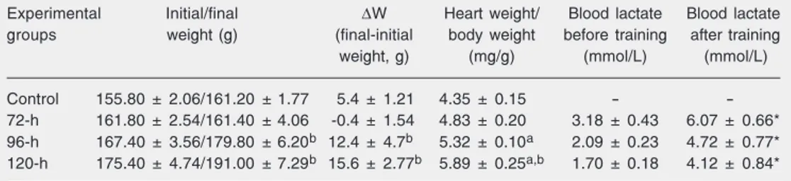
A significant increase in body weight was evident after the fourth and fifth days of training compared with the group that was trained for 3 days but not compared to the control group (Table 1), probably due to a slight weight loss in the 72-h group. The heart weight was also significantly increased after 4 and 5 days of training (96-h and 120-h groups) compared to control. Histological evaluation showed no alterations in the ex-perimental groups compared to the control group (results not shown). Blood lactate lev-els were significantly increased after the training sessions in all trained groups com-pared to values obtained before the sessions for each day (Table 1, Figure 1). Animals seemed to adapt to training since blood lac-tate concentrations were reduced with the increase in the number of days of training before and immediately after all training sessions as well (Figure 1). Indeed, the blood lactate levels after training in the 120-h group were similar to the values of the 24-h group before training (Figure 1).
Table 1. Body and heart weights and blood lactate of rats before and after acute swimming training.
Experimental Initial/final ∆W Heart weight/ Blood lactate Blood lactate
groups weight (g) (final-initial body weight before training after training
weight, g) (mg/g) (mmol/L) (mmol/L)
Control 155.80 ± 2.06/161.20 ± 1.77 5.4 ± 1.21 4.35 ± 0.15 -
-72-h 161.80 ± 2.54/161.40 ± 4.06 -0.4 ± 1.54 4.83 ± 0.20 3.18 ± 0.43 6.07 ± 0.66* 96-h 167.40 ± 3.56/179.80 ± 6.20b 12.4 ± 4.7b 5.32 ± 0.10a 2.09 ± 0.23 4.72 ± 0.77* 120-h 175.40 ± 4.74/191.00 ± 7.29b 15.6 ± 2.77b 5.89 ± 0.25a,b 1.70 ± 0.18 4.12 ± 0.84*
Data are reported as means ± SEM for 5 rats per group.
aP < 0.05 compared to control; bP < 0.05 compared to the 72-h group; *P < 0.05 compared to values before
Myosin heavy chain isoform gene expression measured by mRNA levels
The changes in MHC-ß mRNA levels occurred after 3 days of training and were statistically significant and more evident on the fifth day (120 h). At the end of the training protocol there was a significant in-crease in mRNA MHC-ß (Figure 2). Data for the 24-h, 48-h, and 72-h groups were dispersed and are not shown.
Detection of matrix metallopeptidase activity
MMP activity was detected in the myo-cardium extract as a single ~60-kDa lytic band (Figure 3A). This activity was com-pletely abolished when the gels were incu-bated in substrate buffer with 15 mM EDTA (results not shown). Increased MMP activity became statistically significant in the 72-h group, with a further increase in the 96-h and 120-h groups (Figure 3B). The appearance of lytic bands with molecular weights of approximately 62 and 58 kDa suggested the activation of MMP-2, which was confirmed by Western blotting analysis with anti-MMP-2 antibodies (Figure 3C). Anti-MMP-1 and -3 gave no signals in blots (results not shown); thus, MMP-2 was the only MMP activity detected in this condition.
Discussion
In the present study, we used a training protocol previously reported to induce car-diac hypertrophy (12) in order to identify the molecular mechanisms involved in this pro-cess. In fact, myocardium hypertrophy was suggested by the significant increase in heart weight after 96 and 120 h. Morphological evaluation showed no signs of edema or inflammatory infiltration, which could in-crease tissue weight, even in the absence of cellular growth. The increase of body weight at the end of training in the 96- and 120-h groups compared to the 72-h group
corre-Figure 1. Adaptation of blood lactate concentration to training. Blood samples were taken be-fore and after each training ses-sion. Each point is the mean value (± SEM) for 5 animals. The symbols “a” and “b” represent data significantly different (P < 0.05) from the respective 24-h group after and before sessions, respectively (ANOVA followed by Tukey post-test).
Figure 2. Levels of MHC-ß gene expression in cardiac muscle before training (control), and af-ter 96 and 120 h of training. MHC expression was normal-ized to GAPDH expression. MHC = myosin heavy chain. *P < 0.01 compared to control (ANOVA followed by Dunnett post-test).
2.5
2.0
1.5
1.0
0.5
0.0
Control 96 120
Time (h)
MHC-(arbitrary units)
sponds to the physiological growth of rats. The lack of body weight gain in the first 3 days might be due to the stress during the adaptation period to the training protocol.
At the beginning of training (24 h) blood lactate was high, probably due to the inten-sity of the protocol and to stress. These results indicate that this protocol has an anaerobic component. However, lactate lev-els before and after the sessions were pro-gressively reduced with training, suggesting that animals became adapted.
The present results show that the slow myosin isoform MHC-ß gene was differ-ently expressed in response to acute swim-ming training. Training induced significant up-regulation of the MHC-ß gene expres-sion in the 120-h group compared to con-trols.
Both young and adult rats submitted to endurance training show a discrete increase
a a
a a
b
b b 12
11 10 9
8 7
6 5
4 3 2
1
10 20 30 40 50 60 70 80 90 100 110 120130 Before exercise session After exercise session
Time after the beginning of exercise training (h)
Plasma lactate
in the expression of the MHC-ß gene, which is usually compensated by a similar decrease in the expression of MHC-α (15,16). Al-though these transformations are poorly un-derstood, MacIntosh and colleagues (16) have shown that sympathectomized new-born rats have higher expression levels of MHC-ß compared to controls. This observa-tion suggests that in some way, the decrease in the activity of the sympathetic nervous system, which is a common adaptive
re-sponse to endurance training, may facilitate MHC-ß gene up-regulation.
We have shown here an increase in MHC-ß expression using an acute experimental training protocol. Little is known about the myosin light chain gene expression in acute exercise. Using a different protocol in which rats swam 75 min twice daily, 5 days per week for 6 weeks, Buttrick et al. (17) did not observe modification in the expression of the myosin light chain I gene. However, the expression of MHC-ß and -α genes was not studied in that investigation.
The anaerobic threshold might be achieved and even exceeded in the swim-ming training protocol used here. If this is true, it could affect MHC-α and -ß expres-sion. In fact, the results of blood lactate showed that the present protocol has an anaerobic component at least by the end of each training session, which could be an additional factor modifying gene expression. This is an interesting hypothesis that re-mains to be investigated.
The interventions that modify MHC ex-pression result in economy of energy con-sumption during the process of power pro-duction to supportthe additional mechanical effort. Therefore, the heart can support the increased afterload associated with the hy-pertensive state as well as the volume over-load due to endurance training (18,19).
Myocardial remodeling was also sug-gested by a significant increase in gelatinase activity in the 72-, 96-, and 120-h groups. Gelatinase activity was completely abolished by EDTA, and the presence of MMP-2 was confirmed by Western blotting. The appear-ance of a lytic band of about 60 kDa repre-sents the activated form of this 72-kDa ge-latinase (9). No other proteins having MMP activity were observed in the heart muscle.
Heart remodeling is normally a dynamic process that involves both collagen synthe-sis and degradation (9). It has been demon-strated that MMPs exist in latent forms in the normal myocardium, and they can usually
Figure 3. Detection of gelatinase in activity cardiac muscle extracts by zymography. After tissue extraction and SDS-PAGE with gelatin as a substrate, MMP-2 activity (~60 kDa) was detected as an increase in gelatinase activity with training time.A, Each lane corresponds to an individual sample (30 µg) from 1 animal for each group. B, The same bands were quantified by densitometry. *P < 0.05 compared to control (ANOVA followed by Dunnett post-test) C, Detection of MMP-2 by Western blotting. Proteins of cardiac tissue extracts were separated by 15% SDS-PAGE, transferred to a nitrocellulose membrane and probed with an anti-MMP-2 antibody. Lanes 1-3, Samples from 120-h group (20 µg). Lane 4, Pre-stained SDS-PAGE Standard Low Range (Bio-Rad). MMP = matrix metallopeptidase.
A
Control
24 h
48 h
72 h
96 h
120 h
0 1 2 3 4 5 6 7 8
120 96 72 48 24 Control
*
*
*
Time after the beginning of exercise training (h)
1 2 3 4
107
49.3
36.4
28.5
20 72
B
C
Area (x 10
)
be activated in vitro by trypsin or plasmin
(20). Increased MMP activity is necessary to permit tissue growth in order to support the increased metabolic needs during exer-cise.
The heart of rats can be biochemically and functionally remodeled depending on energy needs imposed not only on this tissue but on the whole body as well. These modi-fications are associated with the plasticity of MHC isoforms. At least in the heart, this early remodeling also involves the up-regu-lation and/or activation of MMP-2.
Acknowledgments
The authors would like to thank Dr. Vilmar Baldissera for critical discussion of the results as well as Jose Carlos Lopes, both from Departamento de Ciências Fisiológicas, Universidade Federal de São Carlos, São Carlos, SP, Brazil, and Antônio Garcia Jun-ior, Departamento de Histologia e Embrio-logia, Instituto de Ciências Biomédicas, Universidade de São Paulo, São Paulo, SP, Brazil, for excellent technical assistance and skills.
References
1. Pette D (2001). Historical perspectives: plasticity of mammalian skeletal muscle. Journal of Applied Physiology, 90: 1119-1124. 2. O’Neill DS, Zheng D, Anderson WK et al. (1999). Effect of
endur-ance exercise on myosin heavy chain gene regulation in human skeletal muscle. American Journal of Physiology, 45: 414-419. 3. Murphy AM (1996). Contractile protein phenotypic variation during
development. Cardiovascular Research, 31: 25-33.
4. Reggiani C, Bottinelli R & Stienen GJM (2000). Sarcomeric myosin isoforms: Fine tuning of a molecular motor. News in Physiological Sciences, 15: 26-33.
5. Eddinger TJ & Meer DP (1997). Myosin isoform heterogeneity in single smooth muscle cells. Comparative Biochemistry and Physiol-ogy. Part B, Biochemistry and Molecular Biology, 117: 29-38. 6. Goldspink G (1998). Selective gene expression during adaptation of
muscle in response to different physiological demands. Compara-tive Biochemistry and Physiology. Part B, Biochemistry and Molecu-lar Biology, 120: 27-39.
7. Baldwin KM & Haddad F (2001). Effects of different activity and inactivity paradigms on myosin heavy chain gene expression in striated muscle. Journal of Applied Physiology, 90: 345-357. 8. Bigard XA, Janmot C, Merino D et al. (1996). Endurance training
affects myosin heavy chain phenotype in regenerating fast-twitch muscle. Journal of Applied Physiology, 81: 2658-2665.
9. Cleutjens JPM, Kandala JC, Guarda E et al. (1995). Collagen re-modeling after myocardial infarction in the rat heart. Journal of Molecular and Cellular Cardiology, 27: 1281-1292.
10. Docherty AJ & Murphy G (1990). The tissue metalloproteinase family and the inhibitor TIMP: a study using cDNAs and recombinant proteins. Annals of the Rheumatic Diseases, 49: 469-479.
11. Scheuer J & Tipton CM (1977). Cardiovascular adaptations to physi-cal training. Annual Review of Physiology, 39: 221-251.
12. Hickson RC, Hammons GT & Holloszy JO (1979). Development and regression of exercise-induced cardiac hypertrophy in rats. Ameri-can Journal of Physiology, 236: 268-272.
13. Bradford MM (1976). A rapid and sensitive method for the quantita-tion of microgram quantities of protein utilizing the principle of pro-tein-dye binding. Analytical Biochemistry, 72: 248-254.
14. Laemmli UK (1970). Cleavage of structural proteins during the as-sembly of the head of bacteriophage T4. Nature, 227: 680-685. 15. Swoap SJ, Haddad F, Bodell P et al. (1994). Effect of chronic energy
deprivation on cardiac thyroid hormone receptor and myosin isoform expression. American Journal of Physiology, 266: 254-260. 16. MacIntosh AM, Mullin WM, Fitzsimons DP et al. (1986). Cardiac
biochemical and functional adaptations to exercise in sympathecto-mized neonatal rats. Journal of Applied Physiology, 60: 991-996. 17. Buttrick PM, Kaplan M, Leinwand LA et al. (1994). Alterations in
gene expression in the rat heart after chronic pathological and physiological loads. Journal of Molecular and Cellular Cardiology, 26: 61-67.
18. Alpert BS, Flood NL, Strong WB et al. (1982). Responses to ergom-eter exercise in a healthy biracial population of children. Journal of Pediatrics, 101: 538-545.
19. Swynghedauw B, Schwartz K, Lauer B et al. (1988). Striated muscle overload. European Heart Journal, 9: 1-6.