CLINICAL SCIENCE
Comparative study of earthquake-related and
non-earthquake-related head traumas using
multidetector computed tomography
Zhi-gang Chu,IZhi-gang Yang,I,IIZhi-hui Dong,ITian-wu Chen,IIIZhi-yu Zhu,IVHeng ShaoI
IDepartment of Radiology, West China Hospital, Sichuan University, 37#Guo Xue Xiang, Chengdu, Sichuan, China.IIState Key Laboratory of Biotherapy, West China Hospital, Sichuan University, 37#Guo Xue Xiang, Chengdu, Sichuan, China.IIISichuan Province Key Laboratory of Medical Imaging and Department of Radiology, Affiliated Hospital of North Sichuan Medical College, Nanchong, Sichuan, China.IVDepartment of Pathology, West China College of Stomatology, Sichuan University, Sichuan, China.
OBJECTIVE:The features of earthquake-related head injuries may be different from those of injuries obtained in daily life because of differences in circumstances. We aim to compare the features of head traumas caused by the Sichuan earthquake with those of other common head traumas using multidetector computed tomography.
METHODS:In total, 221 patients with earthquake-related head traumas (the earthquake group) and 221 patients with other common head traumas (the non-earthquake group) were enrolled in our study, and their computed tomographic findings were compared. We focused the differences between fractures and intracranial injuries and the relationships between extracranial and intracranial injuries.
RESULTS:More earthquake-related cases had only extracranial soft tissue injuries (50.7% vs. 26.2%, RR = 1.9), and fewer cases had intracranial injuries (17.2% vs. 50.7%, RR = 0.3) compared with the non-earthquake group. For patients with fractures and intracranial injuries, there were fewer cases with craniocerebral injuries in the earthquake group (60.6% vs. 77.9%, RR = 0.8), and the earthquake-injured patients had fewer fractures and intracranial injuries overall (1.5¡0.9 vs. 2.5¡1.8; 1.3¡0.5 vs. 2.1¡1.1). Compared with the non-earthquake group, the incidences of soft tissue injuries and cranial fractures combined with intracranial injuries in the earthquake group were significantly lower (9.8% vs. 43.7%, RR = 0.2; 35.1% vs. 82.2%, RR = 0.4).
CONCLUSION:As depicted with computed tomography, the severity of earthquake-related head traumas in survivors was milder, and isolated extracranial injuries were more common in earthquake-related head traumas than in non-earthquake-related injuries, which may have been the result of different injury causes, mechanisms and settings.
KEYWORDS: Earthquake; Cranio-maxillofacial trauma; Tomography; X-ray computed.
Chu ZG, Yang ZG, Dong ZH, Chen TW, Zhu ZY, Shao H. Comparative study of earthquake-related and non-earthquake-related head traumas using multidetector computed tomography. Clinics. 2011;66(10):1735-1742.
Received for publication onMay 8, 2011;First review completed onMay 23, 2011;Accepted for publication onJune 28, 2011
E-mail: [email protected] Tel.: 86 28 8542 3817
INTRODUCTION
Earthquakes are among the most destructive natural disasters and have been the cause of some of the world’s highest death tolls in peacetime.1 They usually occur abruptly, and, therefore, the ability to utilize resources promptly and effectively to reduce casualties is the main measure of a successful response. Earthquake trauma is diverse, but many victims suffer from crush injuries from building collapses.2,3Non-earthquake traumas, by contrast, usually do not include crush injuries. In light of the unique
presentations of earthquake-related trauma, it is necessary to study this type of injury. The features of earthquake-related injuries in abdominal, spinal, and pelvic trauma patients have been thoroughly described in previous studies.4-6
Non-earthquake-related head injuries have contributed significantly to trauma-related deaths, accounting for up to one half of all deaths.7 Similarly, in earthquake-related
traumas, head injuries account for 30% of fatalities.1,8
Although head trauma is common and deadly in both earthquake- and non-earthquake-related traumas, the fea-tures of the injuries may be different because of different injury circumstances. Accordingly, we retrospectively reviewed computed tomography (CT) data and compared the findings of head traumas caused by the Sichuan earthquake with traumas caused by other common factors in daily life with the aim of determining the distinctive features of earthquake-related head traumas.
Copyrightß2011CLINICS– This is an Open Access article distributed under
the terms of the Creative Commons Attribution Non-Commercial License (http:// creativecommons.org/licenses/by-nc/3.0/) which permits unrestricted non-commercial use, distribution, and reproduction in any medium, provided the original work is properly cited.
MATERIALS AND METHODS
Participants
The local institutional ethical review board approved the present study, and informed consent was waived because of the retrospective nature of this study. The study subjects were patients with head traumas caused by the Sichuan earthquake from the 12th to the 25th of May 2008 as well as head traumas caused by typical, non-earthquake-related traumas over a similar time frame from the 1st to the 30th of May 2009. To illustrate the features more clearly, we chose as a control group consecutive patients with non–earthquake-related head traumas at the same university hospital during a similar period one year after the major earthquake to avoid bias arising from the different seasons and the confounding influence of the earthquake–related situations. We applied the following inclusion criteria: 1) the patients should have had a definite history of head trauma, 2) the patients should have had no surgical treatment before undergoing a CT examination, 3) the CT scan should have covered all injured parts of the head, and 4) the CT image should have been of adequate quality for the diagnosis. Among the patients with earthquake-related head traumas, any patient whose trauma resulted from falling or from a traffic accident during the earthquake was excluded. Finally, 221 cases with earthquake-related head traumas and 221 cases with non-earthquake-related traumas were enrolled into this study.
CT Protocols
The head CT scans of 39, 308, and 95 patients were obtained using a Philips Brilliance 16 slice multiple detector
row CT scanner (Philips Medical Systems, Eindhoven, the Netherlands), a Siemens Somatom Sensation 16 and a Siemens Somatom Plus 4, and a multiple detector CT scan (Siemens Medical Systems, Forchheim, Germany), respec-tively. According to the injury site and physical examination, cranial, maxillofacial, and cranio-maxillofacial CT scans were performed in 248, 102, and 91 patients, respectively. The cranial and maxillofacial CT scans were performed from the base to the roof of the skull and from the supraorbital margin to the inferior margin of the chin, respectively. The scanning parameters were 120 kV, 200-440 mAs, 0.5-s rotation time, pitch of 0.891, and collimation of 1660.75 mm for the Philips
Brilliance 16–MDCT scanner; 120 kV, 200-250 mAs, 0.5-s rotation time, pitch of 0.85, and collimation of 1660.75 mm
for the Siemens Somatom Sensation 16-MDCT scan; and 120 KV, 180-260 mAs, 0.5-s rotation time, pitch of 1.5, and collimation of 460.75 mm for the Siemens Somatom Plus
4-MDCT scan. The lower current (200 or 180 mAs) was used for the maxillofacial CT scan. The reconstructed section thick-ness was 8 mm for the cranial CT and 1 mm for the maxillofacial CT. The sagittal and coronary images used to observe maxillofacial and basal fractures were reconstructed with a thickness of 1-3 mm.
Image review
All of the MDCT scans were reviewed by the authors on the Syngo workflow picture archiving and communicating system workstation (Siemens Medical Systems, Forchheim, Germany). The cranio-maxillofacial soft tissue, bones, and brain were reviewed, and the anatomic distributions or types of injuries were recorded. Discrepancies with regard to the interpretations were resolved by consensus. The window width and window level used in reviewing the images were as follows: soft tissue (W: 350 HU, L: 50 HU), bone (W: 3200 HU, L: 700 HU), and brain (W: 80 HU, L: 35 HU).
Injuries only involving the scalp and/or maxillofacial soft tissue were classified as extracranial soft tissue injuries. When the fractures were evaluated, the whole head was divided into cranial and maxillofacial regions. The anato-mical sites of the cranial region included the frontal, parietal, temporal, sphenoid, and occipital bones, and those of the maxillofacial region included the nasal and ethmoid bones, zygoma, orbit, maxilla, and mandible. The diagnosis of the fractures was achieved with transverse plane and
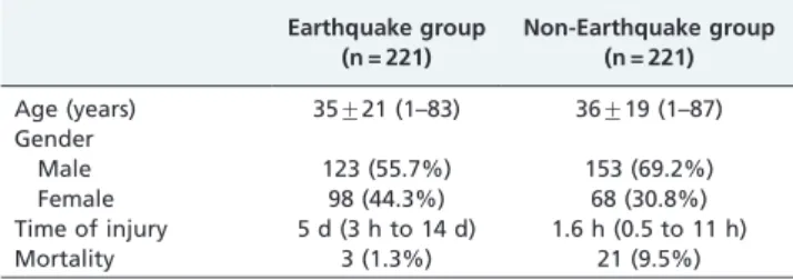
Table 1 -Baseline characteristics of patients in the earthquake and non-earthquake groups.
Earthquake group (n = 221)
Non-Earthquake group (n = 221)
Age (years) 35¡21 (1–83) 36¡19 (1–87)
Gender
Male 123 (55.7%) 153 (69.2%)
Female 98 (44.3%) 68 (30.8%)
Time of injury 5 d (3 h to 14 d) 1.6 h (0.5 to 11 h)
Mortality 3 (1.3%) 21 (9.5%)
Time of injury: the mean time from injury to CT scan.
multi-planar reformation (MPR) as well as with three-dimensional (3D) reconstructions (volume rendering, VR; surface shade display, SSD). Craniocerebral injury was traumatic injury involving the scalp, cranium, or intracra-nial structures (i.e., brain, meninges, and other structures), whereas intracranial injury was injury that only involved the intracranial structures. Intracranial injuries included extradural hematoma, subdural hematoma, subarachnoid hemorrhage, cephalophyma, pneumocephalus, cerebral edema and cerebral contusions, and lacerations.
Statistical analysis
The patients’ ages and sexes and the cause of injury for the patients with non-earthquake traumas were recorded. The numbers of patients with simple head soft tissue injuries, fractures, and intracranial injuries as well as the numbers of fractures and intracranial injuries in the patients were counted. Moreover, the proportions of soft tissue injuries, skull fractures, maxillofacial fractures, and cranio-maxillofacial fractures combined with intracranial injuries in both groups were calculated. The continuous variables were expressed as means¡standard deviation, and the
categorical variables were expressed as numbers and percentages. An independent-sample t-test and chi-square test were used to compare the continuous variables and the
categorical variables in both groups, respectively. Statistical analysis was performed with the SPSS statistical package (version 13.0 for Windows, SPSS Inc., Chicago, IL, USA). A two-tailed p-value of less than 0.05 was accepted as
indicating a statistically significant difference.
RESULTS
The baseline characteristics of the patients in the earth-quake and non-earthearth-quake groups are shown in Table 1. There were fewer male patients in the earthquake group than in the non-earthquake group (p,0.01). The patients’ age distributions in both groups are shown in Figure 1 with no significant differences (p.0.05), but there were more cases with ages ranging from 20 to 50 years in the non-earthquake group (p,0.01). The mean time from injury to CT scan among the earthquake patients was longer than that for the non-earthquake patients. The mortality was rela-tively high in the non-earthquake group. The non-earth-quake trauma causalities are summarized in Table 2.
Comparison of the types and the regions of head injuries
Extracranial soft tissue injuries, fractures, and intracranial injuries were detected in 112 (50.7%), 97 (43.9%), and 38 (17.2%) cases in the earthquake group, respectively, and we found 58 (26.2%), 117 (52.9%), and 112 (50.7%) cases in the non-earthquake group, respectively. The comparisons of different types of injuries between the two groups are shown in Figure 2. Earthquake victims had more extra-cranial soft tissue injury cases (50.7% vs. 26.2%, RR = 1.9,
p,0.001), but fewer patients had intracranial injuries (17.2% vs. 50.7%, RR = 0.3, p,0.001) than in the non-earthquake group. Among the earthquake victims with fractures and/ or intracranial injuries, maxillofacial fractures and cranio-cerebral injuries were detected in 60 (55.0%) and 66 (60.6%)
Table 2 -The reasons for head traumas in the non-earthquake group.
Reasons Patients (n) Percentage (%)
Traffic accident 138 62.4
Fall 34 15.4
Assault 25 11.3
Falling down 13 5.9
Crush 6 2.7
Others 5 2.3
cases, respectively. These same maxillofacial fractures and craniocerebral injuries were detected in 73 (44.8%) and 127 (77.9%) patients, respectively, in the non-earthquake group. Craniocerebral injuries were significantly less frequent in the earthquake group than in the non-earthquake group (60.6% vs. 77.9%, RR = 0.8,p,0.01).
Comparison of cranio-maxillofacial fractures
One hundred forty-one (mean: 1.5¡0.9 per patient,
range: 1–8) fractures were found in 97 patients in the earthquake group, and 294 (mean: 2.5¡1.8 per patient,
range: 1–9) fractures were found in 127 patients in the non-earthquake group. Among these patients, the cases in the earthquake group had fewer fractures than those in the non-earthquake group (p,0.001) (Figure 3). The fracture anatomic distributions in the cranial and maxillofacial regions for both groups are shown in Table 3. In the
earthquake and non-earthquake groups, the temporal bone, orbit, and nasal and ethmoid bones were the most commonly involved sites in the cranial and maxillofacial regions. Compared with the patients with non-earthquake-related fractures, patients with earthquake-non-earthquake-related fractures had more occipital bone and mandible fractures but fewer maxillary fractures (p,0.01).
Comparison of the intracranial injuries
Fifty (mean: 1.3¡0.5 per patient, range: 1–3) intracranial
injuries were found in 38 patients in the earthquake group, and 242 (mean: 2.1¡1.1 per patient, range: 1–5) intracranial
injuries were found in 112 patients in the non-earthquake group. Among these patients, the cases in the earthquake group had fewer intracranial injuries than did the patients in the non-earthquake group (p,0.001). The different intracranial injuries in both groups are shown in Table 4. The principal types of intracranial injuries in the earthquake and non-earthquake groups were subarachnoid hemorrhage versus cerebral contusions and lacerations, respectively. Cerebral contusions and lacerations were more common in the non-earthquake group than in the earthquake group (p,0.05). Moreover, contrecoup injuries (Figure 4) were common in the non-earthquake group, and intracranial injuries were usually close to cranial fractures or scalp injuries (Figure 5) in the earthquake group.
Comparison of the relationship between extracranial and intracranial injuries
In the earthquake group, 12 (9.8%) of 123 patients with soft tissue injuries but no fractures, 6 (12.2%) of 49 patients with maxillofacial fractures, 13 (35.1%) of 37 with cranial fractures, and 7 (58.3%) of 12 with cranio-maxillofacial fractures had intracranial injuries, respectively. In the non-earthquake group, 45 (43.7%) of 103 patients with soft tissue injuries but no fractures, 8 (18.2%) of 44 with maxillofacial fractures, 37 (82.2%) of 45 with cranial fractures and 22 (75.9%) of 29 cases with cranio-maxillofacial fractures had intracranial injuries, respectively. A comparison of intracra-nial injuries among the different types of extracraintracra-nial injuries is shown in Figure 6. Compared with the non-earthquake group, the incidence of soft tissue injuries and cranial fractures combined with intracranial injuries was significantly lower in the earthquake group (9.8% vs. 43.7%, RR = 0.2; 35.1% vs. 82.2%, RR = 0.4, eachp,0.001).
Figure 3 -(A) A 26-year-old man with earthquake-related head trauma caused by a collapsed building in the Sichuan earth-quake. The volume-rendering image shows fractures involving the right orbit, zygomata and maxilla. (B) A 40-year-old man with head trauma caused by a traffic accident. The volume-rendering image shows multiple fractures involving the bilateral orbits, zygomata, nasal bone, left maxilla and mandible, and the bilateral maxillas are detached.
Table 3 -Anatomical distributions of fractures in the cranial and maxillofacial regions.
Anatomic region Earthquake group Non-earthquake group
Cranial region 60 (42.6) 123 (41.8)
Frontal bone 12 (8.5) 27 (9.2)
Parietal bone 12 (8.5) 21 (7.1)
Temporal bone 18 (12.8) 47 (16.3)
Occipital bone* 14 (10.0) 11 (3.8)
Sphenoid bone 4 (2.8) 16 (5.4)
Maxillofacial region 81 (57.4) 171 (58.2)
Mandible * 16 (11.3) 15 (5.1)
Maxilla* 9 (6.4) 40 (13.6)
Zygoma 15 (10.6) 27 (9.2)
Orbit 20 (14.2) 46 (15.7)
Nasal and ethmoid bone 21 (14.9) 43 (14.6)
Data are expressed as n (%).
Comparison of the combined injuries of head traumas
In the earthquake group, 35 (15.8%) patients had a total of 48 combined injuries (mean: 1.4¡0.5 per patient, range: 1–3
injuries). In the non-earthquake group, 46 (20.8%) patients
had a total of 91 combined injuries (mean: 2.0¡0.8 per
patient, range: 1–4 injuries). The combined injuries in both groups are shown in Table 5.
Table 4 -Comparison of the intracranial injuries between both groups.
Types of injury Earthquake group Non-earthquake group
Extradural hematoma (EDH) 8 (16) 35 (14.5)
Subdural hematoma (SDH) 4 (8) 28 (11.6)
Subarachnoid hemorrhage (SAH) 18 (36) 60 (24.8)
Cephalophyma 6 (12) 23 (9.5)
Pneumocephalus 3 (6) 7 (2.9)
Cerebral edema 2 (4) 11 (4.5)
Cerebral contusion and laceration* 9 (18) 78 (32.2)
Data are expressed as n (%).
*p,0.05 between the earthquake and non-earthquake groups.
Figure 4 -A 30-year-old woman with head trauma caused by a collapsed building in the Sichuan earthquake. (A) There is an extradural hematoma in left frontal region. (B) A CT image shows a depressed fracture of the left front bone.
DISCUSSION
According to the clinical features of the patients in the earthquake and non-earthquake groups, we found that the age distributions in both groups were similar, but there were significantly more patients with ages ranging from 20– 50 years old in the non-earthquake group. Additionally, male patients were also more common in the non-earth-quake group than in the earthnon-earth-quake group. These differ-ences were consistent with previous reports.9,10 This
difference possibly reflects the tendency of young and middle-aged male patients to engage in riskier social behaviors.
We found that head traumas caused by the earthquake were possibly different from head traumas with other causes. Patients in the earthquake group had fewer overall fractures and intracranial injuries. Additionally, the earthquake group had more cases with extracranial soft tissue injuries, but fewer of these injuries coexisted with intracranial injuries. Finally, the extracranial injuries, especially the soft tissue injuries and cranial fractures, had a lower incidence of combined intracranial injuries in the earthquake group. Additionally, patients with head traumas in the earthquake group had
fewer combined injuries. Generally, the earthquake-related head injuries as depicted on CT were less serious.
Differences between the head traumas caused by the earthquake and other common traumas had a possibly direct relationship with the severity of the injuries. In an earthquake, head traumas commonly result from building collapses and falling objects,2,11-14whereas non-earthquake-related traumas are frequently caused by traffic accidents and falls.4,10,15,16 The energy of impact is significantly different between these two settings, and high-mass, low-velocity injuries may be more common among earthquake-related injuries than among non-earthquake injuries. In addition, patients in an earthquake have a little time to react to falling buildings. However, patients in traffic accidents or who have fallen are even more vulnerable, because the impact is so instantaneous that they have no time to protect themselves from the trauma, which perhaps makes these traumas more severe. In the present study, the head traumas in the earthquake group mainly resulted from building collapses, and the non-earthquake-related traumas from falls and traffic collisions were more serious than the traumas in the earthquake group.
Similarly, injury mechanisms could also account for the different CT findings between earthquake-related and non-earthquake-related head traumas. The crush injuries caused by building collapses do not involve notable head motion after the initial collision. However, everyday traumas, such as those resulting from traffic collisions, cause deceleration injuries, which can also result in countercoup injuries from the relative motion of the brain.10,17-19 Therefore, decelera-tion-related brain injuries are more common in non-earth-quake-related head traumas and seem to cause more complicated brain injuries, such as the brain contusions and lacerations seen in this study.
In addition, external conditions may also be responsible for the differences in head traumas. In the massive Sichuan earthquake, traffic accidents, complicated infrastructure, and aftershocks all delayed rescue to the outlying areas,
Figure 6 -Comparisons of the incidences of different types of extracranial injuries combined with intracranial injuries between these two groups.*p,0.001;**p.0.05.
Table 5 -Comparison of the combined injuries from head traumas.
Types
Earthquake group
Non-Earthquake group
Cervical vertebral fracture 3 (8.6) 11 (22.9) Thoracic vertebral fracture 8 (22.9) 13 (27.1) Lumbar vertebral fracture 6 (17.1) 9 (18.8)
Rib fracture 10 (28.6) 19 (39.6)
Clavicle fracture 3 (8.6) 5 (10.4)
Shoulder blade fracture – 7 (14.6)
Pulmonary contusion 2 (5.7) 15 (31.3)
Pelvic fracture 9 (25.7) 3 (6.3)
Fractures of extremities 7 (20.0) 9 (18.8)
and patients with severe injuries died before they could receive emergency services. By contrast, rescue in the downtown area of Chengdu city was more prompt, and, therefore, even severely injured patients could be quickly sent to hospitals for treatment. We recognized the signifi-cant difference in the time to treatment and CT scans between the earthquake and non-earthquake groups. Thus, the mortality for the earthquake group was lower because some severely injured patients died before arriving at the hospital, and there were more patients with severe injuries in the non-earthquake group, although some died even-tually.
Understanding the difference between head traumas caused by earthquake- and non-earthquake-related traumas is helpful for directing the specific diagnosis and treatment of injuries after an earthquake. As demonstrated in our study, patients in the non-earthquake group had more serious and complex head injuries. To diagnose their injuries, it was necessary to have them undergo CT examinations as quickly as possible. Treatment in the aftermath of an earthquake presents a special problem. The patient load in a local area will suddenly increase, resulting in a sudden excess demand for CT scanning for head traumas and other injuries. Under these conditions, selecting the patients who need urgent treatment will help improve the survival rate and optimize the use of limited medical resources. This study suggests that patients with suspected maxillofacial fractures have the greatest risk of intracranial injuries and, therefore, might deserve priority among head injury patients.
MDCT to examine the head is advantageous in an earthquake because it can save scanning time. The diagnosis is rapid, which will shorten the time required for lifesaving treatment.20 Additionally, high-quality MPR images can demonstrate extra-intracranial injuries accurately and thor-oughly and exclude the obvious intracranial injuries.21-23
Furthermore, patients with head traumas present with varying levels of consciousness. However, physical examina-tions cannot accurately depict the extent of severe trauma. In such conditions, the CT examination is quite necessary and remains the initial diagnostic modality of choice.
One limitation of this study should be mentioned again. Some of the patients with serious head traumas died before rescue workers could bring these patients to our university hospital. The Sichuan earthquake, the epicenter of which was 92 km (57 miles) from the hospital, damaged much of the infrastructure in the area, hampering rescue efforts directed to these outer suburban areas. The results of our study only address the patients who were stable enough to have survived the rescue effort and transport. Although we could not evaluate the injuries of the victims who died in the earthquake, the study of the survivors’ injuries is more meaningful and reflects how we can make an actual difference in an earthquake’s aftermath. Delayed rescue is common after a massive earthquake, and studying injuries in these conditions provides our medical teams with the practical knowledge that they will need to respond.
CONCLUSION
In the present study, we determined the features of earthquake-related head traumas by comparing the CT findings of head injuries caused by the Sichuan earthquake and by typical, non-earthquake-related events. Compared
with non-earthquake-related head traumas, the severity of earthquake-related traumas in survivors, as depicted on CT, was milder, and isolated extracranial injuries were more frequent, which may have been the result of the different injury causes, mechanisms and settings. A comparative study of the features of earthquake-related head injuries is helpful for understanding this common condition in such a special setting.
ACKNOWLEDGMENTS
This study was supported by the Science Foundation for Distinguished Young Scholars of Sichuan Province in China (Grant N˚2010JQ0039).
REFERENCES
1. Bulut M, Fedakar R, Akkose S, Akgoz S, Ozguc H, Tokyay R. Medical experience of a university hospital in Turkey after the 1999 Marmara earthquake. Emerg Med J. 2005;22:494-8, doi: 10.1136/emj.2004.016295. 2. Peek-Asa C, Kraus JF, Bourque LB, Vimalachandra D, Yu J, Abrams J.
Fatal and hospitalized injuries resulting from the 1994 Northridge earthquake. Int J Epidemiol. 1998;27:459-65, doi: 10.1093/ije/27.3.459. 3. Liao YH, Hwang LC, Chang CC, Hong YJ, Lee IN, Huang JH, et al.
Building collapse and human deaths resulting from the Chi-Chi Earthquake in Taiwan, September 1999. Arch Environ Health. 2003;58:572-8, doi: 10.3200/AEOH.58.9.572-578.
4. Chen TW, Yang ZG, Dong ZH, Chu ZG, Tang SS, Deng W. Earthquake-related crush injury versus non-earthquake injury in abdominal trauma patients on emergency multidetector computed tomography: a com-parative study. J Korean Med Sci. 2011;26:438-43, doi: 10.3346/jkms.2011. 26.3.438.
5. Dong ZH, Yang ZG, Chen TW, Chu ZG, Wang QL, Deng W, Denor JC. Earthquake-related versus non-earthquake-related injuries in spinal injury patients: differentiation with multidetector computed tomogra-phy. Crit Care. 2010;14:R236, doi: 10.1186/cc9391.
6. Chen TW, Yang ZG, Dong ZH, Tang SS, Chu ZG, Shao H. Earthquake-related pelvic crush fracture vs. non-earthquake fracture on digital radiography and MDCT: a comparative study. Clinics. 2011;66:629-34, doi: 10.1590/S1807-59322011000400018.
7. Hieu LC, Vander Sloten J, Bohez E, Phien HN, Vatcharaporn E, An PV, et al. A cheap technical solution for cranioplasty treatments. Technol Health Care. 2004;12:281-92.
8. Liang NJ, Shih YT, Shih FY, Wu HM, Wang HJ, Shi SF, et al. Disaster epidemiology and medical response in the Chi-Chi earthquake in Taiwan. Ann Emerg Med. 2001;38:549-55, doi: 10.1067/mem.2001. 118999.
9. Gassner R, Tuli T, Ha¨chl O, Rudisch A, Ulmer H. Cranio-maxillofacial trauma: a 10 year review of 9,543 cases with 21,067 injuries. J Craniomaxillofac Surg. 2003;31:51-61.
10. Rajendra PB, Mathew TP, Agrawal A, Sabharawal G. Characteristics of associated craniofacial trauma in patients with head injuries: An experience with 100 cases. J Emerg Trauma Shock. 2009;2:89-94, doi: 10.4103/0974-2700.50742.
11. Sheng CY. Medical support in the Tangshan earthquake: a review of the management of mass casualties and certain major injuries. J Trauma. 1987;27:1130-35, doi: 10.1097/00005373-198710000-00007.
12. Noji EK, Kelen GD, Armenian HK, Oganessian A, Jones NP, Sivertson KT. The 1988 earthquake in Soviet Armenia: a case study. Ann Emerg Med. 1990;19:891-7, doi: 10.1016/S0196-0644(05)81563-X.
13. Roces MC, White ME, Dayrit MM, Durkin ME. Risk factors for injuries due to the 1990 earthquake in Luzon, Philippines. Bull WHO. 1992;70:509-14.
14. Papadopoulos IN, Kanakaris N, Triantafillidis A, Stefanakos J, Kainourgios A, Leukidis C. Autopsy findings from 111 deaths in the 1999 Athens earthquake as a basis for auditing the emergency response. Br J Surg. 2004;91:1633-40, doi: 10.1002/bjs.4752.
15. Zargar M, Khaji A, Karbakhsh M, Zarei MR. Epidemiology study of facial injuries during a 13 month of trauma registry in Tehran. Indian J Med Sci. 2004;58:109-14.
16. Kamulegeya A, Lakor F, Kabenge K. Oral maxillofacial fractures seen at a Ugandan tertiary hospital: a six-month prospective study. Clinics. 2009;64:843-8, doi: 10.1590/S1807-59322009000900004.
17. Maas AI, Stocchetti N, Bullock R. Moderate and severe traumatic brain injury in adults. Lancet Neurol. 2008; 7:728-41, doi: 10.1016/S1474-4422(08)70164-9.
18. Hardman JM, Manoukian A. Pathology of head trauma. Neuroimaging Clin N Am. 2002;12:175-87, doi: 10.1016/S1052-5149(02)00009-6. 19. Poirier MP. Concussions: assessment, management, and
20. Gralla J, Spycher F, Pignolet C, Ozdoba C, Vock P, Hoppe H. Evaluation of a 16-MDCT scanner in an emergency department: initial clinical experience and workflow analysis. AJR Am J Roentgenol. 2005;185:232-8.
21. Bar-Am Y, Pollard RE, Kass PH, Verstraete FJ. The Diagnostic Yield of Conventional Radiographs and Computed Tomography in Dogs and Cats with Maxillofacial Trauma. Vet Surg. 2008;37:294-9, doi: 10.1111/j. 1532-950X.2008.00380.x.
22. Dos Santos DT, Costa e Silva AP, Vannier MW, Cavalcanti MG. Validity of multislice computerized tomography for diagnosis of maxillofacial frac-tures using an independent workstation. Oral Surg Oral Med Oral Pathol Oral Radiol Endod. 2004;98:715-20, doi: 10.1016/j.tripleo.2004.09.012. 23. Livingston DH, Lavery RF, Passannante MR, Skurnick JH, Baker S,