Category verbal fl uency performance
may be impaired in amnestic mild
cognitive impairment
Márcio Luiz Figueredo Balthazar
1, Fernando Cendes
2, Benito Pereira Damasceno
3Abstract – To study category verbal fl uency (VF) for animals in patients with amnestic mild cognitive impair-ment (aMCI), mild Alzheimer disease (AD) and normal controls. Method: Fifteen mild AD, 15 aMCI, and 15 normal control subjects were included. Diagnosis of AD was based on DSM-IV and NINCDS-ADRDA criteria, while aMCI was based on the criteria of the International Working Group on Mild Cognitive Impairment, using CDR 0.5 for aMCI and CDR 1 for mild AD. All subjects underwent testing of category VF for animals, lexical semantic function (Boston Naming-BNT, CAMCOG Similarities item), WAIS-R forward and backward digit span, Rey Auditory Verbal Learning (RAVLT), Mini-Mental Status Examination (MMSE), and other task relevant functions such as visual perception, attention, and mood state (with Cornell Scale for Depression in Dementia). Data analysis used ANOVA and a post-hoc Tukey test for intergroup comparisons, and Pearson’s coeffi cient for correlations of memory and FV tests with other task relevant functions (statistical signifi cance level was p<0.05).
Results: aMCI patients had lower performance than controls on category VF for animals and on the backward digit span subtest of WAIS-R but higher scores compared with mild AD patients. Mild AD patients scored sig-nifi cantly worse than aMCI and controls across all tests. Conclusion: aMCI patients may have poor performance in some non-memory tests, specifi cally category VF for animals in our study, where this could be attributable to the infl uence of working memory.
Key Words: verbal fl uency, mild cognitive impairment, Alzheimer disease, neuropsychological tests.
A fl uência verbal para categoria pode estar alterada no comprometimento cognitivo leve amnéstico Resumo – Estudar a fl uência verbal (FV) para a categoria animais no comprometimento cognitivo leve am-néstico (aCCL), doença de Alzheimer (DA) leve e controles normais. Método: Incluímos 15 pacientes com DA leve, 15 com aCCL e 15 controles normais, usando os critérios DSM-IV, NINCDS-ADRDA e CDR 1 para DA, e os do International Working Group on Mild Cognitive Impairment, e CDR 0,5 para aCCL. Todos os sujeitos passaram por avaliação da FV para a categoria animais, função léxico-semântica (Teste de nomeação de Boston - TNB, item de Similaridades do CAMCOG), extensão de dígitos direto e indireto do WAIS-R, aprendizado auditivo-verbal de Rey (TAAVR), Mini-Exame do Estado Mental (MEEM), e de outras funções (contraprovas) capazes de infl uenciar nestes testes, como percepção visual, atenção e estado de humor (este com a Escala Cor-nell para Depressão em Demência). A análise dos dados usou o teste de análise de variância (ANOVA) seguido do teste de Tukey post hoc para comparações entre os grupos e o coefi ciente de Pearson para correlação entre testes e contraprovas (nível de signifi cância p<0,05). Resultados: Os pacientes com aCCL tiveram performance inferior à dos controles nos testes de FV para animais e na extensão de dígitos indireta do WAIS-R. Pacientes com DA leve tiveram performance inferior à de sujeitos com aCCL e controles em todos os testes. Conclusão:
Pacientes com aCCL tiveram desempenho rebaixado em testes de fl uência verbal para animais, o que pode ter sido infl uenciado pela memória operacional.
Palavras-chave: fl uência verbal, comprometimento cognitivo leve, doença de Alzheimer, testes neuropsicoló-gicos.
1Postgraduate Neurologist. 2Associate Professor. 3Professor, Department of Neurology, Medical School, State University of Campinas - SP, Brazil.
Mild cognitive impairment (MCI) is a clinical entity in patients with objective cognitive problems (most often episodic memory) but without impairment in daily life ac-tivities,1 having a greater likelihood of transforming into dementia, most often Alzheimer disease (AD), than in the normal population.2 MCI can be classifi ed according to the clinical presentation of symptoms into amnestic MCI (aMCI), multiple domain or single non-memory domain MCI.1,2 Thus, by defi nition, aMCI presents with exclusive memory defi cit, sparing other cognitive domains such as language, visuospatial perception or executive functions. Nonetheless, aMCI individuals may present some non-memory-related poor performance in specifi c neuropsy-chological tests, following a pattern similar to AD,3 and continue to be classifi ed as amnestic rather than multiple domains MCI. This classifi cation is based on the clinical judgment that poor performance in one test is not enough to consider an entire cognitive domain as impaired.
Verbal fl uency (VF) for animal’s names is a simple and widely used task that can reveal impairment in early phases of AD,4 where a recent study points to impairment even in aMCI.3 Category VF involves several cognitive aspects, such as semantic knowledge, executive function and work-ing memory. Henry et al. suggested that verbal fl uency is “an excellent way of evaluating how subjects organize their thinking and ability to “organize output in terms of clusters of meaningfully related words”.5
Our aim was to compare verbal fl uency (category: ani-mals) in healthy controls and patients diagnosed as aMCI and mild AD, hypothesizing that these two groups of pa-tients have similar performance, because impairment of this function is common even in early stages of AD.
Methods
We studied 45 subjects, comprising 15 with aMCI and 15 with mild AD attended at the Unit for Neuropsychol-ogy and Neurolinguistics (UNICAMP Clinic Hospital), along with 15 controls. Routine laboratory examinations for dementia assessment (including B12 and folate dos-age, sorology for syphilis, thyroid hormones) and brain computed tomography were carried out in all patients. The local ethics committee approved this research.
MCI in our clinic is a clinical diagnosis carried out by trained neurologists using a standardized mental sta-tus battery and was based on the following criteria of the International Working Group on Mild Cognitive Impair-ment:1 (i) the person is neither normal nor demented; (ii) there is evidence of cognitive deterioration shown by either objectively measured decline over time and/or subjective report of decline by self and/or informant in conjunction with objective cognitive defi cits; and (iii) activities of daily
living are preserved and complex instrumental functions are either intact or minimally impaired. We included only patients older than 50 years who had a CDR (Clinical De-mentia Rating)6 of 0.5. This classifi cation was performed by using a semi-structured interview.
For probable AD diagnosis, we used the criteria of the National Institute of Neurological and Communicative Disorders and Stroke (NINCDS) and Alzheimer’s Disease and Related Disorders Association (ADRDA)7, including only patients classifi ed as CDR 1. Exclusion criteria were history of other neurological or psychiatric diseases, head injury with loss of consciousness, use of sedative drugs within 24 hours of the neuropsychological assessment, drug or alcohol addiction and prior exposure to neuro-toxic substances. The control group consisted of subjects with CDR 0 and no previous history of neurological or psychiatric disease, or memory complaints.
Neuropsychological evaluation comprised the follow-ing tests:
1) Verbal fl uency (VF) for animals’ category (the score was the total number of different animal names given the by patient in one minute).
2) Mini Mental Status Examination (MMSE),8 Brazilian version.
3) Episodic memory was evaluated using the Rey auditory verbal learning test (RAVLT).9
4) Boston Naming Test(BNT- translated and culturally adapted version for Brazilian population by Dr. Cân-dida Camargo – Psychiatry Institute, Medicine School, University of São Paulo).10 The BNT score was the sum of spontaneous correct responses plus correct responses following a semantic cue.
5) CAMCOG’s subscale of similarities between pairs of nouns.11 The patients were asked “ In what way are they
alike?” for the pairs apple/banana, chair/table, shirt/ dress and animal/vegetable. The score was calculated as the number of correct responses (zero to two for each pair; maximum score 8).
6) Visual perception subtests of Luria’s Neuropsychologi-cal Investigation12 (LNI; maximum score 20).
7) Attention: The forward and backward digit span subtest of WAIS-R.13
8) Cornell Scale for Depression in Dementia14 (CSDD).
Data analysis by means of Systat software used ANOVA and a post-hoc Tukey tests for intergroup comparisons of demographic and cognitive scores, as well as Pearson coef-fi cient for correlation between tests. Statistical signicoef-fi cance considered was p<0.05.
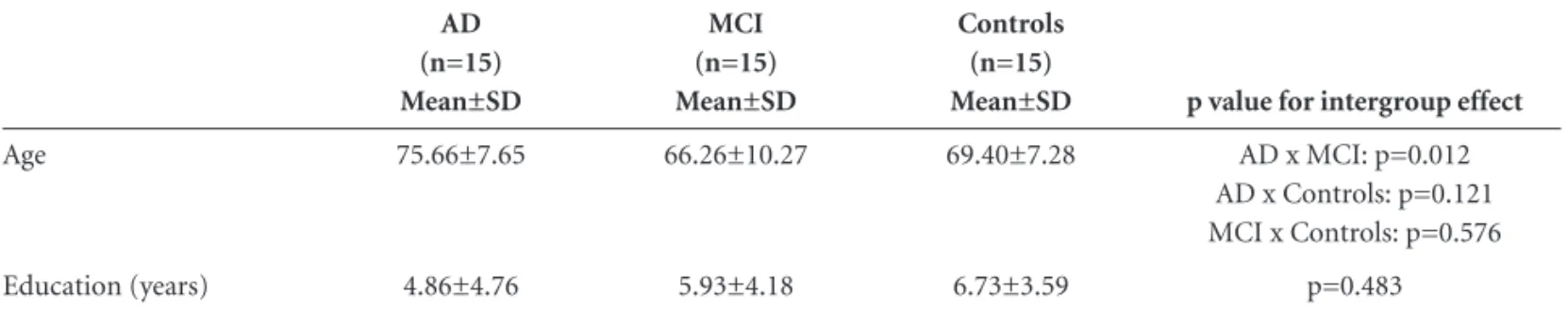
Table 1. Demographics results of amnestic mild cognitive Impairment (AMCI), Alzheimer disease (AD), and normal control subjects.
AD (n=15) Mean±SD
MCI (n=15) Mean±SD
Controls (n=15)
Mean±SD p value for intergroup effect
Age 75.66±7.65 66.26±10.27 69.40±7.28 AD x MCI: p=0.012
AD x Controls: p=0.121 MCI x Controls: p=0.576
Education (years) 4.86±4.76 5.93±4.18 6.73±3.59 p=0.483
Table 2. Neuropsychological results of amnestic mild cognitive Impairment (AMCI), Alzheimer disease (AD), and normal control subjects.
AD (n=15) Mean±SD
MCI (n=15) Mean±SD
Controls (n=15)
Mean±SD P value intergroups
MMSE 22.53±3.06 26.86±2.50 29.06±0.70 AD x MCI: p<0.001
AD x Controls: p<0.001 MCI x Controls: p=0.034
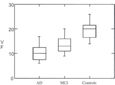
VF 10.20±3.44 13.86±3.85 19.46±3.31 AD x MCI: p=0.019
AD x Controls: p<0.001 MCI x Controls: p<0.001
BNT 38.73±8.64 51.06±7.78 53.66±4.11 AD x MCI: p=0.001
AD x Controls: p<0.001 MCI x Controls: p=0.582
A7- RAVLT 1.00±1.25 4.26±2.54 9.40±3.20 AD x MCI: p=0.002
AD x Controls: p<0.001 MCI x Controls: p<0.001
Similarities 4.86±1.80 7.00±1.19 7.33±1.04 AD x MCI: p<0.001
AD x Controls: p<0.001 MCI x Controls: p=0.789
fDS 4.73±1.03 4.60±0.82 4.93±0.79 p=0.583
bDS 3.13±0.51 3.13±0.99 3.93±1.09 AD x MCI: p=1.000
AD x Controls: p<0.05 MCI x Controls: p<0.05
Visuo-spatial LNI 17.33±1.39 18.80±1.01 18.66±1.11 AD x MCI: p=0.004
AD x Controls: p=0.01 MCI x Controls: p=0.949
Results
The results of demographic data are shown in Table 1 and neuropsychological evaluation in Table 2. aMCI subjects were similar to controls in age (p=0.576) and education (p=0.483). aMCI subjects performed similar to controls in CAMCOG’s item of similarities (p=0.789) and Boston Naming Test (p=0.582) but performed worse than controls in verbal fl uency (p<0.001), MMSE (p=0.034), backward digit span (p<0.05), delayed recall (p<0.001) of RAVLT, CAMCOG’s item of similarities (p=0.789) and Boston Naming Test (p=0.582).
AD patients were older than aMCI (p=0.012) but not control subjects (p=0.121). The educational level of the AD group was lower than that of controls (though not statisti-cally signifi cant). These patients scored lower than controls and aMCI subjects on all tests, except the forward digit span. The cognitive performance of mild AD was worse than aMCI, which in turn was poorer than controls.
The analysis of relationships between tests in the groups showed statistically signifi cant correlations only between VF and RAVLT delayed recall in the AD group (r=0.545; p<0.05) and between VF and BNT in the aMCI group (r= 0.540; p<0.05). In AD group, FV tended to correlate to BNT, but not reaching statistical signifi cance (p=0.066). Scores on the Cornell Scale for Depression did not corre-late to any of the cognitive tests: F (2,42)=0.929; p=0.403.
Discussion
Our fi ndings showed that aMCI patients performed worse than controls but better than mild AD on the cat-egory VF task. This task involves not only speed and ease of word production, but also lexical-semantic fi eld selection, executive function and working memory, in keeping track of what words have already been said. Some authors have found poor performance on category VF in MCI patients, and have interpreted this fi nding as a degradation of se-mantic networks.15-17 We suggest that working memory and attention, rather than semantic or executive function defi -cits, may have infl uenced VF in our patients, since aMCI subjects had signifi cantly lower scores on backward digit span test yet normal performance in semantic and executive tasks (neither anamnesis nor objective cognitive tests used in our diagnostic process showed executive dysfunction in any patients classifi ed as aMCI). In fact, Perry et al.18 have shown that defi cits in attention are more prevalent than defi cits in semantic memory in early AD. Similarly, our re-sults on lexical semantic tests such as BNT and CAMCOG’s similarities, showed no difference between aMCI and con-trols. Thus, our fi ndings suggest that semantic knowledge is not impaired and cannot explain the poor performance of this group of patients in category VF.
AD patients’ low VF was correlated to their impaired RAVLT delayed recall. A plausible explanation for this fi nd-ing could be that our VF task partly depends on active re-trieval (lexical-semantic selection) of animals’ names from long-term declarative memory, also the case in the RAVLT delayed recall task. On the other hand, it is diffi cult to explain why VF was correlated to BNT in the aMCI group, since this group performed as well on the BNT as did controls. Never-theless, the fact that FV was correlated to BNT in the aMCI group and also tended to correlate in the AD group, suggests that both groups may have impairment of some linguistic competence involved in lexical-semantic selection, although this was not specifi cally tested in our study.
We have found that aMCI patients may have poor per-formance in some non-memory tests, specifi cally category VF for animals, and that this could be attributable to the infl uence of working memory. However, further studies using more comprehensive testing of VF, including a pho-nemic task, as well as more specifi c tests for executive func-tion and lexical-semantic selecfunc-tion in a larger sample are needed for more robust conclusions to be drawn.
Supported by grants from CAPES (Brazil).
References
1. Winblad B, Palmer K, Kivipelto M, et al. Mild cognitive im-pairment-beyond controversies, towards a consensus: report of the International Working Group on Mild Cognitive Im-pairment. J Intern Med 2004;256:240-246.
2. Petersen RC. Mild cognitive impairment as a diagnostic entity. J Intern Med 2004;256:183-194.
3. Murphy KJ, Rich JB, Troyer AK. Verbal fl uency patterns in am-nestic mild cognitive impairment are characteristic of Alzheim-er’s type dementia. J Int Neuropsychol Soc 2006;12:570-574. 4. Henry JD, Crawford JR, Phillips LH. Verbal fl uency
perfor-mance in dementia of the Alzheimer’s type: A metanalysis. Neuropsychologia 2004;42:1212-1222.
5. Estes WK. Learning theory and intelligence. Am Psychol 1974;29:740-749.
6. Morris JC. The clinical dementia rating (CDR): Current ver-sion and scoring rules. Neurology 1993;43:2412-2414. 7. McKhann G, Drachman D, Folstein M, Katzman R, Price D,
Stadlan EM. Clinical diagnosis of Alzheimer’s disease: report of the NINCDS-ADRDA Work Group under the auspices of Department of Health and Human Services Task Force on Alzheimer’s Disease. Neurology 1984;34:939-944.
8. Brucki SMD, Nitrini R, Caramelli P, Bertolucci PHF, Okamoto IH. Sugestões para o uso do mini-exame do estado mental no Brasil. Arq Neuropsiquiatr 2003;61:777-781.
10. Lezak MD. Neuropsychological Assessment. 3rd ed. New York:
Oxford University Press; 2004.
11. Roth M, Huppert FA, Tym E, Mountjoy CQ. The Cambridge Examination for Mental Disorders of the Elderly: CAMDEX. Cambridge University Press; 1988.
12. Christensen A-L. Luria’s Neuropsychological Investigation. 2nd ed. Copenhagen: Munksgaard; 1979.
13. Wechsler D. Wechsler Memory Scale - Revised: Manual. The Psychological Corporation. Hartcourt Brace Jovanovich Inc; 1987.
14. Alexopoulos GS, Abrams RC, Young RC, Shamoian CA. Cornell Scale for Depression in Dementia. Biol Psychiatry 1988;23:271-284.
15. Dudas RB, Clague F, Thompson SA, et al. Episodic and se-mantic memory in mild cognitive impairment. Neuropsy-chologia 2005;43:1266-1276.
16. Adlam AL, Bozeat S, Arnold R et al. Semantic knowledge in mild cognitive impairment and mild Alzheimer’s disease. Cortex 2006;42:675-684.
17. Radanovic M, Carthery-Goulart MT, Charchat-Fichman H, et al. Analysis of brief language tests in the detection of cognitive decline and dementia. Dement Neuropsychol 2007;1:37-45. 18. Perry RH, Watson P, Hodges JR. The nature and staging of