Computer-assisted 3D reconstruction
of the human basal forebrain complex
Lea Tenenholz Grinberg
1, Helmut Heinsen
2Abstract – The basal forebrain complex (BFC) is a small but intricate structure. Its organization and function is hard to investigate using conventional methods, especially in humans. By combining new methods of research we present a comprehensive overview of this complex, in order to better understand its function in normal and diseased brains. Methods: The right and left BFC of a 29-year-old male were reconstructed from gallcocyanin (Nissl) stained 440 µm-thick serial horizontal sections by using advanced computer-assisted 3D reconstruction
software. Results: The reconstructed components in the present case include Ch2, Ch3, Ch4am-al, Ch4i, Ch4p, juxtacommissural, Ayala’s medial (subpallidal) and lateral (periputaminal) subnuclei. These components are arranged in an arch-like course mainly beneath the anterior commissure. The bilateral volume of all subnuclei was 99.06 mm³, the left side accounting for 48.05 mm³. Some of the subnuclei exhibited volume asymmetry indices varying from 28.3 to 12.9%.The volume of Ayalas’ lateral or periputaminal nucleus was 9.7% higher on the right, than on the left side. Conclusions: Our methodological approach promises to be highly effi cient and reproducible in studying morphofunctional correlations in complex cognitive features
Key words: human, adults, models/ structural, substantia innominata, nucleus basalis of Meynert, quantitative analysis, neurons/cytology.
Reconstrução computadorizada tridimensional do complexo prosencefálico basal humano
Resumo – O complexo prosencefálico basal (CPB) é uma estrutura complicada, apesar de pequena. É difícil estudar sua organização e função por métodos convencionais, especialmente em humanos. Ao combinar novos métodos de investigação, apresentamos uma abordagem completa deste complexo, com objetivo de auxiliar a compreensão de sua função em cérebros controles e acometidos por doenças. Métodos: Os CPBs direito e esquerdo de um homem de 29 anos de idade foram reconstruídos tridimensionalmente com uso de um sof-tware potente, à partir de secções histológicas horizontais de 440 µm de espessura. Resultados: Os núcleos
reconstruídos no presente caso são: Ch2, Ch3, Ch4am-al, Ch4i, Ch4p, e os subnúcleos juxtacommissural, Ayala medial (ou subpalidal) e lateral (periputaminal). Essas estruturas se arranjam em forma de arco, praticamente acompanhando a comissura anterior. O volume bilateral de todas as estruturas é de 99.06 mm³, sendo que o lado esquerdo responde por 48.05 mm³. Alguns dos subnúcleos exibem índices de assimetria variando de 28.3 a 12.9%. O volume do subnúcleo de Ayala lateral ou periputaminal é 9.7% maior à direita. Conclusão: O método apresentado tem grande potencial de ser bastante efi ciente e reprodutível para estudos de correlação morfofun-cional relacionadas a características cognitivas complexas.
Palavras-chave: humano, adulto, modelos/estruturas, substância innominata, núcleo basal de Meynert, análise quantitativa, neurônios; citologia.
Large chromophilic neurons of the basal forebrain complex (BFC) are the major source of cholinergic in-nervation to isocortical and allocortical regions as well as to subcortical nuclei. The BFC is implicated in attention, memory and learning processes.1,2 Classically, this complex is subdivided into four cell groups: Ch1 corresponding to the medial septal nucleus; Ch2 and Ch3 corresponding to
the nucleus of vertical and horizontal limb of the diagonal band of Broca, respectively; and Ch4 also referred to as the nucleus basalis of Meynert.3,5 More recently, emphasis has been placed on another group of cells, the so-called Ayala´s nucleus.6,7
The BFC is affected in several neurodegenerative dis-orders including Alzheimer’s disease, Parkinson’s disease,
1LTG, MD, PhD, Department of Pathology of Faculty of Medical Sciences of University of Sao Paulo, Brazil; Israeli Institute of Education and Research Albert
Einstein of São Paulo, Brazil. 2HH, MD, Prof., Labor fuer Morphologische Hirnforschung der Klinik und Poliklinik fuer Psychiatrie und Psychotherapie.
Dr. Helmut Heinsen – Labor fuer Morphologische Hirnforschung der Klinik und Poliklinik fuer Psychiatrie und Psychotherapie, Oberduerrbacher Str.
carried out to date have been based on animal models com-prising mainly rodents and to a lesser extent, primates.12,13 Although these studies depict considerable details regard-ing neuroanatomy, connections and biochemical features, the human BFC is likely to display an even more specifi c degree of organization.14 In addition, considering the se-lective vulnerability of the cells of this complex shown in neurodegeneration, it is important to have a more detailed understanding of the human BFC in order to allow more specifi c investigations into this complex.
A 3D reconstruction of the human BFC was previ-ously published in 2006.7 Currently, further data based on bilateral horizontal sections will be presented in order to provide more detailed aspects on size, shape, parcellation and asymmetry of this complex.
Methods
The brain of a 29-year-old male, whose cause of death was pulmonary arrest, was formalin-fi xed within the fi rst 24 hours after death. The detailed procedure of fi xation, dehydration, celloidin mounting and gallocyanin staining of the brain was described by Heinsen et al.15 In brief, the frontal, parieto-occipital and most lateral part of the tem-poral lobes of the present case, were severed leaving the central parts of the tel- and diencephalon intact. This cen-tral part/block was dehydrated in graded series of ethanol solutions (70, 80, 96%) for 1 week per stage and soaked in celloidin. This hardened celloidin-embedded block was sectioned horizontally on a sliding microtome at a thick-ness of 440 µm. Every slice was stained with gallocyanin, dehydrated, coverslipped and mounted with Permount®, as outlined in detail by Heinsen et al.15
In addition, both hemispheres of a 66-year-old male were processed in an identical manner and serially cut in the coronal plane. The brain was removed 8hrs after death and the preservation of the tissue was excellent. However, bilateral artifacts at the caudal level of the substantia in-nominata prevented a 3D reconstruction of the BFC in this case.
Computer assisted 3D reconstruction
A total number of 50 consecutive gallocyanin stained sections of the central block containing all parts of the BFC were photographed with a digital SLR-camera with close-up lenses mounted. These pictures were imported into a
respectively, and Ch4 to the basal nucleus of Meynert.3,12,16 Ch1, the medial septal nucleus, was not reconstructed in this case. Furthermore, we adopted the terminology pro-posed by Simic et al.6 and Boban et al.17 for the medial and lateral parts of Ayala’s nucleus. Finally, we favored the term anterior commissure – juxtacommissural cells (for the sake of simplicity, called just juxtacommissural) for clus-ters of large basophilic neurons closely arranged around the anterior commissure.7 Subsequently, the outlines of all subnuclear components of the BFC, as well as the profi les of both fornices and anterior commissure were identifi ed in each gallocyanin stained section and traced manually on the digital pictures with the help of a graphic tablet. Amira® converts all the outlines into digital coordinates for generating a surface based upon the individual outlines. Moreover, the in-built modules of Amira® can calculate surface areas and volumes of 3D reconstructed objects.
The volumetric side differences of each subnucleus were expressed by the asymmetry index of Eidelberg and Gala-burda.18 Negative values represent right-sided volume pre-dominance, whereas positive values indicate the opposite.
Results
a.c.al., central anterolateral arteries; acc. ncl., accumbens.; Ayala lat., lateral or periputaminal part of Ayala’s nucleus; c.a., anterior commissure; Ch4am,
basal ncl. of Meynert, anteromedial part; Ch4i, basal ncl. of Meynert, intermediate part; Ch4p, basal ncl. of Meynert, posterior part; c.i., internal capsule;
cl, claustrum; fo, fornix; g.p., globus pallidus; g.p.e., external globus pallidus; g.p.i., internal globus pallidus; i.c., interstitial cells; III, third ventricle; i.o.,
olfactory islands; pt, paraterminal (subcallosal) gyrus; put, putamen; v.p., ventral pallidum; v.s., ventral striatum.
Figure 1. Horizontal gallocyanin stained section through the fornix and dorsomedial parts of the anterior commissure. Red line in Figure 6 in-dicates plane of section in Figure 1.
Figure 2. Horizontal gallocyanin stained section 4.4 mm ventral to plane of section indicated in Figure 1. Plane of section is indicated in Figure 6 by the white line.
Figure 3. Coronal gallocyanin stained section through the right hemisphere of a 66-year-old male. Figure 1-3, gal-locyanin-stained 440 µm-thick
celloi-din mounted sections.
paraterminal gyrus. The plane of section is slightly inclined to the right side; therefore the profi les of the anterior com-missure do not completely match. On both sides, the ante-rior commissure is abutting the ventral striatum rostrally and the globus pallidus laterally and caudally. The plane of section in Figure 2 is parallel to that in Figure 1, however, it is exactly 4.4 mm more ventral than the former one (see also white line in Figure 6). Mainly the right hemisphere is depicted in Figure 2 at a higher magnifi cation than in Fig-ure 1. The right fornical column and the lateral parts of the anterior commissure can be easily identifi ed. At this plane of section, the paraterminal gyrus is no longer visible since its ventrolateral extension, called the horizontal limb of the diagonal band of Broca, is following another direction, traversing the surface of the anterior perforate substance. The region between the caudal rim of the ventral striatum (or nucleus accumbens at this plane of section) and the ventral parts of the internal capsule, as well as the regions lateral to the fornix or medial to the outlines of the anterior commissure is called substantia innominata.
The substantia innominata comprises: conspicuous cell islands mainly composed of small-sized neurons (i.e., ol-factory islands), nuclei of the lateral hypothalamus that are cell-sparse and diffi cult to delineate, and irregular aggre-gates of rather large darkly stained (chromophilic) neurons that constitute the BFC. Some of these neuronal aggregates are closely associated with perforating branches of the cen-tral anterolateral arteries (Figure 2, a.c.a.l.). At this plane of section the latter are easily identifi able by conspicuous perivascular spaces.
The subnuclei of the BFC are parcellated according to
size, shape, density, and staining characteristics of their con-stituent neurons and by their topography. Three examples of cytological criteria are given in Figure 4A-C. From medial to lateral, the outlines of Ch3, Ch4am, lateral or peripu-taminal Ayala´s nucleus, Ch4i and Ch4p can be delineated in Figure 2. Juxtacommissural cells cannot be seen at both horizontal levels (Figures 1 and 2), however they can be identifi ed in the coronal plane of section in Figure 3.
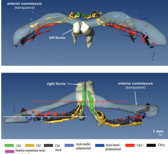
The complicated spatial arrangement of the BFC can be only perceived after a computer-assisted 3D reconstruction of the subnuclear profi les (Figures 5 and 6). In our 3D re-construction the most rostral parts of the lateral or peripu-taminal Ayala’s nucleus are found about 1.5 mm frontal to the plane of the most rostral parts of Ch4am. In a dorso-ventral view, the components of the BFC are mainly con-fi ned to the trajectory of the anterior commissure (Figure 5). Exceptions to this rule are Ch2 and dorsal Ch3 nuclei, as well as most of the components of Ayala´s nucleus, which are located rostral to the anterior commissure. Parts of the Ch4i and Ch4p are likewise not completely covered by the caudal rim of the anterior commissure. In a rostrocaudal view (Figure 6) it is clearly recognizable that the interme-diate parts of the anterior commissure and the Ch4am-al subnucleus take a disparate course. Consequently, the intermediate parts of these structures are separated by a wide cleft.
The juxtacommissural cells (Figure 3) are characterized by their close association with the anterior commissure. However, they are arranged in widely dispersed patches and do not form a continuous sheath around the internal capsule.
Ayala lateral 0.25 0.37 0.62 –0.097
Pericommisural 0.10 0.36 0.46 –0.283
Ch2* 3.9 2.27 6.17 0.132
Ch3† 17.27 14.86 32.13 0.038
Ch4am_al‡ 17.91 23.22 41.13 –0.129
Ch4i§ 4.45 3.92 8.37 0.032
Ch4p|| 3.65 5.44 9.09 0.099
Total 48.05 51.01 99.06 –0.015
*nucleus of the vertical limb of the diagonal band of Broca; †nucleus of the horizontal limb of the diagonal band of Broca; ‡ anteromedial-anterolateral parts of the nucleus basalis of Meynert; § intermediate part of the nucleus basalis of Meynert pars intermedia; ||posterior part of the nucleus
Figure 6. Basal forebrain complex in a fronto-occiptal perspective. Figure 5. Basal forebrain complex seen from dorsal after reconstruction with Amira.
We have excluded from our 3D reconstruction, large basophilic neurons that cluster apart from the Ch1 to Ch4 continuum. These interstitial (12;16) or outlying cells (19) can be easily recognized in coronal sections (Figure 3, i.c.). In a 66-year-old male a perivascular aggregate of large ba-sophilic cells proved to be continuous in serial sections with a more dorsal cell cluster at the ventrolateral side of the internal capsule.
The total volume of all components of the BFC is 99.06 mm³. Some of the components exhibit quite conspicuous side differences, e.g., the right juxtacommissural complex is 28.3% larger than the left one (Figure 4, Table 1). The left-sided Ch2 subnucleus is longer in the vertical axis (Figure 6) and its volume is 13.9% higher on the left, than on the right side (Table 1). The volumes of the Ch4am-al sub-nuclei are also asymmetric with 12.9% predominance of the right side. The asymmetry between the lateral (peripu-taminal) Ayala´s subnuclei are less marked. In the present case, the volume of the right-sided subnucleus was larger by 9.7% (Table 1).
Discussion
The subnuclei of the BFC are subject to age-related nerve cell loss, as well as early appearance of neurofi
bril-lary tangles in Alzheimer’s disease.20-26 In addition, neuro-nal loss in the BFC has been hypothesized to represent a common mechanism leading to dementia in some neuro-degenerative diseases.8 Therefore, its integrity is crucial for cognition, attention, and memory.
In a recent publication, we succeeded in correlating MRI signal changes with the coordinates of the Ch4am-al–Ch4p subnuclear continuum.27 This kind of point-to-point MRI-neuroanatomy correlation may be a signifi cant method-ological achievement to verify the integrity of the BFC and can be used for advanced in-vivo imaging methods aiming to diagnose early Alzheimer’s disease, as well as to monitor its progression or to study the effects of new drugs to slow down or to halt progression of Alzheimer’s disease.
of Ayala are located conspicuously apart from the Ch4 complex. Together with its particular cytoarchitectonics features (Figure 4B) and specifi c connections with Broca’s area,6 this spatial separation would be a further argument to categorize Ayala’s nucleus as an entity.
Several authors have described asymmetries in the neu-ronal number of the BFC.21,28-32 Special focus was directed on Ayala’s nucleus whose asymmetry was explained by its cholinergic axons to Broca’s speech area.6,17 We succeeded in visualizing size and shape differences of the periputam-inal or lateral Ayala’s subnucleus by computer-assisted 3D reconstruction.7 While only 3D reconstructions from the left hemisphere were available in our previous publication, both hemispheres were available for the current study. The lateral Ayala´s subnucleus is an inconspicuous nuclear group compared to the other subnuclei (Figures 5 and 6). In contrast with the observations of Simic et al.6, the right Ayala´s nucleus was 9.7% bigger than left nucleus. On the other hand, the volume differences between the Ch2 and Ch4am-al subnuclei were far more expressed, ranging from 28.3 to 12.9% (Table 1). These asymmetries could either represent an extreme example of the well-known individual variability of the human CNS or result from uncertainties in the cytoarchitectonic parcellation of the outlines. Further studies involving a larger number of subjects are necessary to confi rm these asymmetry fi ndings. Lowes-Hummel et al.30 described a higher neuron number in the right BFC in their samples. Furthermore, such asymmetries could refl ect size differences in the cortical projection areas.21,22
We were unable to fi nd published volume data for the complete BFC in humans. However, Halliday et al.33 pub-lished bilateral Ch4 volumes varying from 76 to 154 mm³. This is in line with our present data of 58.6 mm³ in the case investigated. Previous authors have shown that the com-ponents of the BFC receive multivariate afferents and send their cholinergic axons to different brain regions.2,34-36,36-41 Therefore, considering our preliminary results, we believe it necessary to study the subnuclei of the BFC together with the regions connected to them. A combination of in-situ post-mortem MRI, computer-assisted 3D reconstructions and classical stereological analyses will help to avoid meth-odological errors and to correct shrinkage factors due to histological procedures. In addition, a statistical analysis of the quantitative results correlated with clinicofunctional data of the patients will facilitate unraveling the
morpho-Broschk for her excellent technical assistance.
References
1. Mesulam MM. The cholinergic contribution to neuromodula-tion in the cerebral cortex. Semin Neurosci 1995;7:297-307. 2. Heimer L, van Hoesen GW. The limbic lobe and its output
channels: implications for emotional functions and adaptive behavior. Neurosci Biobehav Rev 2006;30:126-147.
3. Mesulam MM, Mufson EJ, Wainer BH, Levey AI. Central cho-linergic pathways in the rat: an overview based on an alternative nomenclature (Ch1-Ch6). Neuroscience 1983;10:1185-1201. 4. Selden NR, Gitelman DR, Salamon-Murayama N, Parrish
TB, Mesulam MM. Trajectories of cholinergic pathways within the cerebral hemispheres of the human brain. Brain 1998;121:2249-2257.
5. Alonso JR, U HS, Amaral DG. Cholinergic innervation of the primate hippocampal formation: II. Effects of fi mbria/fornix transection. J Comp Neurol 1996;375:527-551.
6. Simic G, Mrzljak L, Fucic A, Winblad B, Lovric H, Kostovic I. Nucleus subputaminalis (Ayala): the still disregarded mag-nocellular component of the basal forebrain may be human specifi c and connected with the cortical speech area. Neuro-science 1999;89:73-89.
7. Heinsen H, Hampel H, Teipel SJ. Nucleus subputaminalis: neglected part of the basal nucleus of Meynert - Response to Boban et al: computer-assisted 3D reconstruction of the nucleus basalis complex, including the nucleus subputami-nalis (Ayala’s nucleus). Brain 2006;129:U1-U4.
8. Arendt T, Bigl V, Arendt A, Tennstedt A. Loss of neurons in the nucleus basalis of Meynert in Alzheimer’s disease, pa-ralysis agitans and Korsakoff ’s Disease. Acta Neuropathol 1983;61:101-108.
9. Sassin I, Schultz C, Thal DR, et al. Evolution of Alzheimer’s disease-related cytoskeletal changes in the basal nucleus of Meynert. Acta Neuropathol 2000;100:259-269.
10. Hauw JJ, Agid Y. Progressive supranuclear palsy (PSP) or Steele-Richardson-Olszewski disease. In: Dickson DW, edi-tor. Neurodegeneration: the molecular pathology of dementia and movement disorders Neurodegeneration: the molecular pathology of dementia and movement disorders. Basel: ISN Neuropath Press; 2003:103-114.
11. Dickson DW, Bergeron C, Chin SS, et al. Offi ce of rare dis-eases neuropathologic criteria for corticobasal degeneration. J Neuropathol Exp Neurol 2002;61:935-946.
innervation of cortex by the basal forebrain: cytochemistry and cortical connections of the septal area, diagonal band nu-clei, nucleus basalis (substantia innominata), and hypothala-mus in the rhesus monkey. J Comp Neurol 1983;214:170-197.
13. Geula C, Schatz CR, Mesulam MM. Differential localization of NADPH-diaphorase and calbindin- d(28k) within the cho-linergic neurons of the basal forebrain, striatum and brain-stem in the rat, monkey, baboon and human. Neuroscience 1993;54:461-476.
14. Mesulam MM, Hersh LB, Mash DC, Geula C. Differential cholinergic innervation within functional subdivisions of the human cerebral cortex: a choline acetyltransferase study. J Comp Neurol 1992;318:316-328.
15. Heinsen H, Arzberger T, Schmitz C. Celloidin mounting (embedding without infi ltration) - a new, simple and reli-able method for producing serial sections of high thickness through complete human brains and its application to ste-reological and immunohistochemical investigations. J Chem Neuroanat 2000;20:49-59.
16. Mesulam MM. Cholinergic pathways and the ascending re-ticular activating system of the human brain. Ann N Y Acad Sci 1995;757:169-179.
17. Boban M, Kostovic I, Simic G. Nucleus subputamina-lis: neglected part of the basal nucleus of Meynert. Brain 2006;129:2005-2006.
18. Eidelberg D, Galaburda AM. Symmetry and asymmetry in the human posterior thalamus. Arch Neurol 1982;39:325-332. 19. Hedreen JC, Struble RG, Whitehouse PJ, Price DL.
Topogra-phy of the magnocellular basal forebrain system in human brain. J Neuropathol Exp Neurol 1984;43:1-21.
20. Arendt T, Bigl V, Tennstedt A, Arendt A. Correlation between cortical plaque count and neuronal loss in the nucleus basalis in Alzheimer’s disease. Neurosci Lett 1984;48:81-85. 21. Arendt T, Bigl V, Tennstedt A, Arendt A. Neuronal loss in
dif-ferent parts of the nucleus basalis is related to neuritic plaque formation in cortical target areas in Alzheimer’s disease. Neu-roscience 1985;14:1-14.
22. Cullen KM, Halliday GM, Double KL, Brooks WS, Creasey H, Broe GA. Cell loss in the nucleus basalis is related to re-gional cortical atrophy in Alzheimer’s disease. Neuroscience 1997;78:641-652.
23. Cullen KM, Halliday GM. Neurofi brillary degeneration and cell loss in the nucleus basalis in comparison to cortical Al-zheimer pathology. Neurobiol Aging 1998;19:297-306. 24. Lehericy S, Hirsch EC, Cerverapierot P, et al. Heterogeneity
and selectivity of the degeneration of cholinergic neurons in the basal forebrain of patients with Alzheimer’s disease. J Comp Neurol 1993;330:15-31.
25. McGeer PL, McGeer EG, Suzuki J, Dolman CE, Nagai T. Ag-ing, Alzheimer’s disease, and the cholinergic system of the basal forebrain. Neurology 1984;34:741-745.
26. Whitehouse PJ, Price DL, Clark AW, Coyle JT, DeLong MR.
Alzheimer disease: evidence for selective loss of cholinergic neurons in the nucleus basalis. Ann Neurol 1981;10:122-126. 27. Teipel SJ, Flatz WH, Heinsen H, et al. Measurement of basal
forebrain atrophy in Alzheimer‘s disease using MRI. Brain 2005;128:2626-2644.
28. Amunts VV. Structural asymmetry of the basal nucleus of Meynert in men and women. Zh Nevrol Psikhiatr Im S S Kor-sakova 2006;106:50-54.
29. Doucette R, Ball MJ. Left-right symmetry of neuronal cell counts in the nucleus basalis of Meynert of control and of Alzheimer-diseased brains. Brain Res 1987;422:357-360. 30. Lowes-Hummel P, Gertz HJ, Ferszt R, Cervos-Navarro J. The
basal nucleus of Meynert revised: the nerve cell number de-creases with age. Arch Gerontol Geriatr 1989;8:21-27. 31. Vogels OJ, Broere CA, Ter Laak HJ, Ten Donkelaar HJ,
Nieu-wenhuys R, Schulte BP. Cell loss and shrinkage in the nucleus basalis Meynert complex in Alzheimer’s disease. Neurobiol Aging 1990;11:3-13.
32. Zubenko GS, Moossy J, Hanin I, Martinez AJ, Rao GR, Kopp U. Bilateral symmetry of cholinergic defi cits in Alzheimer’s disease. Arch Neurol 1988;45:255-259.
33. Halliday GM, Cullen K, Cairns MJ. Quantitation and three-dimensional reconstruction of Ch4 nucleus in the human basal forebrain. Synapse 1993;15:1-16.
34. Lewis PR, Shute CC. The cholinergic limbic system: projec-tions to hippocampal formation, medial cortex, nuclei of the ascending cholinergic reticular system, and the subfornical organ and supra-optic crest. Brain 1967;90:521-540. 35. Kievit J, Kuypers HG. Subcortical afferents to the frontal lobe
in the rhesus monkey studied by means of retrograde horse-radish peroxidase transport. Brain Res 1975;85:261-266. 36. Mesulam MM, Mufson EJ. Neural inputs into the nucleus
basalis of the substantia innominata (Ch4) in the rhesus monkey. Brain 1984;107:253-274.
37. Rye DB, Wainer BH, Mesulam MM, Mufson EJ, Saper CB. Cortical projections arising from the basal forebrain: a study of cholinergic and noncholinergic components employ-ing combined retrograde tracemploy-ing and immunohistochemi-cal loimmunohistochemi-calization of choline acetyltransferase. Neuroscience 1984;13:627-643.
38. Jones BE, Cuello AC. Afferents to the basal forebrain cholinergic cell area from pontomesencephalic - catecholamine, serotonin, and acetylcholine - neurons. Neuroscience 1989;31:37-61. 39. Russchen FT, Amaral DG, Price JL. The afferent connections
of the substantia innominata in the monkey, Macaca fascicu-laris. J Comp Neurol 1985;242:1-27.
40. Mesulam MM. The systems-level organization of cholinergic innervation in the human cerebral cortex and its alterations in Alzheimer’s disease. Prog Brain Res 1996;109:285-297. 41. Gaykema RP, Zaborszky L. Direct