Research Division, Heart Institute (InCor), São Paulo University Medical School - São Paulo/SP, Brazil.
E-mail: [email protected]
Received for publication on April 25, 2006. Accepted for publication on June 20, 2006
BASIC RESEARCH
LOW HEMATOCRIT IMPAIRS GASTRIC MUCOSAL
CO
2
REMOVAL DURING EXPERIMENTAL SEVERE
NORMOVOLEMIC HEMODILUTION
Daniel Perin, Ruy J. Cruz Jr, Eliezer Silva, and Luiz Francisco Poli-de-Figueiredo
Perin D, Cruz Jr. RJ, Silva E, Rocha-e-Silva M, Poli-de-Figueiredo LF. Low hematocrit impairs gastric mucosal co2
removal during experimental severe normovolemic hemodilution. CLINICS. 2006(5):445-52.
OBJECTIVE: The net effects of acute normovolemic hemodilution with different hemoglobin levels on splanchnic perfusion
have not been elucidated. The hypothesis that during moderate and severe normovolemic hemodilution, systemic and splanchnic hemodynamic parameters, oxygen-derived variables, and biochemical markers of anaerobic metabolism do not reflect the adequacy of gastric mucosa, was tested in this study.
METHODS: Twenty one anesthetized mongrel dogs (16 ± 1 kg) were randomized to controls (CT, n = 7, no hemodilution),
moderate hemodilution (hematocrit 2 5% ± 3%, n = 7) or severe hemodilution (severe hemodilution, hematocrit 15% ± 3%, n = 7), through an isovolemic exchange of whole blood and 6% hydroxyethyl starch, at a 20 mL/min rate, to the target hematocrit. The animals were followed for 120 min after hemodilution. Cardiac output (CO, L/min), portal vein blood flow (PVF, mL/min), portal vein-arterial and gastric mucosa-arterial CO2 gradients (PV-artCO2 and PCO2 gap, mm Hg, respectively) were measured throughout the experiment.
RESULTS: Exchange blood volumes were 33.9 ± 3.3 and 61.5 ± 5.8 mL/kg for moderate hemodilution and severe hemodilution,
respectively. Arterial pressure and systemic and regional lactate levels remained stable in all groups. There were initial increases in cardiac output and portal vein blood flow in both moderate hemodilution and severe hemodilution; systemic and regional oxygen consumption remained stable largely due to increases in oxygen extraction rate. There was a significant increase in the PCO2-gap value only in severe hemodilution animals.
CONCLUSION: Global and regional hemodynamic stability were maintained after moderate and severe hemodilution. However,
a very low hematocrit induced gastric mucosal acidosis, suggesting that gastric mucosal CO2 monitoring may be useful during major surgery or following trauma.
KEYWORDS: Gas tonometry. Hemodilution. Oxygen metabolism. Regional hemodynamics. Splanchnic perfusion.
INTRODUCTION
The increasing frequency of complex surgical proce-dures with extensive blood loss, the high costs of transfu-sion therapy, the shortages in blood banks and the risk of transmission of diseases such as acquired immune
defi-ciency syndrome and hepatitis have increased interest for the intraoperative use of acute normovolemic hemodilution (ANH).1,2
Acute normovolemic hemodilution refers to blood re-moval and simultaneous replacement with acellular fluids immediately before surgical procedures. Hemodilution could be classified according to the target hematocrit (Ht) as mild (hematocrit ≥ 30%), moderate (30 < hematocrit
≥ 20%), or severe (hematocrit < 20%).3 A decrease in
car-diac output. However, a decrease in tissue oxygen deliv-ery, particularly to splanchnic organs, is a possible unde-sirable by-product.
Hypoperfusion of the splanchnic bed has been impli-cated as the trigger of systemic inflammatory response due to bacterial or toxin translocation from the gut lumen to blood stream, and this phenomenon has been associated with the development of postoperative multiple organ dys-function and to a worsened outcome.4 The net effects of
ANH with different hemoglobin levels on splanchnic perfusion have not been elucidated. We tested the hypoth-esis that during moderate and severe normovolemic hemodilution, systemic and splanchnic hemodynamic pa-rameters, oxygen-derived variables, and biochemical mark-ers of anaerobic metabolism do not necessarily reflect the adequacy of gastric mucosal perfusion, which may be im-paired.
MATERIALS AND METHODS
The experimental protocol was approved by the Ethi-cal Review Board of the Institution with adherence to the “Principles of Laboratory Animal Care” formulated by the National Society for Medical Research and to the “Guide for the Care and Use of Animals” by the National Insti-tutes of Health.
Animal preparation
The present study was performed using 21 male mon-grel dogs, weighing 16.7 ± 0.7 kg. The animals were fasted for 12 hours before the study, with free access to water. Animals with a hematocrit below 30% were excluded. Anesthesia was induced with an intravenous injection of sodium pentobarbital, 25 mg/kg. After endotracheal intu-bation, the animals were mechanically ventilated. The ven-tilator was adjusted to achieve an initial arterial PCO2 be-tween 35 and 45 mm Hg, with a 1.0 inspired oxygen frac-tion throughout the experiment. Anesthesia was maintained with intravenous injections of fentanyl (0.005 mg/kg/h) and vecuronium bromide (0.15 mg/kg/h).
The right common femoral vein was catheterized for measurement of inferior vena cava pressure, for infusion of Ringer’s solution (4 mL/kg/h) during the experiment, and for volume replacement during hemodilution. A polyethylene cannula was inserted into the right common femoral artery for blood removal. The left common femo-ral artery was catheterized to measure the mean arterial pressure (MAP) and to collect samples for blood gas, pH, bicarbonate, base deficit, hematocrit, and hemoglobin analysis.
A 7.5 Fr flow-directed thermodilution fiberoptic pulmo-nary artery catheter with thermal filament (CCOmbo 744H7.5F, Edwards Swan-Ganz, Edwards Life and Sci-ences, Irvine, CA, USA) was introduced through the right external jugular vein and its tip placed into the pulmonary artery, guided by pressure monitoring and wave tracings. This catheter was connected to a cardiac computer (Vigi-lance, Edwards Life and Sciences) to measure mean pul-monary arterial pressure and cardiac output and to collect mixed venous samples for blood gas analysis. All pressure-measuring catheters were connected to pressure transduc-ers (Transpac Disposable Transducer, Abbott, Chicago, IL, USA) and then to a Biopac Data Acquisition System (Model MP100, Biopac Systems, Goleta, CA, USA) for continuous recording of systemic and pulmonary artery pressures.
Through a median laparotomy, the spleen was removed, and a polyethylene catheter was placed into the portal vein, through the splenic vein, to draw blood samples and moni-tor portal vein pressure. A transit time ultrasonic flowprobe (Transonic System Inc., Ithaca, NY, USA) was positioned around the portal vein and connected to a flowmeter (T206 Transonic Volume Flowmeter, Transonic Systems, Inc, Ithaca, NY, USA).
An orogastric tube was inserted into the stomach, and gastric lavage was performed using warm isotonic saline solution, until a clear fluid solution was obtained. A 16F TRIP® tonometry catheter was then introduced orally and
manually placed along the greater gastric curvature. This catheter was connected to a gas capnometer (Tonocap, model TC-200, Tonometrics, Datex-Egstrom, Finland) for gastric PCO2 measurement.
Hemodilution was produced progressively by removing blood from the right common femoral artery at a rate of 20 mL/min while replacing it with 6% hydroxyethyl starch (Plasmasteril, Fresenius Laboratórios Ltda., São Paulo, Bra-zil) pumped into the left common femoral vein at the same rate.
The total volume removed (TVR) necessary to obtain a given hematocrit (dHct) was calculated using the initial hematocrit (iHct) according to the following formula: TVR (mL) = [70 mL x weight (kg)] x [(iHct-dHct)/ (iHctc+dHct/2)]
A microhematocrit sample was obtained every 5 min-utes during the hemodilution to check exactly whether the desired hematócrito was attained.
Experimental design
measurements were obtained, and the animals were ran-domly assigned to 3 experimental groups: CT (control, n = 7), MHD (moderate normovolemic hemodilution, n = 7, hematocrit = 25% ± 3%) and SHD (severe normovolemic
hemodilution, n = 7, hematocrit = 15% ± 3%).
The animals were followed for 120 minutes after hemodilution and were euthanized by anesthesia overdose and hypertonic potassium chloride solution injection.
Measured variables
Mean systemic and pulmonary arterial pressures, heart rate, and mesenteric artery blood flow were continuously recorded. Cardiac output was determined using 3-mL bo-lus injections of isotonic saline at 20oC. Each
determina-tion was the arithmetic mean of 3 consecutive measure-ments when their differences did not exceed 10%.
Arterial, portal, and mixed venous base deficit, pH,
PCO2, oxygen tension, oxygen saturation, hemoglobin,
hematocrit, and bicarbonate levels, were obtained at base-line (BL), immediately after the end of hemodilution (HD0), and 30, 60, 90, and 120 minutes after hemodilution. All arterial, venous, and portal blood samples were analyzed by a Stat Profile Ultra Analyzer (Nova Biomedi-cal, Waltham, MA, USA).
Systemic and pulmonary vascular resistance (SVR and
PVR), oxygen delivery and consumption (DO2 andVO2),
and the systemic and splanchnic oxygen extraction ratios (O2ERsyst and O2ERsplanc) were calculated using stand-ard formulae.
Gastric mucosal PCO2 (PrCO2) was evaluated every 10
minutes. PCO2 gap was calculated as the difference
be-tween gastric mucosal and arterial PCO2.
Statistical methodology
Results are expressed as mean ± standard error of mean (SEM). Statistical analysis was performed using a Statis-tic Package for Social Sciences for Windows software (ver-sion 6.0, SPSS Inc., Chicago, IL, USA).
Differences between groups were analyzed using re-peated measures analysis of variance and post hoc Tukey’s test. Statistical significance was considered for P values less than 0.05.
RESULTS
Baseline measurements were similar between groups. Both moderate and severe acute normovolemic hemodilution promoted a sustained and significant reduc-tion in hematocrit levels, as planned (Figure 1). Mean
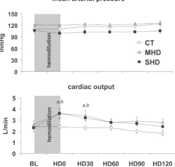
ar-terial pressure (Figure 2A), central venous, and pulmonary wedge pressures (not shown) showed no significant changes throughout the experimental protocol in all groups.
There was a significant increase in cardiac output after hemodilution in both MHD and SHD up to time point HD30; thereafter, this difference was no longer significant (Figure 2B).
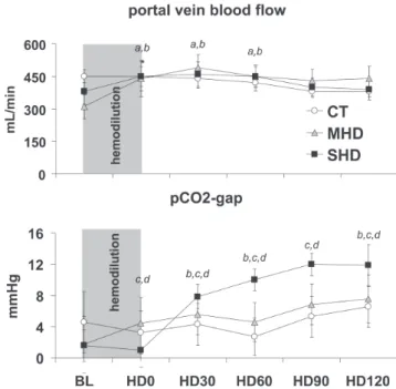
Hemodilution promoted a transient significant increase in portal vein blood flow in comparison to baseline values in the MHD and SHD groups at HD0, HD30, and HD60. However, there was no significant difference between groups when considered for the entire experimental time. (Figure 3A).
Figure 2 - (A) Mean arterial pressure during the experiment in the control (CT), moderate hemodilution (MHD), and severe hemodilution (SHD) groups. No significant changes between and within groups were observed. (B) Cardiac output during the experiment in the control (CT), moderate hemodilution (MHD) and severe hemodilution (SHD) groups. (a) MHD vs CT, P < 0.01; (b) SHD vs CT, P < 0.01.
cant changes during the entire experiment. Hemodilution promoted a sustained decrease in systemic and regional oxygen delivery, which was significantly greater in the SHD than in the MHD group. Systemic and regional oxygen con-sumption remained stable through increases in oxygen ex-traction rate. Additionally, systemic and regional lactate levels did not rise in the 3 groups.
There was no significant increase in the PCO2 gap in
the CT and MHD groups (P = 0.207). In contrast, there
was a significant increase in the PCO2 gap in the SHD
group (Figure 3B).
DISCUSSION
The main finding of this study, which is part of a gen-eral program of experimental analysis of fluid replace-ment5,6 was that both moderate and severe hemodilution are
associated with regional and systemic hemodynamic sta-bility. However, severe hemodilution, to a hematocrit of 15%, induces moderate gastric mucosal acidosis and an in-creased PCO2 gap. These findings may become relevant in the presence of a transient hemodynamic instability that may be induced by an intraoperative blood loss or surgi-cal maneuvers such as aortic unclamping and major can-cer resections.
Our experimental protocol promoted a predicted hematocrit reduction to the desired target levels, while
Table 1 - Systemic oxygen delivery, consumption and extraction (DO2, VO2 and O2ER, respectively) and arterial pH, base excess and lactate, in control (CT) moderate and severe hemodilution (MHD and SHD) groups
GROUPS BL HD0 HD30 HD60 HD120
Systemic DO2 CT 433± 26 442±32 398±43 414±49 318±30
MHD 535± 86 416±55a,b 384±63a,b 314±27a,b 304±32a,b
SHD 518±111 326±52a,b 271±22a,b 236±23a,b 202±30a,b
Systemic VO2 CT 75±11 80± 9 55± 3 73± 7 61± 9
MHD 68± 8 55±17 79±13 69±10 50±18
SHD 87±14 76±10 83± 6 70±10 78± 8
Systemic O2ER CT 18±3 18±2 15±1 18±2 20±3
MHD 14±2 14±5 21±4 23±4 20±7
SHD 19±4 27±4a,b,c 32±3 a,b,c 30±3 a,b,c 41±5 a,b,c
arterial pH CT 7.43±0.02 7.44±0.01 7.42±0.01 7.43±0.01 7.45±0.01
MHD 7.40±0.03 7.41±0.01 7.41±0.01 7.43±0.01 7.43±0.01
SHD 7.40±0.02 7.42±0.01 7.43±0.01 7.43±0.01 7.43±0.01
arterial BE CT 1.86±0.62 1.36±0.40 2.14±0.46 1.87±0.65 1.26±0.41
MHD 1.03±0.34 0.80±0.25 0.61±0.13 0.91±0.35 0.84±0.28
SHD 1.14±0.19 1.76±0.50 1.30±0.35 0.93±0.30 1.41±0.16
Systemic Lactate CT 1.2±0.1 1.1±0,1 1±0.2 0.8±0.2 0.9±0.2
MHD 1.3±0.4 1.1±0.3 1.3±0.2 1.1±0.3 1.1±0.3
SHD 1±0.3 0.5±0.2 0.7±0.2 0.8±0.2 0.8±0.3
Legends: (a) p<0.05 vs. CT; (b) p<0.05 vs. BL; (c) p<0.05 vs. MHD
Figure 3 – (A) Portal vein blood flow during the experiment in the control (CT), moderate hemodilution (MHD), and severe hemodilution (SHD) groups. (a) MHD vs CT, P < 0.05; (b) SHD vs CT, P < 0.05. (B) PCO2 gap during the experiment in the control (CT), moderate hemodilution (MHD), and severe hemodilution (SHD) groups. (d) SHD vs CT, P < 0.05; (c) MHD vs SHD, P < 0.05; (b) SHD vs CT, P < 0.05.
signifi-isovolemia was adequately maintained, as indicated by cen-tral venous and pulmonary wedge pressures. With un-changed cardiac filling pressures, arterial pressure was kept stable due to an increase in cardiac output, largely related to the reduction in systemic vascular resistance by the lower viscosity. These compensatory hemodynamic changes are expected.7 However, if a more severe anemia is induced,
impairment of myocardial oxygenation could jeopardize cardiac performance.8 In this situation, the compensatory
increment of cardiac output may not occur and conse-quently arterial pressure falls.
The mechanisms that increase cardiac output are also responsible for the increased portal venous blood flow, which was able to parallel cardiac output only because there was no hypovolemia. This fact is important in terms of blood flow distribution during normovolemic hemodilution. In our experimental model, the critical oxygen deliv-ery level to the various regional territories was probably not achieved. While systemic oxygen consumption did not vary significantly in the splanchnic region, it showed a trend toward reduction, from 12 to 6 mL/kg/min. As ex-tensively demonstrated, oxygen consumption decreases whenever critical oxygen delivery levels are not achieved.9
However, systemic and portal vein lactate levels were kept below 2 mmol/L, suggesting that anaerobic metabolism was not present. Lactate levels only increase when anaerobic metabolism is the main source of cellular energy and/or when there is an impairment of lactate clearance.10 As the
portal vein blood flow was normal or even increased, lac-tate clearance was maintained and, as already mentioned, the level of hemoglobin was not sufficient to trigger anaero-bic metabolism. Indeed, the level of venous oxygen satu-ration was higher than those related to critical oxygen
de-livery in models of normovolemic hemodilution.11,12 By the
end of the experiment, the oxygen extraction rate was around 40% (or oxygen venous saturation around 60%) in severe hemodilution. In a model of decreased cardiac out-put, the mean oxygen venous saturation that corresponded to the critical point in which VO2 becomes dependent on DO2 was around 25%.13 Hence, hemoglobin levels of about
5 mg/dL are still tolerated without evidence of anaerobic metabolism.11
Hypoperfusion of the splanchnic vascular bed has been implicated as the trigger of systemic inflammatory response and multiple organ dysfunction syndrome, which com-monly compromise the outcome after trauma or extensive surgeries.14,15 A decrease in splanchnic DO
2 caused by
hemodilution could amplify the harmful consequences of tissue perfusion heterogeneity during complex surgeries. The balance between oxygen delivery and consumption in the splanchnic area could be assessed by the arterial and portal oxygen saturation gradients, as well as by the por-tal lactate levels. Alternatively, the porpor-tal-arterial pCO2 gra-dient could be a surrogate marker of splanchnic blood flow and/or perfusion.16,17 As the portal blood flow was
main-tained throughout the experiment, the splanchnic oxygen delivery decreased exclusively because hemoglobin dropped. However, as we have observed in the systemic oxygen-derived variables, these hemoglobin levels were not low enough to induce anaerobic metabolism. Additional support for this evidence is the fact that splanchnic oxy-gen consumption was maintained and portal lactate levels did not increase.
In contrast, the behavior of the gastric mucosal-arterial PCO2 gradient was different. Indeed, the PCO2 gap did not increase significantly during the moderate isovolemic
Table 2 - Splanchnic oxygen delivery, consumption and extraction (DO2, VO2 and O2ER, respectively) and portal vein lactate levels, in control (CT) moderate and severe hemodilution (MHD and SHD) groups.
GROUPS BL HD0 HD30 HD60 HD120
Splanchnic DO2 CT 82±9.0 83±11.3 78±9.4 75±7.5 66±4.6
MHD 62±9.3 51±9.6a,b 54±7.3a,b 50±6.6a,b 48±6.6a,b
SHD 84±17.9 41±6.8a,b 40±5.6a,b,c 38±4.5a,b,c 32±4.9a,b,c
Splanchnic VO2 CT 8±1 9±1 6±1 8±1 10±1
MHD 10±3 6±2 5±2 11±3 8±1
SHD 12±3 6±2 6±2 7±1 6±1
Splanchnic O2ER CT 10±2 11±2 9±1 11±1 16±2
MHD 16±3 17±7 11±4 22±5 19±5
SHD 16±4 16±5 16±4 20±3 22±5
Portal vein Lactate CT 1.3±0.1 1.2±0.1 0.8±0.2 1±0.1 1.1±0.2
MHD 1.4±0.4 1.1±0.3 1.4±0.3 1.4±0.3 1.2±0.3
SHD 1.1±0.3 0.7±0.3 0.8±0.3 0.8±0.2 0.9±0.3
hemodilution, but did during severe isovolemic hemodilution. Additionally, after HD30, all subsequent time points showed significant increases in comparison to base-line. This alteration in the SHD group was not paralleled by splanchnic DO2, VO2, nor by portal vein blood flow, or by portal lactate levels. There are some explanations for this finding. Tissue PCO2 is linearly related to tissue blood flow over a large range of flows, especially if hematocrit remains stable. When tissue blood flow decreases, tissue PCO2 increases simply because the CO2 removal rate de-creases, particularly during aerobic metabolism.18,19
How-ever, if tissue blood flow is kept normal, or even if it is
increased, tissue CO2 removal can be affected by the
hemoglobin level. For the same blood flow, but in the
pres-ence of a lower tissue hematocrit, CO2 removal can
de-crease. This is the first explanation for the greater PCO2 gap observed in the present experiment. Secondly, severe isovolemic hemodilution may have induced anaerobic me-tabolism at the mucosal layer. Although portal lactate lev-els have not increased, this fact does not rule out the par-ticipation of anaerobic tissue CO2 production. In fact, there is no relationship between splanchnic metabolism and mu-cosal perfusion.20 Even when the gastric mucosal perfusion
is severely compromised, portal vein lactate levels could be normal, basically because the other tissues (muscular and/or serosa) could maintain aerobic metabolism. How-ever, when tissue DO2 falls below the critical point, there is a decrease in CO2 production by aerobic tissue, which counterbalances the anaerobically generated CO2. From our data, it is not possible to exclude the anaerobic CO2 gen-eration, since we did not measure mucosal PO2. However,
it has been shown that the main component of the PCO2
gap is flow, since in models of hypoxic hypoxia, the PCO2 gap is normal.21
Dubin et al performed a similar analysis in a sheep
model of isovolemic hemodilution or hemorrhage.22 They
induced a more severe isovolemic hemodilution, down to a 5% hematocrit. At this point, the PCO2 gap reached circa
15 mm Hg. As the authors compared this PCO2 gap with a
40 mm Hg PCO2 gap after hemorrhage, thereby reducing
blood flows, they concluded that severe isovolemic hemodilution is unable to reflect intestinal dysoxia. These results are in agreement with ours. Although we did not induce such an extreme decrease in hematocrit (15% vs. 5%), both models resulted in increased tissue PCO2, sug-gesting that low hemoglobin levels do not induce tissue dysoxia, but even though there microvascular derangement is serious enough to impair tissue CO2 clearance. In a clini-cal scenario, there are studies showing that a PCO2 gap greater 15 mm Hg is an early marker of poor prognosis.23,24
In a model of shock and resuscitation, Diebel et al have
shown that there is no substantial increase in mucosal PCO2 when a 18% hematocrit was induced after volume resusci-tation.25 Although these data apparently are in disagreement
to ours, we have noted that it is quite difficult to establish
the exact point at which tissue CO2 removal becomes
im-paired. Different experimental models and resuscitation protocols do interfere with the relationship between flow and hemoglobin. The combination of these factors will probably determine the point at which tissue CO2 accumu-lation begins. We believe that a direct comparison between these studies is difficult to accomplish due to marked dif-ferences between models, ie, normal versus hemorrhaged animals.
Care must be exercised in extrapolating our results to clinical practice. We used normal dogs submitted to a laparotomy and splenectomy and placement of flow probes and catheters. Our follow-up was very short. A long-term study performed in unanesthetized baboons evaluated the effects of withholding transfusion or lowering the transfu-sion trigger to a hematocrit of 15%, similar to our severe hemodilution model.26 These baboons adapted well to
se-vere anemia, and no adverse effects on long-term survival occurred. We believe that their findings are in agreement to ours, since they used normal baboons submitted to a sur-gical stress (laparotomy)26 which was similar to that in our
dogs. In our acute model, we observed systemic and re-gional hemodynamic stability, as well as the maintenance of the metabolic status. Only gastric perfusion was mod-erately affected. We consider this fact our major contribu-tion. We expect that, under a greater surgical stress or trauma, a more significant compromise of splanchnic perfusion may occur in our model as well as in the baboon model.
In spite of these limitations, we believe that our find-ings may be clinically relevant. Restrictive transfusion poli-cies have been widely recommended to avoid
transfusion-related reactions and immunoparalysis.27 Isovolemic
hemodilution may be used as a blood-saving technique for several procedures, usually those not commonly associated with hemodynamic instability, such as orthopedic surger-ies. Vascular surgeries could be an ideal scenario for isovolemic hemodilution. However, major vascular proce-dures are associated with abrupt hemodynamic changes and ischemia-reperfusion of large territories. In this scenario, isovolemic hemodilution has been associated with greater hemodynamic instability only during reperfusion.2
experiment whether this phenomenon may be also associated with tissue oxygen compromise that could lead to cellular de-rangement. Additionally, systemic and splanchnic hemodynamic parameters and biochemical markers of
anaero-bic metabolism did not reflect the adequacy of gastric mu-cosal perfusion. These data reinforce the need to monitor re-gional tissue perfusion in settings of severe hemodilution when larger surgical proceedings are performed.
RESUMO
Perin D, Cruz Jr. RJ, Silva E, Rocha-e-Silva M, Poli-de-Figueiredo LF. Hematócrito baixo compromete a remoção de CO2 da mucosa gástrica na hemodiluição normovolêmica intensa experimental. CLINICS. 2006(5):445-52.
OBJETIVO: Os efeitos da hemodiluição normovolêmica com diferentes níveis de hemoglobina na perfusão esplâncnica são pouco conhecidos. Testamos a hipótese que durante a hemodiluição moderada e acentuada, os parâmetros hemodinâmicos sistêmicos e regionais e as va-riáveis relacionadas ao metabolismo de oxigênio não re-fletem a adequação da perfusão da mucosa gástrica.
MÉTODOS: Vinte e um cães anestesiados com fentanil e vecurônio (16±1 kg) foram randomizados como controles (CT, n=7, sem hemodiluição normovolêmica), hemo-diluição normovolêmica moderada (Ht 25±3%, n=7) ou hemodiluição normovolêmica acentuada (Ht 15±3%, n=7), pela troca isovolêmica entre o sangue total e hidroxietil
amido a 6%, 20 mL/min até o hematócrito
pré-estabeleci-do para cada grupo. Os animais foram acompanhapré-estabeleci-dos por 120 min após a hemodiluição normovolêmica. Durante todo o experimento foram medidos o débito cardíaco (CO, L/ min), o fluxo de veia porta (PVF, mL/min), e os gradien-tes de CO2 veia porta-arterial e mucosa gástrica-arterial
(PV-artCO2 and PCO2-gap, mmHg, respectivamente).
RESULTADOS: O volume de sangue trocado foi de 33,9±3,3 mL/kg para hemodiluição normovolêmica mode-rada e de 61,5±5,8 mL/kg para a hemodiluição normovolêmica acentuada. A pressão arterial e os níveis de lactato sistêmico e regional permaneceram estáveis em to-dos os grupos. Houve aumentos iniciais de débito cardíaco e de fluxo de veia porta, tanto na hemodiluição normovolêmica moderada quanto na hemodiluição normovolêmica acentuada; o consumo de oxigênio sistêmico e regional permaneceram estáveis, principalmente por con-ta de aumentos na con-taxa de extração de oxigênio. O PCO2 -gap apresentou aumento significativo apenas nos animais submetidos a hemodiluição normovolêmica acentuada.
CONCLUSÃO: Ocorre estabilidade hemodinâmica global e regional tanto na hemodiluição normovolêmica modera-da quanto na acentuamodera-da. Entretanto, o hematócrito de 15% induziu acidose moderada de mucosa gástrica, o que pode ser relevante em procedimentos cirúrgicos de grande porte ou no trauma.
UNITERMOS: Hemodiluição. Hemodinâmica regional. Metabolismo do oxigênio. Perfusão esplâncnica. Tonometria a gás.
REFERENCES
1. Monk TG, Goodnough LT, Brecher ME, Pulley DD, Colberg JW, Andriole GL, et al. Acute normovolemic hemodilution can replace preoperative autologous blood donation as a standard of care for autologous blood procurement in radical prostatectomy. Anesth Analg. 1997;85:953-8.
2. Poli de Figueiredo LF, Mathru M, Tao W, Solanki D, Uchida T, Kramer GC. Hemodynamic effects of isovolemic hemodilution during descending thoracic aortic cross clamping and lower torso reperfusion. Surgery. 1997;122:32-8.
3. Stehling L, Zauder HL. Acute normovolemic hemodilution. Transfusion. 1991;31:857-68.
4. Swank GM, Deitch EA. Role of the gut in multiple organ failure: bacterial translocation and permeability changes. World J Surg. 1996;20:411-417.
5. Rocha-e-Silva R, Mansur AP, Fabri Junior J, Ramos RB, Cunha Filho CEC, Dallan LAO et al. Coronary revascularization with the left internal thoracic artery and radial artery: comparison of short-term clinical evolution between elective and emergency surgery. Clinics. 2005;60:227-232.
6. Rocha-e-Silva M, Poli de Figueiredo LF. Small volume hypertonic resuscitation of circulatory shock. Clinics. 2005; 60(2):159-172. 7. Spahn DR, Leone BJ, Reves JG, Pasch T. Cardiovascular and coronary
physiology of acute isovolemic hemodilution: a review of nonoxygen-carrying and oxygen-nonoxygen-carrying solutions. Anesth Analg. 1994;78:1000-21.
9. Connett RJ, Honig CR, Gayeski TE, Brooks GA. Defining hypoxia: a systems view of VO2, energetics and intracellular PO2. J Appl Physiol.
1990;68:833-842.
10. Severin PN, Uhing MR, Beno DW, Kimura RE. Endotoxin-induced hyperlactatemia results from decreased lactate clearance in hemodynamically stable rats. Crit Care Med. 2002;30:2509-14. 11. van Bommel J, Siegemund M, Henny CP, Trouwborst A, Ince C. Critical
hematocrit in intestinal tissue oxygenation during severe normovolemic hemodilution. Anesthesiology. 2001;94:152-60.
12. Räsänen J: Supply-dependent oxygen consumption and mixed venous oxyhemoglobin saturation during isovolemic hemodilution in pigs. Chest. 1992;101:1121-4.
13. Ronco JJ, Fenwick JC, Tweeddale MG, Wiggs BR, Phang PT, Cooper DJ, et al. Identification of the critical oxygen delivery for anaerobic metabolism in critically ill septic and nonseptic humans. JAMA. 1993;270:1724-30.
14. Ivatury RR, Simon RJ, Islam S, Fueg A, Rohman M, Stahl WM. A prospective randomized study of end points of resuscitation after major trauma: Global oxygen transport indices versus organ-specific gastric mucosal pH. J Am Coll Surg. 1996;183:145-54.
15. Donati A, Cornacchini O, Loggi S, Caporelli S, Conti G, Falcetta S, et al.. A comparison among portal lactate, intramucosal sigmoid Ph, and deltaCO2 (PaCO2 - regional Pco2) as indices of complications in patients
undergoing abdominal aortic aneurysm surgery. Anesth Analg. 2004;99:1024-31.
16. Poli de Figueiredo LF, Silva E, Cruz Jr RJ, Rocha e Silva M. Gas tonometry for evaluation of gastrointestinal mucosal perfusion. Experimental models of trauma, shock and complex surgical maneuvers – Part 1. Acta Cir Bras. 2002;17:281-8.
17. Garrido AG, Poli de Figueiredo LF, Cruz Jr RJ, Silva E, Rocha E Silva M. Short-lasting systemic and regional benefits of early crystalloid infusion after intravenous inoculation of dogs with live Escherichia coli. Braz J Med Biol Res. 2005;38:873-84.
18. Neviere R, Chagnon JL, Teboul JL, Vallet B, Wattel F. Small intestine intramucosal PCO(2) and microvascular blood flow during hypoxic and ischemic hypoxia. Crit Care Med. 2002;30:379-84.
19. Dubin A, Murias G, Estenssoro E, Canales H, Badie J, Pozo M, et al. Intramucosal-arterial PCO2 gap fails to reflect intestinal dysoxia in
hypoxic hypoxia. Crit Care. 2002;6:514-20.
20. Thoren A, Jakob SM, Pradl R, Elam M, Ricksten SE, Takala J. Jejunal and gastric mucosal perfusion versus splanchnic blood flow and metabolism: an observational study on postcardiac surgical patients. Crit Care Med. 2000;28:3649-54.
21. Vallet B, Teboul JL, Cain S, Curtis S. Venoarterial CO (2) difference during regional ischemic or hypoxic hypoxia. J Appl Physiol. 2000;89:1317-21.
22. Dubin A, Estenssoro E, Murias G, Pozo MO, Sottile JP, Baran M, et al. Intramucosal-arterial PCO2 gradient does not reflect intestinal dysoxia
in anemic hypoxia. J Trauma. 2004;57:1211-7.
23. Silva E, Kai M, Garrido AG, Blecher S, Martins PS, Assumpcao MS, et al. Gastric-arterial PCO2 gradient as an early marker of outcome in septic patients. Crit Care. 2005 (in press).
24. Levy B, Gawalkiewicz P, Vallet B, Briancon S, Nace L, Bollaert PE. Gastric capnometry with air-automated tonometry predicts outcome in critically ill patients. Crit Care Med. 2003; 31:474-80.
25. Diebel LN, Tyburski JG, Dulchavsky SA. Effect of acute hemodilution on intestinal perfusion and intramucosal pH after shock. J Trauma. 2000;49:800-05.
26. Levine E, Rosen A, Sehgal L, Gould S, Sehgal H, Moss G. Physiologic effects of acute anemia: implications for a reduced transfusion trigger. Transfusion. 1990;30:11-4.