The decreased oxygen uptake during
progressive exercise in
ischemia-induced heart failure is due to reduced
cardiac output rate
1Instituto do Coração, Faculdade de Medicina, 2Escola de Educação Física e Esporte, 3Instituto de Ciências Biomédicas, Universidade de São Paulo, São Paulo, SP, Brasil 4Department of Cardiology, Los Angeles Medical School, University of California,
Los Angeles, CA, USA N.P.L. Rolim3, K.C.Mattos2,
P.C. Brum2, M.V.C. Baldo3,
H.R. Middlekauff4
and C.E. Negrão1,2
Abstract
We tested the hypothesis that the inability to increase cardiac output during exercise would explain the decreased rate of oxygen uptake (VO2)in recent onset, ischemia-induced heart failure rats. Nine
nor-mal control rats and 6 rats with ischemic heart failure were studied. Myocardial infarction was induced by coronary ligation. VO2 was
measured during a ramp protocol test on a treadmill using a metabolic mask. Cardiac output was measured with a flow probe placed around the ascending aorta. Left ventricular end-diastolic pressure was higher in ischemic heart failure rats compared with normal control rats (17 ± 0.4 vs 8 ± 0.8 mmHg, P = 0.0001). Resting cardiac index (CI) tended to be lower in ischemic heart failure rats (P = 0.07). Resting heart rate (HR) and stroke volume index (SVI) did not differ significantly between ischemic heart failure rats and normal control rats. Peak VO2
was lower in ischemic heart failure rats (73.72 ± 7.37 vs 109.02 ± 27.87 mL min-1 kg-1, P = 0.005). The VO
2 and CI responses during
exercise were significantly lower in ischemic heart failure rats than in normal control rats. The temporal response of SVI, but not of HR, was significantly lower in ischemic heart failure rats than in normal control rats. Peak CI, HR, and SVI were lower in ischemic heart failure rats. The reduction in VO2 response during incremental exercise in an
ischemic model of heart failure is due to the decreased cardiac output response, largely caused by depressed stroke volume kinetics.
Correspondence
C.E. Negrão
Unidade de Reabilitação Cardiovascular e Fisiologia do Exercício, InCor, FM, USP Av. Dr. Enéas C. Aguiar, 44 05403-000 São Paulo, SP Brasil
Fax: +55-11-3069-5043 E-mail: [email protected] Research supported by FAPESP (No. 01/00009-0), and in part by Fundação Zerbini.
Received March 10, 2005 Accepted September 30, 2005
Key words
•Heart failure
•Oxygen uptake rate
•Cardiac index
•Coronary ligation
•Ischemia
Introduction
Heart failure is characterized by dyspnea on exertion and exercise intolerance (1-3). In acute heart failure, such as in the setting of acute myocardial ischemia, or hypertensive
ca-pacity in chronic heart failure has been at-tributed to abnormalities of the periphery, specifically the skeletal myopathy of heart failure, and abnormal muscle metabolism (5). A chain of events beginning with car-diac dysfunction and decreased carcar-diac out-put, which in turn leads to neurohumoral activation, cytokine release, and decreased muscle blood flow, all probably contribute to the skeletal myopathy of chronic heart failure. In patients with recent myocardial infarction and exercise intolerance, it is not known whether abnormal hemodynamic re-sponses during exercise, or skeletal myopa-thy, or both, contribute to exercise intoler-ance (6). That is, it is not known at what point central mechanisms of exercise dys-function (low cardiac output, and elevated filling pressures) give way to peripheral ab-normalities.
In the present study, we tested the hypo-thesis that at 5 weeks after myocardial in-farction, diminished cardiac output still con-tributes to exercise dysfunction. In previous studies of exercise and heart failure, cardiac output has not been directly evaluated nor continuously measured (7,8). In the present study, we measured oxygen uptake (VO2)
and cardiac output throughout progressive exercise until exhaustion in rats with ische-mia-induced heart failure, 5 weeks post-myocardial infarction. We reasoned that di-rect cardiac output measurements with a flow probe placed around the aortic ascend-ing portion on a beat-to-beat basis would further elucidate the actual role of cardiac output in the decreased VO2 in heart failure.
Material and Methods
Animals
The study was performed according to the Ethics Principles in Animal Research adopted by the Brazilian College of Animal Experimentation, and was approved by the Committee on Animal Research of the
Bio-medical Sciences Institute of the University of São Paulo. A total of 112 male Wistar rats were selected for the study. Seventy-eight rats (Medical School, University of São Paulo, Brazil; 330 g body weight, 5 rats/ cage) were used for the experimental proto-col, but only fifteen completed the study. The other rats did not complete the study because of death during the experimental procedures and the recordings were not ob-tained. The rats were fed standard laboratory chow and water ad libitum in a temperature-controlled room (22ºC) with a 12:12-h dark-light cycle.
Measurements and procedures
Induction of myocardial infarction. Myo-cardial infarction was induced by a coronary ligation technique described elsewhere (9). Briefly, the 3-month-old Wistar rats were anesthetized with ketamine (50 mg/kg, ip) and xylazine (10 mg/kg, ip), endotracheally intubated, and mechanically ventilated with room air (respiratory rate of 60-70 breaths/ min and tidal volume of 2.5 mL). Left thora-cotomy was performed and the left anterior descending coronary artery was ligated.
Measurements of cardiac output. Four weeks after myocardial infarction induction, the rats were submitted to a second thora-cotomy. Rats were anesthetized with keta-mine (50 mg/kg, ip) and xylazine (10 mg/kg,
1). Body temperature was maintained at 37ºC by heating with an infrared lamp. After sur-gery, rats received an antibiotic, 20,000 U penicillin, and were returned to their cages. During the experimental session, the micro-probe was connected to an ultrasonic flow-meter and then to a microcomputer for direct aortic flow velocity measurement. Cardiac output was recorded at 1000 Hz on a beat-to-beat basis using the AT/CODAS program (DataQ Instruments, Inc., Akron, OH, USA).
Measurement of oxygen consumption.
Exercise capacity is a common method to characterize cardiac reserve and function. In addition, the ramp exercise test avoids sud-den alterations in neuromuscular motor unit recruitment or metabolic changes associated with incremental protocols by employing a constant and continuous increase in external work. VO2 was measured by means of
ex-pired gas analysis during a ramp protocol of progressive exercise on a treadmill with 1-m/min increments every 40 s and no grade until exhaustion. In a previous study from our laboratory, the time spent by the sample gas in the mask to reach the gas analyzer was 35 s. In addition, we observed that the steady state during submaximal workloads was achieved at about 90 s. These findings justi-fied the use of the ramp protocol in the present study. Therefore, a metabolic mask adapted from Russell et al. (10) was used (Figure 1). The mask fit over the face of the rat and was attached behind the ears with a latex collar. A small length of polyethylene tubing (PE-280, 3 mm ID) was attached to the top portion of the mask in such a way that ambient air was drawn unidirectionally into the mask from around the rat’s head and exhausted through the tubing just above the rat’s nose. The rats were conditioned to the metabolic mask from 6 weeks of age to the time of the experimental procedure. Gas anal-ysis was performed using an oxygen (S-3A/ I) analyzer (Ametek, Pittsburgh, PA, USA). VO2 was calculated using the measured flow
through the metabolic mask, the expired
fraction of effluent oxygen and the fraction of oxygen in room air.
Left ventricular pressure measurement.
Rats were anesthetized with ketamine (50 mg/kg, ip) and xylazine (10 mg/kg, ip) for measurements of left ventricular end-dia-stolic pressure (LVEDP). A fluid-filled cath-eter (PE10) was inserted into the right ca-rotid artery and advanced retrogradely into the left ventricle. LVEDP signals were re-corded at 1000 Hz on a beat-to-beat basis using AT/CODAS (DataQ Instruments). A strain gauge transducer (Statham P23Db, Hato Rey, Puerto Rico) was used for arterial pressure measurement. The transducer sig-nal was fed to an amplifier (GPA-4 model Z Stemtech, Inc., Wood Dale, IL, USA) and further to a 10-byte analog-to-digital con-verter. Only animals with LVEDP ≥15 mmHg
were included in the ischemic heart failure group. These values of LVEDP were associ-ated with an infarcted area of 40% (data not shown). Contractile function was studied by directly monitoring left ventricular dP/dt.
Experimental protocol
One month after surgery for the induc-tion of myocardial infarcinduc-tion, an ultrasonic perivascular flow probe was placed around the ascending aorta according to the proce-dure described above. Five days after sur-gery, one cannula was inserted into the left
ventricle for LVEDP measurements and the study was started 24 h after cannula implan-tation. VO2 and cardiac output were then
continuously evaluated during a progressive exercise test until exhaustion. Heart rate (HR) was counted from cardiac output pulses. Stroke volume was calculated from the divi-sion of cardiac output by HR, and the arte-riovenous oxygen difference (a-vO2 diff.)
was determined by the Fick principle.
Numerical and statistical analysis
Given the nonlinearity in the VO2 and
car-diac index (CI) temporal responses during continuous measurements, we employed the Marquardt-Levenberg algorithm (imple-mented by means of the SigmaPlot for Win-dows software, version 4.01, SPSS Inc., Point Richmond, CA, USA) to fit the sigmoidal equation to the experimental data points. The physiological meaning of this curve can be determined on he basis of the four param-eters that characterize the equation, where:
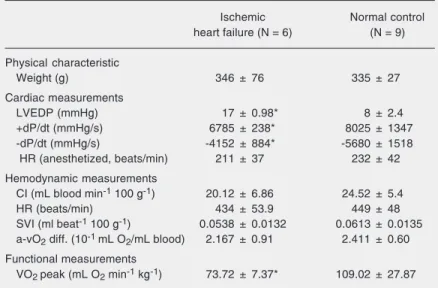
Table 1. Baseline cardiovascular parameters of rats with ischemic heart failure before treadmill exercise.
Ischemic Normal control
heart failure (N = 6) (N = 9)
Physical characteristic
Weight (g) 346 ± 76 335 ± 27
Cardiac measurements
LVEDP (mmHg) 17 ± 0.98* 8 ± 2.4
+dP/dt (mmHg/s) 6785 ± 238* 8025 ± 1347
-dP/dt (mmHg/s) -4152 ± 884* -5680 ± 1518
HR (anesthetized, beats/min) 211 ± 37 232 ± 42
Hemodynamic measurements
CI (mL blood min-1 100 g-1) 20.12 ± 6.86 24.52 ± 5.4
HR (beats/min) 434 ± 53.9 449 ± 48
SVI (ml beat-1 100 g-1) 0.0538 ± 0.0132 0.0613 ± 0.0135
a-vO2 diff. (10-1 mL O2/mL blood) 2.167 ± 0.91 2.411 ± 0.60
Functional measurements
VO2 peak (mL O2 min-1 kg-1) 73.72 ± 7.37* 109.02 ± 27.87
Data are reported as mean ± SD. LVEDP = left ventricular end-diastolic pressure; HR = heart rate during measurements of LVEDP; CI = cardiac index; SVI = stroke volume index; a-vO2 diff. = arterial-mixed venous oxygen difference; VO2 = oxygen
consump-tion.
*P ≤ 0.05 compared to control (unpaired Student t-test).
y0is the baseline (lower asymptote) of the
variable under study (i.e., VO2, CI), x0is the
time the variable takes to achieve its maxi-mal slope (latency measurement), a is calcu-lated by the difference between upper and lower asymptote (∆ of change) and b is a parameter used to calculate the curve’s gain (a/4b). This sigmoidal function is given by the equation:
In the present study, the four parameters defining a sigmoidal curve were obtained from the best fit to each individual set of VO2 and CI experimental points. A mean
sigmoidal curve, representing an experimen-tal group, was obtained by averaging its parameters over the individual curves be-longing to that group. Regarding the HR and stroke volume index (SVI) measures, their temporal profiles were rectilinear enough to yield a simple linear regression. For each parameter, obtained from either linear or non-linear regression, the statistical comparison between heart failure and normal control groups was done by the unpaired Student t-test, with the level of significance set at α = 0.05. Data are reported as means ± SD.
Results
Baseline measurements
0.07). HR and SVI were not significantly different in ischemic heart failure rats and normal controls. a-vO2 diff. was similar for
the two groups.
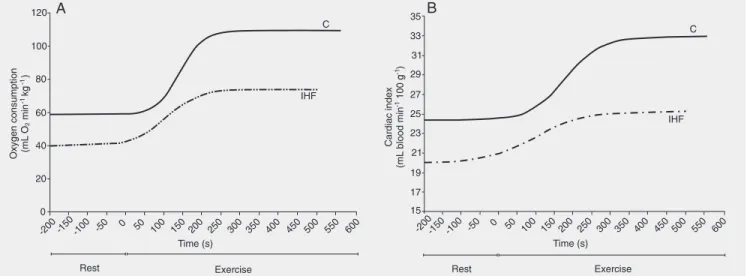
Temporal responses of oxygen consumption during exercise
The temporal responses of VO2 during
progressive exercise in rats with ischemic heart failure and normal control rats are shown in Figure 2. In both groups, the VO2
response during the incremental exercise test displayed a sigmoidal pattern, which could be observed by a delayed VO2 increase at the
onset of exercise paralleled by VO2 leveling
off at the offset of exercise. The VO2 slope
during progressive exercise was significant-ly lower in ischemic heart failure rats than in normal control rats (0.29 ± 0.15 vs 0.66 ± 0.48 mL O2 min-1 kg-1 s-1, P = 0.049). In
addition, the VO2 peak was significantly
decreased in rats with ischemic heart failure (73.72 ± 7.37 vs 109.02 ± 27.87 mL O2 min-1
kg-1, P = 0.005). Indeed, ∆VO
2 (a
param-eter) was significantly decreased in ischemic heart failure rats compared to normal con-trols (33.97 ± 7.64 vs 50.73 ± 17.31 mL O2 min-1 kg-1, P < 0.05).
Temporal hemodynamic responses during exercise
The temporal responses of CI during ex-ercise in ischemic heart failure rats and nor-mal controls are shown in Figure 2. The relationship between CI and workload throughout progressive exercise also dis-played a sigmoidal behavior for both groups. Similar to VO2, theincrement of CI was
attenuated at the onset and at the offset of exercise. The CI slope during progressive exercise was significantly lower in ischemic heart failure rats (0.08 ± 0.05 vs 0.25 ± 0.27 mL blood min-1 100 g-1 s-1, P = 0.051). In
addition, peak CI was significantly lower in ischemic heart failure rats than in normal controls (25.42 ± 4.21 vs 33.03 ± 6.75 mL blood min-1 100 g-1, P = 0.018).
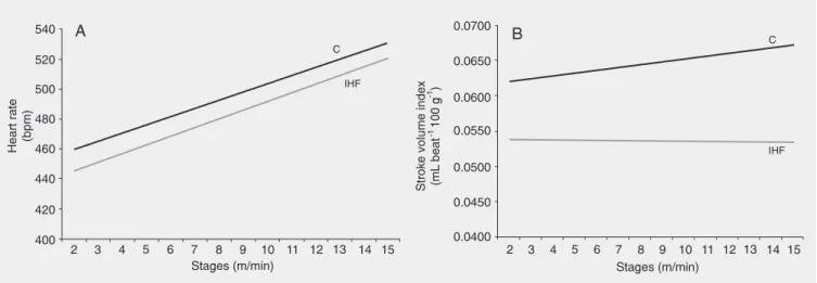
The temporal responses of HR and SVI are shown in Figure 3. In both groups stud-ied, HR showed a linear regression pattern during progressive exercise. The HR slope was not significantly different in ischemic heart failure rats and normal control rats (5.77 ± 2.9 vs 5.49 ± 3.6 beat min-1 s-1, P =
0.449). However, peak HR was significantly lower in ischemic heart failure rats (490 ± 56.3 vs 530 ± 23.1 beats/min, P = 0.040).
Figure 3. Time course of heart rate (HR, panel A) and stroke volume index (panel B) during progressive exercise in normal control (C) and ischemic heart failure (IHF) rats. Note that the HR responses of both groups were similar but, in contrast, stroke volume index responses were reduced in ischemic heart failure rats compared to normal controls. Panel A: C (y = 5.4922x + 454.05, r2 = 0.955), IHF (y = 5.767x + 439.67, r2 = 0.932). Panel B:
C (y = 0.0004x + 0.0616, r2 = 0.797), IHF (y = -0.00003x + 0.0538, r2 = 0.117) (P ≤ 0.05, unpaired Student t-test).
Similar to HR, the SVI slope was signifi-cantly attenuated in ischemic heart failure rats (-0.00003 ± 0.0002 vs 0.0004 ± 0.0003 mL beat-1 100 g-1 s-1, P = 0.022). In addition,
peak SVI was significantly lower in ischemic heart failure rats compared to normal con-trols (0.0534 ± 0.0113 vs 0.0675 ± 0.0153 mL beat-1 100 g-1, P = 0.038).
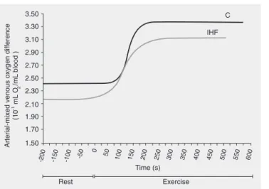
The temporal responses of a-vO2 diff. in
ischemic heart failure rats and normal con-trol rats are shown in Figure 4. In both groups, a-vO2 diff. showed a non-linear
pat-tern during progressive exercise. The a-vO2
diff. slope did not differ between ischemic heart failure and normal control rats (0.009 ± 0.005 vs 0.034 ± 0.048 mL O2 mL blood-1
100 g-1 s-1, P = 0.117). Similarly, no
signifi-cant difference was found in peak a-vO2 diff.
between ischemic heart failure rats and nor-mal controls (3.12 ± 1.13 vs 3.37 ± 0.57 10-1
mL O2/mL blood, P = 0.295).
Discussion
In this study of the rat-infarct model of heart failure 5 weeks after the infarct, we found that the cardiac output response was blunted during exercise, and this was largely
due to a fixed stroke volume. The SVI dem-onstrated markedly blunted kinetics during exercise, whereas HR kinetics was relatively preserved. Although abnormal cardiac out-put kinetics has been previously implicated in the decreased VO2 kinetics during
incre-mental exercise (7,8), we believe that the present methodological strategy provides evidence for the role of CI-mediated VO2
kinetics reduction in heart failure. First, car-diac output was directly and continuously measured in the aortic ascending portion on a beat-to-beat basis in exercising heart fail-ure rats. Second, cardiac output and VO2
were simultaneously evaluated during pro-gressive exercise until exhaustion.
Our methodological strategy permits a more detailed analysis of cardiac output and VO2 kinetics during progressive exercise. In
contrast to previous reports, our findings demonstrate that the temporal responses of cardiac output and VO2 show a non-linear
pa-rameters such as slope, amplitude of change, and latency of the variable under study can be obtained. The sigmoidal behavior of VO2
can contribute to our understanding of cer-tain physiological responses during exer-cise. For instance, the highest VO2 at
sub-maximal exercise may indicate the time when recruitment of oxidative skeletal muscle fi-bers is highest. Since the cardiac response during exercise is modulated by skeletal muscle energy signaling (needed), the non-linearity of CI could be well explained by the VO2 response. The latency at the onset of
exercise may reflect the delay of oxygen consumption by the skeletal muscle. At the onset of exercise the energy supply is pro-vided by stored ATP and creatine phosphate and the leveling off of VO2 at the peak
exercise may be due to the increased recruit-ment of glycolytic skeletal muscle fibers.
Besides the decreased VO2 kinetics, the
amplitude of change (∆VO2 from rest to
maximal exercise) and the maximal VO2
were reduced in exercising heart failure rats. These physiological alterations could be at-tributed to both central and peripheral dys-functions. The reduced cardiac output limits the arterial oxygen transport during exercise in heart failure, which, in turn, provokes a mismatch between oxygen supplied and oxy-gen needed in skeletal muscle to respond to workload increments. Evidence accumulated in the last years has strongly indicated that heart failure provokes a muscle fiber shift from type I to type II, reduction in muscle mass, and blunted muscle vasodilatation, a phenomenon defined as skeletal muscle myopathy (12,13). These alterations can ex-plain, at least in part, the reduced maximal VO2 in heart failure.
The absence of a difference in a-vO2 diff.
between ischemic heart failure rats and nor-mal control rats suggests that the oxygen uptake from the blood vessels is not impli-cated in the reduced VO2 kinetics during
exercise in rats with ischemia-induced heart failure. Tanabe et al. (8) showed that the rate
of O2 extraction by peripheral muscle
rela-tive to work rate increment increased with decreasing cardiac output response to exer-cise. But the decreased O2 supply due to
severely reduced cardiac output could not be fully compensated for by the increased O2
extraction in advanced congestive heart fail-ure (7). Although skeletal myopathy may occur within weeks following myocardial infarction (14), our findings suggest that these muscle abnormalities are not yet se-vere enough to play a role in the diminished oxygen consumption after recent myocar-dial infarction.
Limitations
We recognize several limitations of the present study. a-vO2 diff. was not measured
directly. However, we are confident that our a-vO2 diff. findings were accurate, because
a-vO2 diff. was directly and continuously
calculated from VO2 and cardiac output, and
simultaneously measured from the mask and aortic ascending portion, respectively. The LVEDP measurements suggest that our rats were not in the advanced stage of heart failure. On the other hand, it is likely that the VO2 and CI kinetics was even more
de-pressed in sicker ischemic heart failure rats.
References
1. Colucci WS (1998). The effects of norepinephrine on myocardial biology: implications for the therapy of heart failure. Clinical Cardiol-ogy, 21: I-20-I-24.
2. Lipkin DP, Canepa-Anson R, Stephens MR et al. (1986). Factors determining symptoms in heart failure: comparison of fast and slow exercise tests. British Heart Journal, 55: 439-445.
3. Lunde PK, Sjaastad I, Schiotz Thorud HM et al. (2001). Skeletal muscle disorders in heart failure. Acta Physiologica Scandinavica, 171: 277-294.
4. Franciosa JA, Park M & Levine TB (1981). Lack of correlation between exercise capacity and indexes of resting left ventricular performance in heart failure. American Journal of Cardiology, 47: 33-39.
5. Vescovo G (2002). Skeletal muscle response to exercise and treat-ment: another sibyl in the heart failure syndrome? International Journal of Cardiology, 83: 33-34.
6. Clark AL, Poole-Wilson PA & Coats AJ (1996). Exercise limitation in chronic heart failure: central role of the periphery. Journal of the American College of Cardiology, 28: 1092-1102.
7. Koike A, Hiroe M, Adachi H et al. (1992). Cardiac output-O2 uptake
relation during incremental exercise in patients with previous myo-cardial infarction. Circulation, 85: 1713-1719.
8. Tanabe Y, Nakagawa I, Ito E et al. (2002). Hemodynamic basis of
the reduced oxygen uptake relative to work rate during incremental exercise in patients with chronic heart failure. International Journal of Cardiology, 83: 57-62.
9. Johns TN & Olson BJ (1954). Experimental myocardial infarction. I. A method of coronary occlusion in small animals. Annals of Surgery, 140: 675-682.
10. Russell JC, Campagna PD & Wenger HA (1980). Small-animal ergometer. Journal of Applied Physiology, 48: 394-398.
11. Matsumoto A, Itoh H, Yokoyama I et al. (1999). Kinetics of oxygen uptake at onset of exercise related to cardiac output, but not to arteriovenous oxygen difference in patients with chronic heart fail-ure. American Journal of Cardiology,83: 1573-1576.
12. Hambrecht R, Fiehn E, Yu J et al. (1997). Effects of endurance training on mitochondrial ultrastructure and fiber type distribution in skeletal muscle of patients with stable chronic heart failure. Journal of the American College of Cardiology, 29: 1067-1073.
13. Negrao CE, Rondon MU, Tinucci T et al. (2001). Abnormal neu-rovascular control during exercise is linked to heart failure severity. American Journal of Physiology, 280: H1286-H1292.
14. Vescovo G, Zennaro R, Sandri M et al. (1998). Apoptosis of skeletal muscle myofibers and interstitial cells in experimental heart failure. Journal of Molecular and Cellular Cardiology, 30: 2449-2459.
Although our rats were accustomed to tread-mill running, we cannot guarantee that they reached the maximal exercise point during the cardiopulmonary exercise test.
Perspectives
The direct and continuous measurement of cardiovascular variables on a beat-to-beat basis, and their assessment by means of more refined mathematical methods (as il-lustrated in the present study), may add an important tool to the analytical apparatus currently employed to study the pathophysi-ology of the cardiovascular system.
The important and clinically relevant question that can be raised from the present
findings is whether the decreased temporal responses of VO2 in heart failure can be
reversed by a therapy that increases stroke volume and cardiac output. To accomplish this, it would be of great interest to associate pharmacological therapy that certainly im-proves cardiac output, such as ß-blockers, angiotensin converting enzyme, AT1 blockers,
with non-pharmacological therapy like aero-bic exercise training to reduce skeletal my-opathy in heart failure subjects. In summary, the reduction in VO2 kinetics during