Arq Bras Cardiol 2003; 81: 611-3.
Corso et al Spontaneous rupture of a right atrial angiosarcoma
611
Serviço de Cirurgia Cardíaca Cardiocirúrgica, Serviço de Cardiologia Clínica Cardiointensiva, Hospital Santa Izabel da Santa Casa de Misericórdia Mailing address: Ricardo Barros Corso - Rua Altino Serberto de Barros, 345/1302 Cep 41810-570 - Salvador, BA, Brazil - E-mail: ricardocorso@ig.com.br. English version by Stela Maris C. e Gandour
Arq Bras Cardiol, volume 81 (nº 6), 611-3, 2003
Ricardo Barros Corso, Nadja Kraychete, Sidnei Nardeli, Rilson Moitinho, Cristiano Ourives, Rosenbert Mamedio da Silva, Ricardo Eloy Pereira
Salvador, BA - Brazil
Spontaneous Rupture of a Right Atrial Angiosarcoma and
Cardiac Tamponade
Case Report
Primary cardiac angiosarcoma is a rare disease of difficult diagnosis and poor prognosis frequently associa-ted with recurring hemopericardium. We report the case of a 30-year-old female with a right atrial angiosarcoma and spontaneous rupture to the pericardial cavity, who was diagnosed during an emergency exploratory thoracoto-my, whose indication was cardiac tamponade. This is the 8th case reported in the literature. Clinical findings are discussed and a literature review is provided.
Angiosarcoma is a tumor of mesenchymal origin and accounts for approximately 25% of the malignant tumors of the heart. It preferably appears in the right atrium between the 3rd and 5th decades of life. It is characterized by early local and systemic dissemination, which restricts the indi-cation for surgical resection to a small number of patients. The use of adjuvant chemotherapy and radiation therapy is controversial, due to the poor patient prognosis because of the disease, mean life expectancy being just 6 months 1.
Spontaneous rupture of an angiosarcoma is extremely rare, and only 7 other cases have been reported in the litera-ture. We report a case with this severe complication, prece-ded by repeated pericardial effusion and no preoperative suspicion of tumor. The clinical evolution is described and a literature review provided 2,3.
Case Report
The patient was a 30-year-old black female reporting de-terioration of her general condition, weight loss, and daily recurring fever for 9 months. On admission to our service, she also reported having dyspnea in the 4 preceding months. The clinical and laboratory investigation showed
ane-mia, enlargement of the cardiac area on simple chest radio-graphy, and voluminous pericardial effusion on transthora-cic echocardiography. The other examinations were within normal parameters.
The patient underwent pericardiocentesis, and 2000 mL of a frankly hemorrhagic fluid were withdrawn. During the same admission, open pericardial drainage was required due to occurrence of new effusion with hemodynamic reper-cussions. Laboratory and cytological analysis of the peri-cardial fluid was inconclusive. The patient was discharged from the hospital for follow-up and ambulatory investiga-tion with improvement in her clinical condiinvestiga-tion, but with no diagnostic confirmation of the cause of the disease.
The patient was readmitted 4 months later with signifi-cant worsening of her general condition and continuation of her previous complaints, among which, dyspnea and iso-lated episodes of hemoptysis predominated. On physical examination, the patient was extremely pale, with signs of decompensated right heart failure and multiple rales in both lungs. A slight systolic murmur in the mitral area and a pre-cordial thrill could be heard.
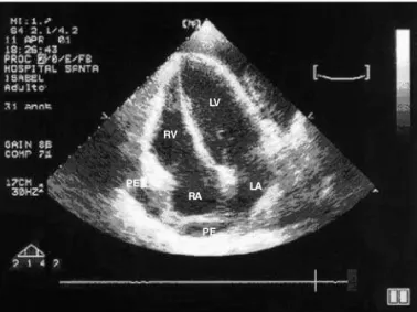
The laboratory tests showed hypochromic microcytic anemia, anisocytosis, polychromatophilia, presence of schi-zocytes and target cells, severe reduction in the number of platelets, slight leukocytosis with no shift to the left, hypoal-buminemia, hypoprothrombinemia, and generalized coagu-lation disorder. The electrocardiogram showed sinus tachy-cardia and right ventricular overload. The chest radiogra-phy showed multiple nodular images in both lungs and bila-teral pleural effusion. The transthoracic echocardiogram re-vealed moderate septate pericardial effusion with no other intracardiac findings.
612
Corso et al
Spontaneous rupture of a right atrial angiosarcoma
Arq Bras Cardiol 2003; 81: 611-3.
anesthesia, and a voluminous hemorrhage was observed after a pericardial opening through a subxiphoid incision, which was followed by hypovolemic shock. Immediate median sternotomy was performed with extension of the first incision followed by longitudinal pericardiotomy. A rupture in the right atrial free wall measuring 3x3 cm was identified, close to the junction of the atrium and the superior vena cava in an area of necrotic tumoral tissue extending throughout most of the atrium. Multiple pericardial implants with the same appearance were identified. The rupture was sutured with temporary clamping of the venae cava, and massive volemic replacement and cardiopulmonary resuscitation were provided. After transitory recovery of the vital signs, the patient experienced refractory shock and died (fig. 1).
Intracardiac inspection revealed extensive tumoral in-volvement in most of the right atrium with the same wall thi-ckness. The interatrial septum, valves, and other cardiac chambers had no abnormalities.
The pleural cavities were opened for pulmonary biop-sy, and multiple subpleural hemorrhagic implants were iden-tified in both lungs, in addition to bilateral voluminous he-morrhagic pleural effusion.
The histopathologic study revealed disseminated angio-sarcoma involving the heart, pericardium, and lungs (fig. 2).
Discussion
Primary heart tumors are extremely rare, with an inci-dence of 0.0017% in autopsy studies reported by the Ameri-can Medical Association. Cardiac metastases are 20 to 40 ti-mes more frequent than are primary heart tumors. Only 25% of the cardiac tumors are malignant, sarcomas being the most common. Angiosarcomas account for 25-30% of those tumors. Of the 24 cases reported by Donsbeck et al 4 with im-munohistochemical confirmation, 9 were undifferentiated sarcomas, 6 were angiosarcomas, 6 were leiomyosarcomas, and 3 had other diagnoses.
Angiosarcomas have already been called
hemangio-sarcoma, hemangioendothelioma, malignant hemangioen-dothelioma, angioendothelial sarcoma, hemangioendothe-lial sarcoma, malignant hemangioma, and hemangioendo-thelial blastoma.
An angiosarcoma of the heart is considered primary when no history or concomitant evidence is present of tu-mors in the soft tissue, bone, or subcutaneous tissue. In a case series of 366 cases, 3% of the tumors originated from the heart or great vessels, or both. Angiosarcoma of the heart involves almost exclusively the right atrium, but has already been reported in other cardiac chambers 5,6.
The symptoms resulting from primary tumors of the heart are usually late and more related to their location than to their histological type, which makes the early diagnosis difficult, impairing the efficacy of treatment. Myocardial in-filtration by the tumor triggers arrhythmias and, more rarely, conduction disorders. Impairment of myocardial contracti-lity may simulate restrictive or dilated hypertrophic cardio-myopathy. Pulmonary arterial hypertension secondary to repetitive tumoral embolization and compression of the right ventricular outflow tract may trigger right heart failure.
Angiosarcomas of the heart grow rapidly, usually within the myocardial wall, which makes their diagnosis through different imaging methods difficult. They are cha-racterized by friability and a tendency towards bleeding. They are often associated with pericardial effusion and car-diac tamponade. Myocardial rupture due to tumoral infiltra-tion and necrosis of the wall may occur, multiseptate hemo-pericardium being the most frequent echocardiographic fin-ding in the few cases reported 2,6.
The symptoms resulting from mechanical cardiac im-pairment are usually preceded by malaise, fever, weight loss, fatigue, and anemia that occur weeks or months before symp-tom onset. Dyspnea, productive cough, and hemoptysis suggest pulmonary metastases. The most frequent sites of metastases are the pericardium, lungs, mediastinal lymph nodes, and vertebrae, and metastases are usually present in 66 to 89% of the patients at the time of diagnosis 7,8.
The diagnosis of angiosarcoma is usually difficult and late, despite the different methods used. Many cases have been confirmed only on thoracotomy for the treatment
Fig. 2 - Histological sections of a myocardial fragment stained with hematoxylin and eosin. Note the fusiform cells with atypical vesicular nuclei delimiting slit vascular structures characteristic of angiosarcoma.
Fig. 1 - Transthoracic echocardiographic image of a voluminous pericardial effusion with septations and signs of cardiac tamponade. RV- right ventricle; LV- left ventricle; LA- left atrium; RA- right atrium; PE- pericardial effusion.
LV
RV
RA LA PE
Arq Bras Cardiol 2003; 81: 611-3.
Corso et al Spontaneous rupture of a right atrial angiosarcoma
613
1. Frota FJD, Luchese FA, Leães P, Valente LA, Vieira MS, Blacher C. Angiossarco-ma cardíaco primário: um dileAngiossarco-ma terapêutico. Arq Bras Cardiol 2002;78:586-8. 2. Ohri SK, Nihoyannopoulos P, Taylor KM, Keogh BE. Angiosarcoma of the heart causing cardiac rupture: a cause of hemopericardium. Ann Thorac Surg 1993;55:525-8.
3. Mukohara N, Tobe S, Azami T. Angiosarcoma causing cardiac rupture. Jpn J Thorac Cardiovasc Surg 2001;49:516-8.
4. Donsbeck AV, Ranchere D, Coindre JM, Le Gall F, Cordier JF, Loire R. Primary cardiac sarcomas: an immunohistochemical and grading study with long-term follow-up of 24 cases. Histopathology 1999;34:295-304.
5. Herrmann MA, Shakermen RA, Edwards WD, Shub C, Schaff HV. Primary cardiac angiosarcoma: a clinicopathologic study of six cases. J Thorac Cardiovasc Surg 1992;103:655-65.
References
6. Oshima K, Ohtaki A, Motoi K, et al. Primary cardiac angiosarcoma associated with cardiac tamponade. Jpn Circ J 1999;63:822-4.
7. Biniwale RM, Pathare HP, Aggrawal N, Tendolkar AG, Deshpande J, Sivaraman A. Cardiac sarcomas: is tumor debulking justifiable therapy? Asian Cardiovasc Thorac Ann 1999;7:52-5.
8. Shapiro S, Scott J, Kaufman K. Metastatic cardiac angiosarcoma of the cervical spine: case report. Spine 1999;24:1156-9
9. Mcfadden PM, Ochsner JL. Atrial replacement and tricuspid valve reconstrution after angiosarcoma resection. Ann Thorac Surg 1997;64:1164-6.
10. Nakamichi T, Fukuda T, Suzuki T, Kaneko T, Morikawa Y. Primary cardiac angio-sarcoma: 53 months’ survival after multidisciplinary therapy. Ann Thorac Surg 1997;63:1160-1.
of repetitive pericardial effusion or on autopsy. Echocardio-graphy is the most frequently used method, the transeso-phageal technique being the preferred one. The presence of cavitary-pericardial fistula in cases of cardiac rupture has al-ready been identified by that method 2.
Patients with the tumoral form may have their diagno-sis suggested by computed tomography or nuclear magne-tic resonance. No method of myocardial biopsy is advisable, due to the characteristic friability of angiosarcoma and its predisposition to hemorrhaging 6.
The treatment of angiosarcoma is controversial due to the poor prognosis in most patients. Surgical resection is indicated when no evidence of metastases exists and when myocardial resection is reparative 9.
Chemotherapy and radiation therapy may be indicated as adjuvant or preferential therapies, but their use is usually
