ARTICLE
Quantitative sensory testing in trigeminal
traumatic neuropathic pain and persistent
idiopathic facial pain
Teste sensitivo quantitativo em dor neuropática trigeminal pós-traumática
e dor facial idiopática persistente
Silvia R. D. T. de Siqueira1, Mariana Siviero2, Fábio K. Alvarez3, Manoel J. Teixeira4, José T. T. de Siqueira5
he persistent idiopathic facial pain (PIFP) is character-ized by a chronic intraoral pain located in the teeth and jaws without major pathology1, 30. It has been described as atypical
odontalgia, atypical facial pain, persistent chronic facial pain, however it still has undeined criteria2,3. he International
Association for the Study of Pain (IASP) describes it as atypi-cal odontalgia, and associates it with previous history of teeth extraction at the area of pain, in the absence of clinical and
radiographic evidence of tooth pathology or other relevant orofacial hard or soft tissue lesion4. However, there is no
ev-idence in many patients of previous dental trauma and, al-though in some cases dental procedures are involved in its development, it still has an idiopathic unclear etiology5,6. On
the other hand, trigeminal traumatic neuropathic pain (TTN) is associated with major or minor nerve damage during oral surgery ( for example, third molar extraction) with clear
1DDS, PhD, Associate Professor, School of Arts, Science and Humanities, University of São Paulo, São Paulo SP, Brazil;
2DDS, Postgraduation student, Orofacial Pain Team, Hospital das Clínicas, Medical School, University of São Paulo, São Paulo SP, Brazil; 3DDS, MSci, Orofacial Pain Team, Hospital das Clínicas, Medical School, University of São Paulo, São Paulo SP, Brazil;
4MD, PhD, Neurology Department, Medical School, University of Sao Paulo, São Paulo SP, Brazil;
5DDS, PhD, Head of the Orofacial Pain Team, Hospital das Clínicas, Medical School, University of São Paulo, São Paulo SP, Brazil.
Correspondence: Silvia Regina Dowgan Tesseroli de Siqueira; Rua Maria Candida 135; 02071-010 São Paulo SP - Brasil; E-mail: [email protected]
Conflict of interest: There is no conflict of interest to declare.
Received 26 February 2012; Received in final form 30 July 2012; Accepted 06 August 2012.
ABSTRACT
The objective of this article was to investigate, with a systematic protocol of quantitative sensory testing, patients with persistent idiopathic facial pain (PIFP) and others with trigeminal traumatic neuropathic pain (TTN) compared to controls. Thirty patients with PIFP, 19 with TTN, and 30 controls were evaluated on subjective numbness and dysesthesia and with a systematic protocol of quantitative sensory testing for thermal evaluation (cold and warm), mechanical detection (touch and pinpricks for mechanical pain), superficial pain thresholds, and corneal reflex. We found that PIFP and TTN had numbness and dysesthesia higher than controls (p<0.001 and p=0.003), and that in both of them mechanical pain by pinpricks detection was abnormal intra and extra orally at the mandibular branch (p<0.001). Cold, warm, and tactile de-tections and pain thresholds were similar among the groups. Corneal reflex was abnormal in TTN (p=0.005). This study supports neuropathic mechanisms involving pain processing in PIFP and that the criterion on absence of sensorial variations in PIFP should be revised.
Key words: facial pain, facial neuralgia, odontalgia, sensory thresholds.
RESUMO
O objetivo deste artigo foi investigar, com um protocolo sistemático de testes sensitivos quantitativos, pacientes com dor facial idiopática persistente (DFIP) e outros com dor neuropática trigeminal traumática (DNTT) comparado aos controles. Trinta pacientes com DFIP, 19 com DNTT e 30 controles foram avaliados quanto à dormência e à disestesia subjetiva e por meio de um protocolo sistemático de testes sensi-tivos quantitasensi-tivos, que incluiu avaliação térmica (frio e quente), detecção mecânica (táctil e alfinetes), limites de dor superficial e reflexo córneo-palpebral. Foi observado que os pacientes apresentaram mais dormência e disestesia do que os controles (p<0,001 e p=0,003), além de mais anormalidades intra e extraorais no ramo mandibular (p<0,001). As alterações de calor, frio, dor e tato foram semelhantes entre os grupos. O reflexo córneo-palpebral foi anormal somente no grupo com DNTT (p=0,005). Este estudo suporta mecanismos de dor neuropática envolvidos no processamento da DFIP, e o critério de ausência de variações sensoriais nesta deve ser revisto.
etiological factor and evidence of abnormal sensory func-tion7,8. Many patients get complete recovery after oral nerve
damage9, but a few persist with pain associated with thermal
hypoesthesia and warm allodynia7.
he sensory indings in the scientiic literature on these conditions remain controversial9. PIFP does not difer from
controls in quantitative sensory testing (QST)10, atypical
fa-cial pain showed to be a heterogeneous group7, and atypical
odontalgia that included only patients with previous history of dental procedures as etiological factors, showing abnormal sensory processing11.
No studies had investigated separately patients with a clear history of trauma and those with idiopathic pain and spontaneous initial about their somatosensory impairment3,
and QST can help in the elucidation of mechanisms involved in sensory processing12. hus, the objective of this study was
to investigate with a systematic protocol of QST patients with PIFP and TTN compared to controls.
MATERIAL AND METHODS Subjects
Seventy-nine subjects were included in this study. Forty-nine consecutive patients (30 with PIFP and 19 with TTN) were evaluated between January 2007 and March 2009. hey corresponded to patients diagnosed with PIFP and TTN that were referred to the Orofacial Pain Clinic of the Hospital das Clínicas, Medical School, University of São Paulo and that fulilled the criteria of the International Headache Society (IHS)1. PIFP is deined as a persistent facial pain that does not
have the classical characteristics of cranial neuralgias and is not attributed to another disorder. he pain is deep and well localized, typically reported as continuous with constant or luctuating intensity and described as burning, aching, or cramping. In this study, only patients with no history of den-tal or facial trauma or any oral procedures within a period of six months before the evaluation were included. In the TTN Group, only patients with history of nerve damage after oral surgery (inferior alveolar or lingual nerve) and evidence of sensory loss and persistent pain within three months after the surgery were included.
We enrolled 30 healthy subjects for the Control Group, with no fulillment of diagnosis for any type of orofacial pain. No subject was excluded. hese subjects were relatives and caregivers from patients and employees from the hospital, which volunteered for this study.
At the time of the study, no controls or patients were hav-ing any analgesic medications or in a period of six months before the evaluation. hey were not under any form of treat-ment. he subjects were evaluated by a trained neurologist, and complaints of generalized body pain and diagnoses of neurodegenerative, neuroinfectious, metabolic or rheuma-tologic diseases meant exclusion. he diagnoses of TTN and
PIFP were made by a trained dentist in orofacial pain, and other painful craniofacial conditions including musculoskel-etal pain were excluded, according to the criteria of the IHS1.
No subject was excluded by these criteria.
All patients and controls were informed about the purpos-es of the study, and signed the informed consent. he protocol had been approved by the local Ethics Committee (0751/10).
Subjective evaluation of sensitivity
All patients and controls were interviewed about their fa-cial sensory perception including numbness and dysesthesia. he frequency of sensorial abnormalities (absent, eventual, frequent, or constant) and intensity of sensation (0 to 10 by the Visual Analogue Scale – VAS) were collected.
Quantitative sensory testing
All subjects underwent a standardized protocol of QST, which consisted of ive tests grouped as follows: thermal de-tection (cold and warm perception); mechanical dede-tection (touch perception and pinpricks for mechanical pain); and supericial pain thresholds.
he body regions evaluated were the three trigeminal branches: front, cheek, and chin. he mechanical pain per-ception with pinpricks was also performed intra-orally at the mandibular and maxillary branches (vestibular gingival tis-sue at the premolar area). he evaluation was performed bi-laterally in all subjects13.
All subjects were evaluated in the sitting position, with the head resting at a lat surface, and in a silent room with acous-tic protection at the walls and the door closed. Only the pa-tient and the researcher were in the room. All subjects were evaluated by the same researcher, who was a dentist trained in orofacial pain and trigeminal sensory testing. he patients and controls were oriented to keep their eyes closed during the exam and to be concentrated in the stimuli. Only the re-searcher knew the order of the stimuli, which was determined by randomization and was repeated in all subjects. here were no training sections but detailed explanations about the pro-cedure of sensory testing, in order to avoid expectation about the sequence of stimuli by the subjects. he interval between each type of stimuli was from two to ive minutes.
hermal detection
hermal testing14 was performed using the hermosensi II
Mechanical/tactile and pain perception
Touch perception14 was assessed using a set of
standard-ized vonFrey ilaments with rounded tips of 0.5 mm diameter (g/mm2) – IITC, WoodlandHills, USA. he mechanical pain
detection was performed with standard pinpricks with 24 mm of length and 0.40 mm of diameter applied with an electronic device (g/mm2) – Micromar, Diadema, São Paulo, Brazil. he
detection or not of the stimuli was considered in the evalu-ation. hree measurements were performed and the means and standard deviations were considered for the analysis.
Supericial pain thresholds
Supericial algometry14 was tested using disposable needles
of 8x10x0.5 mm, which were applied with an electronic device with increasing force (Micromar, Diadema, São Paulo, Brazil). hree measurements in g/mm2 were performed and the means
and standard deviations were considered for the analysis.
Corneal reflex
Corneal relex was tested using a vonFrey (IITC, Woodland Hills, EUA) ilament with a rounded tip of 0.5 mm diameter in both eyes, and the presence or absence of relex was noticed.
Statistical analysis
All data were tabled and the frequencies and percent-ages, means, standard deviations, and ranges were com-pared among the groups. After the initial descriptive evalu-ation, variables were tested about normal distribution with Shapiro-Wilk’s test and Q-Q plots. For the quantitative vari-ables with normal distribution (cold, warm, tactile, pin-prick, and pain detections), the ANOVA 1 factor was used. Post-hoc comparisons were calculated using Tukey’s test. Non-parametric test included Pearson’s χ2 and Fisher’s
ex-act tests. Correlations among the variables were tested with Pearson’s test. he analysis included the comparisons among the sites evaluated, and between the groups. he side afect-ed was comparafect-ed to the opposite one in patients with unilat-eral pain, rather than the comparison between right and left.
All statistical calculations were performed using SPSS 17.0 (SPSS Inc., Illinois, USA). he level of signiicance was 5%.
RESULTS
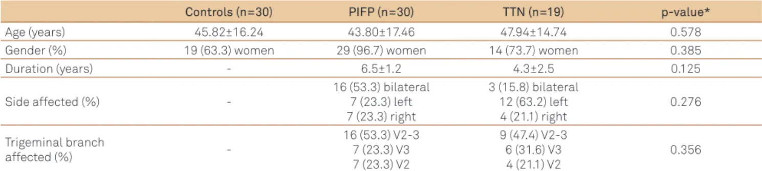
In the group of patients with PFIP, 29 (96.7%) were wom-en and 1 (3.3%) was a man, being their mean age 43.80±17.46 years-old (mean±SD). In the group of patients with TTN, 14 (73.7%) were women and 5 (26.3%) men, and the mean age was 47.94±14.74 years-old (mean±SD). he Control Group had 19 (63.3%) women and 11 (36.7%) men, and the mean age was 45.82±16.24 years-old (mean±SD). Clinical charac-teristics of patients and controls can be observed in Table 1.
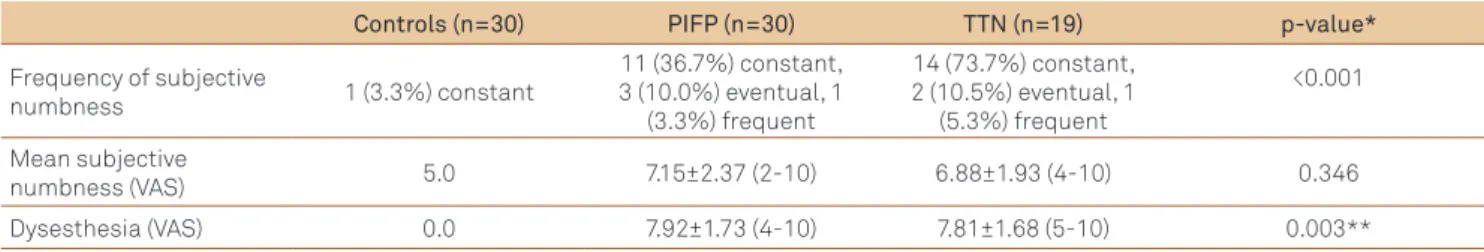
In this study, we observed that the patients had higher frequency of numbness and dysesthesia than the controls (Table 2).
he thermal testing presented no diferences among the groups. Five (8.3%) patients with PIFP had low cold detec-tion at all trigeminal branches, 4 (10.5%) with TTN had low cold detection at all trigeminal branches, and 1 (1.7%) con-trol had low cold detection at the maxillary and mandibular trigeminal branches. Four (6.7%) patients with PIFP had low warm detection at the maxillary and mandibular branches, 5 (13.2%) with TTN had low warm detection at all trigeminal branches, and 1 (1.7%) control presented low warm detection at the mandibular branch.
here were also no diferences in the tactile evaluation; 7 (11.7%) patients with PIFP had low tactile detection in all tri-geminal branches, 7 (18.4%) with TTN had low tactile detec-tion at the maxillary and mandibular branches, and 2 (3.3%) controls had low tactile detection at the maxillary branch.
he mechanical pain detection showed diferences at the maxillary and mandibular branches. he extraoral evaluation showed eight (13.3%) patients with PIFP and eight (21.1%) with TTN including low detection at the maxillary and man-dibular branches. At the intraoral exam, 12 (20.0%) patients with PIFP and 11 (28.9%) with TTN had low detection at the maxillary and mandibular branches.
Controls (n=30) PIFP (n=30) TTN (n=19) p-value*
Age (years) 45.82±16.24 43.80±17.46 47.94±14.74 0.578
Gender (%) 19 (63.3) women 29 (96.7) women 14 (73.7) women 0.385
Duration (years) - 6.5±1.2 4.3±2.5 0.125
Side affected (%)
-16 (53.3) bilateral 7 (23.3) left 7 (23.3) right
3 (15.8) bilateral 12 (63.2) left 4 (21.1) right
0.276
Trigeminal branch
affected (%)
-16 (53.3) V2-3 7 (23.3) V3 7 (23.3) V2
9 (47.4) V2-3 6 (31.6) V3 4 (21.1) V2
0.356
*Pearson’s χ2 test; PIFP: persistent idiopathic facial pain; TTN: trigeminal traumatic neuropathic pain; V2: maxillary branch; V3: mandibular branch; V2-3:
maxillary and mandibular branches.
he supericial pain thresholds were similar among the groups and can be observed in Figure. Only two patients with TTN presented abnormal corneal relex at the afected side, which was statistically diferent from the other groups (p=0.005, Pearson’s χ2 test).
There were positive correlations among the trigeminal branches in all sensorial modalities (p<0.005). The posi-tive ones were found between tactile and warm detections (moderate Pearson’s correlation 0.532; p=0.003), tactile and cold detections (moderate Pearson’s correlation 0.472; p=0.008), pinprick and cold detections (moderate Pearson’s correlation 0.694; p<0.001), pinprick and warm detections (strong Pearson’s correlation 0.788; p=0.002). There were no associations of sensorial findings at the tri-geminal area of pain.
DISCUSSION
In this study, both groups of patients had higher frequen-cy of numbness and dysesthesia and lower mechanical pain perception than the controls, however there were no difer-ences in thermal, supericial pain, and tactile evaluation. Only patients with TTN had abnormalities in corneal relex. his appears to be the irst study that investigated QST tri-geminal neuropathic pain with or not previous history of oral trauma, and suggests that patients with PIFP and TTN have similar sensorial deicits and that, as in TTN, neuropathic pain mechanisms may be underlying PIFP.
Sensorial abnormalities were similar in persistent idiopathic facial pain and trigeminal traumatic neuropathic pain
PIFP, which corresponds to atypical facial pain and atyp-ical odontalgia according to the classiications2-5, can be
as-sociated with previous trauma history. hus, several stud-ies that had investigated somatosensory abnormalitstud-ies in these patients had not separated them accordingly to the etiology of their pain. It is possible that the controversial results in diferent samples can be due to that aspect9,11-13.
In this paper, both patients’ groups (with or with no trau-matic etiology) showed low mechanical pain detection at
Controls (n=30) PIFP (n=30) TTN (n=19) p-value*
Frequency of subjective
numbness 1 (3.3%) constant
11 (36.7%) constant, 3 (10.0%) eventual, 1
(3.3%) frequent
14 (73.7%) constant, 2 (10.5%) eventual, 1
(5.3%) frequent
<0.001
Mean subjective
numbness (VAS) 5.0 7.15±2.37 (2-10) 6.88±1.93 (4-10) 0.346
Dysesthesia (VAS) 0.0 7.92±1.73 (4-10) 7.81±1.68 (5-10) 0.003**
*Pearson’s χ2; Fisher’s exact test; Statistical differences are: PIFP versus controls and TTN versus controls (numbness and dysesthesia); PIFP: persistent
idiopathic facial pain; TTN: trigeminal traumatic neuropathic pain; VAS: visual analogue scale; **F=26.801.
Table 2. Subjective evaluation of sensitivity: patients had more numbness and dysesthesia than the controls (n=79).
Figure. Means of superficial algometry (superficial pain thresholds): there were no differences among the groups in any trigeminal branches (n=79).
g
/m
m
2
g
/m
m
2
g
/m
m
2
p=0.176
p=0.529
p=0.517
Error Bars: 95%CI
Error Bars: 95%CI
Error Bars: 95%CI
Controls PIFP TTN
Controls PIFP TTN
Controls PIFP TTN
Mean Superficial A
lg
ome
try - Oph
thalmic br
anc
h
Mean Superficial A
lg
ome
try - Maxillary br
anc
h
Mean Superficial A
lg
ome
try - Mandibular br
anc
h
60
50
40
30
20
10
0
50
40
30
20
10
0
Facial side
Right Left
Facial side
Right Left
Facial side
Right Left
50
40
30
20
10
0
the afected maxillary and mandibular branches, and the only diference between TTN and PIFP was that the corne-al relex was abnormcorne-al only in TTN. he cornecorne-al relex had been reported as afected in other neuropathic conditions, such as burning mouth syndrome14. hese patients also
have many abnormalities in somatosensory processing, and burning mouth is an idiopathic condition with no etiologi-cal factor that is currently been considered neuropathic due to the QST indings14-18. Our results indicate that the
crite-rion about absence of sensorial abnormality in PIFP should be revised19-20.
Neuropathic mechanisms in idiopathic trigeminal pain
Abnormal somatosensory indings are considered in the current classiication of neuropathic pain of the IASP as im-portant for evaluation and diagnosis21. In the current
deini-tion, neuropathic pain is diagnosed according to sensorial deicits, which were observed in both of our samples, and thus these results support neuropathic pathophysiology for both conditions19,21. he structural integrity of the nervous
system diferentiates nociceptive pain from neuropathic pain22, however, for idiopathic conditions, with no clear
evi-dence of inlammatory/nociceptive etiology, the absence of neurological abnormalities still makes them undeined23.
On the other hand, phenomena that are typical from neuro-pathic pain, such as allodynia, also occur in central sensiti-zation of inlammatory conditions, and there is also periph-eral and central hypperiph-eralgesia in them24.
For trigeminal neuropathic pain with traumatic origin, there are many evidences of central impairment of the so-matosensory and motor processing12,25 involving deicits in
the descending inhibition26. Motor-sensory integration is
evi-denced by the recovery of abnormal thermal processing by the stimulation of motor areas27. he neuroplastic
phenom-ena involve the activation of glial cells surrounding afected neurons at the central nervous system28,29, in many subtypes
of trigeminal pain, including neuropathic and inlammatory etiologies, and thus their physiopathology share many simi-larities when gets chronic.
Correlations among variables
We did not found any association between the location of pain and the sensorial indings. However, there were sever-al positive correlations among the diferent modsever-alities (cold, tactile, warm, and pain); the mechanisms underlying it might involve central processing of sensorial modalities, discrete impairment or sensory losses can occur in association30.
Limitations of the study
his study presented some limitations, including a great variability in sensorial detection that would make necessary a larger sample. On the other hand, these patients represent those looking for treatment in a specialized clinic and not the population, and thus the demographic data may not be gen-eralized. However, they were carefully evaluated by an experi-enced dentist and their diagnosis was very accurate, with no concomitancy of other pain causes as temporomandibular disorders, which could compromise results.
All patients were evaluated in the same sites represent-ing the trigeminal branches in order to standardize the stud-ied groups and to allow comparison among them. It is pos-sible that eventual abnormal sensory testing restricted to the site of the pain was missed in this evaluation. However, the objectives of this study were to investigate and to com-pare the abnormal sensations in the trigeminal territories, but not to verify the extension of sensorial indings in these patients. Besides, the qualitative evaluation of the corneal re-lex did not allow the investigation of the part of the rere-lex that was found abnormal in TTN patients.
Another limitation of the study is the fact that patients were not investigated about their psychological characteris-tics and it is known that chronic pain is often associated with anxiety or depression. Both could interfere in the accuracy of answer during QST and must be considered in the interpre-tation of these results.
In conclusion, patients with PIFP and TTN had lower de-tection of mechanical pain stimuli (pinpricks) than the con-trols at the maxillary and mandibular trigeminal branches. hese results support neuropathic mechanisms involving pain processing in PIFP and TTN.
1. International Headache Society Classification Subcommittee. International classification of headache disorders, 2nd edition. Cephalalgia 2004;24:1-160.
2. Graff-Radford SB. Facial pain. Neurologist 2009;15:171-177.
3. Sardella A, Demarosi F, Barbieri C, Lodi G. An up-to-date view on persistent idiopathic facial pain. Minerva Stomatol 2009;58:289-299.
4. Merskey H, Bogduk N. Classification of chronic pain. Seattle, WA: IASP Press; 1994.
5. Evans RW, Agostoni E. Persistent idiopathic facial pain. Headache 2006;46:1298-1300.
6. Koratkar H, Koratkar S. Atypical odontalgia: a case report. Gen Dent 2008;56:353-355.
7. Forssell H, Tenovuo O, Silvoniemi P, Jääskeläinen SK. Differences and similarities between atypical facial pain and trigeminal neuropathic pain. Neurology 2007;69:1451-1459.
8. Niemi M, Laaksonen JP, Forssell H, Jääskeläinen S, Aaltonen O, Happonen RP. Acoustic and neurophysiologic observations related to lingual nerve impairment. Int J Oral Maxillofac Surg 2009; 38:758-765.
9. Jääskeläinen SK, Teerijoki-Oksa T, Forssell H. Neurophysiologic and quantitative sensory testing in the diagnosis of trigeminal neuropathy and neuropathic pain. Pain 2005;117:349-357.
10. Zakrzewska JM. Facial pain: an update. Curr Opin Support Palliat Care 2009;3:125-130.
11. Lang E, Kaltenhäuser M, Seidler S, Mattenklodt P, Neundörfer B. Persistent idiopathic facial pain exists independent of somatosensory input from the painful region: findings from quantitative sensory functions and somatotopy of the primary somatosensory cortex. Pain 2005;118:80-91.
12. List T, Leijon G, Svensson P. Somatosensory abnormalities in atypical odontalgia: A case-control study. Pain 2008;139:333-341.
13. Eliav E, Gracely RH, Nahlieli O, Benoliel R. Quantitative sensory testing in trigeminal nerve damage assessment. J Orofac Pain 2004; 18:339-344.
14. Siviero M, Teixeira MJ, Siqueira JTT, Siqueira SRDT. Somesthetic, gustatory, olfactory function and salivary flow in patients with trigeminal neuropathic pain. Oral Diseases 2010;16:482-487.
15. Jääskeläinen SK, Forssell H, Tenovuo O. Abnormalities of the blink reflex in burning mouth syndrome. Pain 1997;73:455-460.
16. Femiano F, Lanza A, Buonaiuto C, Gombos F, Cirillo N. Burning mouth disorder (BMD) and taste: a hypothesis. Med Oral Pathol Oral Cir Bucal 2008;13:470-474.
17. Grushka M, Epstein JB, Gorsky M. Burning mouth syndrome and other oral sensory disorders: a unifying hypothesis. Pain Res Manag 2003;8:133-135.
18. Grushka M, Sessle B. Taste dysfunction in burning mouth syndrome. Gerodontics 1988;4:256-258.
19. Zebenholzer K, Wöber C, Vigl M, Wessely P, Wöber-Bingöl C. Facial pain and the second edition of the International Classification of Headache Disorders. Headache 2006;46:259-263.
20. Cornelissen P, van Kleef M, Mekhail N, Day M, van Zundert J. Evidence-based interventional pain medicine according to clinical diagnoses. Persistent idiopathic facial pain. Pain Pract 2009;9:443-448.
21. Geber C, Baumgärtner U, Schwab R, et al. Revised definition of neuropathic pain and its grading system: an open case series illustrating its use in clinical practice. Am J Med 2009;122:S3-S12.
22. Chakravarty A, Sen A. Migraine, neuropathic pain and nociceptive pain: Towards a unifying concept. Med Hypotheses 2010;74:225-231.
23. Woda A. A “dysfunctional” pain group in addition to the “neuropathic” and “nociception/inflammatory” groups of orofacial pain entities? J Orofac Pain 2009;23:89-90.
24. Borsook D, Burstein R, Becerra L. Functional imaging of the human trigeminal system: opportunities for new insights into pain processing in health and disease. J Neurobiol 2004;61:107-125.
25. DaSilva AF, Becerra L, Pendse G, Chizh B, Tully S, Borsook D. Colocalized structural and functional changes in the cortex of patients with trigeminal neuropathic pain. PLoS One 2008;3:3396.
26. Sardella A, Demarosi F, Barbieri C, Lodi G. An up-to-date view on persistent idiopathic facial pain. Minerva Stomatol 2009;58:289-299.
27. Fontaine D, Bruneto JL, El Fakir H, Paquis P, Lanteri-Minet M. Short-term restoration of facial sensory loss by motor cortex stimulation in peripheral post-traumatic neuropathic pain. J Headache Pain 2009;10:203-206.
28. Okada-Ogawa A, Suzuki I, Sessle BJ, et al. Astroglia in medullary dorsal horn (trigeminal spinal subnucleus caudalis) are involved in trigeminal neuropathic pain mechanisms. J Neurosci 2009;29:11161-11171.
29. Upadhyay J, Knudsen J, Anderson J, Becerra L, Borsook D. Noninvasive mapping of human trigeminal brainstem pathways. Magn Reson Med 2008;60:1037-1046.