AR
TIGO ORIGINAL / ORIGINAL AR
TICLE
INTRODUCTION
Short bowel syndrome (SBS) occurs when the functional mass of the small intestine is insuficient for adequate digestion of food and absorption of nutrients and luids(21). In the initial phase of SBS, par-enteral nutrition (PN) is usually necessary and there is not an absolute criterion for weaning off PN. In the intermediate phase, the efforts are primarily devoted to intestinal adaptation to oral feeding(29). The inability to maintain an adequate nutrition makes the patient dependent on PN therapy(34, 45). In this situation, the therapy is focused on nutrient replacement, patient rehabilitation and the return to social activities, pro-viding an improved quality of life(44). Since home PN is not a therapeutic option provided by the Brazilian public health system, patients with severe malabsorp-tive states receive intermittent PN in our service at the Clinical Hospital of Ribeirão Preto of the University of São Paulo(7, 32). These patients are hospitalized every
COPPER AND MAGNESIUM DEFICIENCIES IN
PATIENTS WITH SHORT BOWEL SYNDROME
RECEIVING PARENTERAL NUTRITION OR
ORAL FEEDING
Camila Bitu Moreno
BRAGA
, Iahel Manon de Lima
FERREIRA
, Júlio Sérgio
MARCHINI
and
Selma Freire de Carvalho da
CUNHA
ABSTRACT - Background - Patients with short bowel syndrome havesigniicant luid and electrolytes loss. Objective- Evaluate the mineral and electrolyte status in short bowel syndrome patients receiving intermittent parenteral nutrition or oral feeding. Methods - Twenty two adults with short bowel syndrome, of whom 11 were parenteral nutrition dependent (PN group), and the 11 remaining had been weaned off parenteral nutrition for at least 1 year and received all nutrients by oral feeding (OF group). The study also included 14 healthy volunteers paired by age and gender (control group). Food ingestion, anthropometry, serum or plasma levels of sodium, potassium, phosphorus, magnesium, calcium, zinc, iron and copper were evaluated. PN group subjects were evaluated before starting a new parenteral nutrition cycle. Results - The levels of sodium, potassium, phosphorus, calcium and zinc were similar between the groups. The magnesium value was lower in the PN group (1.0 ± 0.4 mEq /L) than other groups. Furthermore, this electrolyte was lower in the OF group (1.4 ± 0.3 mEq /L) when compared to the Control group (1.8 ± 0.1 mEq/L). Lower values of copper (69±24 vs 73±26 vs 109±16 µg/dL) were documented, respectively, for the PN and OF groups when compared to the control group. Con-clusion - Hypomagnesemia and hypocupremia are electrolyte disturbances commonly observed in short bowel syndrome. Patients with massive intestinal resection require monitoring and supplementation in order to prevent magnesium and copper deiciencies. HEADINGS - Mineral deiciency. Electrolytes. Parenteral nutrition. Short bowel syndrome.
Declared conflict of interest of all authors: none
Source of Funding: Fundacão de Amparo à Pesquisa do Estado de São Paulo (FAPESP), Brazil (Grant # 2007/52058-1).
Division of Nutrology, Department of Internal Medicine, School of Medicine of Ribeirão Preto, University of São Paulo, Ribeirão Preto, SP, Brazil.
Correspondence: Prof.ª Selma Freire de Carvalho da Cunha. Av. Bandeirantes, 3900, 6° andar, Hospital das Clínicas, Faculdade de Medicina de Ribeirão Preto - CEP: 14048-900 - Ribeirão Preto, SP. E-mail: sfreire@fmrp.usp.br
10-40 days to receive PN cycles (3-8 days), depending on symptoms such as diarrhea, dehydration, and poor nutritional status(7).
METHODS
This study was conducted in a university hospital from December 2007 to November 2008. The research was approved by the Institutional Ethics Committee, and all volunteers signed an informed consent. Twenty-two SBS patients were divided into two groups. The irst group (PN group, n=11) consisted of PN-dependent subjects, and the second (OF group, n=11) included patients who were under oral feeding. Hemodynamically unstable subjects and those with intestinal inlammatory diseases, neoplasia, hepatic failure, and diagnoses of active infectious diseases were excluded.
The PN group consisted of SBS patients who were under intermittent PN, ive women and six men, aged 50±12 years, with a median length of remaining small intestine of 25 (10-100) cm and an intestinal transit time up to 15 minutes. Intestinal resection had been performed 15 (6-88) months before, and the colon was present in six patients. For each hospitalization period, the PN group subjects received PN infused by using a totally implantable central venous catheter according to the current recommendations(1). PN provided 1386±272 kcal (31±6 kcal/kg) daily of energy, and 51±5 g amino acids (1.1±0.2 g/kg), 222±38 g glucose (4.5±1.7g/kg) and 29±18 g lipids (0.8±0.3 g/kg) daily. A mixture of multiple trace elements containing 2.5 mg of Zn, 0.8 mg of Cu, 0.4 mg of manganese and 10 µg of chromium (Ad-element®, Darrow, Rio de Janeiro, Brazil) was added to the PN formula-tions. The electrolytes were calculated according to individual requirements which take into account body weight and serum levels (Na: 1.4±0.1 mEq/kg/day; K: 1.7±0.2 mEq/kg/day; Mg: 20±3 mEq/day; P: 38±2 mmol/day; Ca: 8.8±0.9 mEq/day).
Eight women and three men, aged 56±12 years, were included in the OF group. They had median length of re-maining small intestine of 110 (40-210) cm, and the colon was present in nine patients. These patients had undergone intestinal resection 72 (18-180) months before, and received PN during 2 to 6 months after the intestinal resection. PN was discontinued when patients presented satisfactory evolution, at least one year prior to the study.
All SBS patients (PN and OF groups) were followed-up by a specialized outpatient service, which advised them to follow an oral diet at home and to take vitamin and mineral tablets (Centrum®, Wyeth, Richmond, VA, USA) on a daily basis. Each tablet contained 162 mg of Ca, 2 mg of Cu, 18 mg of Fe, 125 mg of P, 100 mg of Mg, 40 mg of K, 15 mg of Zn, and others.
This study also included healthy volunteers (control group, n=14), composed of ive men and nine women, aged 51±8 years, who worked in the institution.
Food questionnaire and anthropometry
Composition of the habitual oral diet was assessed by
a Semi-Quantitative Food Frequency Questionnaire(46). In determining the amount of Na intake, additional table salt was not counted. Weight and height measurements were obtained by using standardized techniques, and the body mass index (BMI, kg/m2) was determined.
Laboratorial analysis
Blood samples were collected from all volunteers after a 12-hour fasting period. PN-dependent patient arrived at the hospital after an overnight fast, and anthropometric data and blood samples were collected prior to the start of PN infusion. Serum levels of Na(2), K(2), P(16), Fe(38), Zn(38) and Cu(3) were analyzed by atomic absorption lame spectrophotometry. Mg(5) and total Ca(41) serum levels were determined by colorimetry.
Statistical analysis
Data are reported as mean ±SD or median and range, according to the distribution of the variables. The differ-ences between groups were determined by ANOVA-F and the Tukey test for variables with normal distribution and by the Kruskal-Wallis and Dunn tests for non-parametric variables. Fisher test was used to compare categorical variables (presence or absence of deiciency). Statistical sig-niicance was set at P<0.05. Data analyses were performed with Statistica® software (version 8.0, StatSoft Inc, Tulsa, Oklahoma).
RESULTS
Despite similar macronutrient intake between the all groups (Table 1), PN-dependent SBS subjects presented lower body weight (46±6 kg in PN and 66±13 kg in OF) (P<0.01) and lower BMI (18±22 kg/m2 in PN and 25±4 vs 29±5 kg/m2 in OF,
P<0.01), as compared to controls (76±15 kg and BMI 25±4 kg/m2).
The PN group showed lower values of hemoglobin, mean corpuscular volume (MCV), plasma values of total protein, transferrin and total cholesterol (Table 2). No vol-unteer presented increased levels C-reactive protein (CRP). On the other hand, ferritin plasma levels were increased in four patients in the PN group and in three patients in the OF group. In addition, all participants presenting plasma ferritin levels above reference values also presented low plas-ma transferrin levels. Alpha-1-acid glycoprotein (α1-AGP) was increased in three individuals (151, 162 e 184 mg/dL ) of the PN group, who did not present increased ferritin or CRP, resulting in greater mean values of α1-AGP in the PN group as compared to OF and control groups.
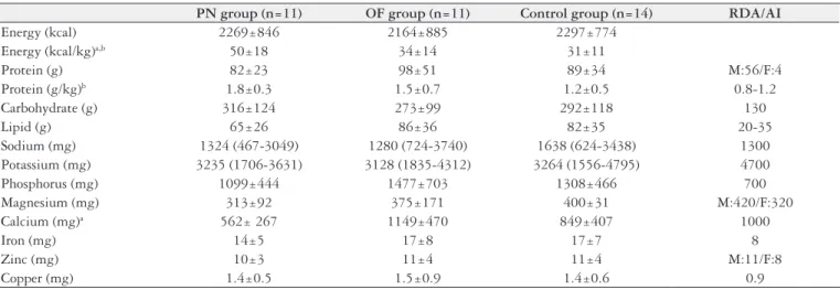
TABLE 1. Daily oral nutritional intake of patients with short bowel syndrome on intermittent parenteral nutrition (PN group), receiving oral feeding (OF group) and healthy volunteers (Control group)
PN group (n=11) OF group (n=11) Control group (n=14) RDA/AI
Energy (kcal) 2269±846 2164±885 2297±774 Energy (kcal/kg)a,b 50±18 34±14 31±11
Protein (g) 82±23 98±51 89±34 M:56/F:4
Protein (g/kg)b 1.8±0.3 1.5±0.7 1.2±0.5 0.8-1.2
Carbohydrate (g) 316±124 273±99 292±118 130
Lipid (g) 65±26 86±36 82±35 20-35
Sodium (mg) 1324 (467-3049) 1280 (724-3740) 1638 (624-3438) 1300 Potassium (mg) 3235 (1706-3631) 3128 (1835-4312) 3264 (1556-4795) 4700 Phosphorus (mg) 1099±444 1477±703 1308±466 700 Magnesium (mg) 313±92 375±171 400±31 M:420/F:320
Calcium (mg)a 562± 267 1149±470 849±407 1000
Iron (mg) 14±5 17±8 17±7 8
Zinc (mg) 10±3 11±4 11±4 M:11/F:8
Copper (mg) 1.4±0.5 1.5±0.9 1.4±0.6 0.9
a: P< 0.05 between PN group and OF group; b: P< 0.05 between PN group and Control group; RDA: recommended dietary allowances; AI: adequate intakes; M: male; F: female
TABLE 3. Serum concentration of minerals and electrolytes in patients with short bowel syndrome on intermittent parenteral nutrition (PN group),
receiving oral feeding (OF group) and healthy volunteers (Control group)
PN group (n=11) OF group (n=11) Control group (n=14) Reference range
Sodium (µmol/L) 140±3 141±3 139±2 135-145
Potassium(µmol/L) 3.7±0.6 4.2±0.6 4.1±0.5 3.5-5.0 Phosphorus(mg/dL) 3.4±1 3.6±1.1 3.6±0.4 2.5-4.9 Magnesium (mEq/L)a,b,c 1.0±0.4 1.4±0.3 1.8±0.1 1.5-2.3
Calcium total (mg/dL) 8.8±1.0 9.2±0.9 9.4±0.3 8.5-10.1
Iron(mg/dL) 102±37 83±27 88±24 35-140
Zinc (µg/dL) 68±25 84±37 90±9 50-120
Copper (µg/dL)b,c 69±24 72±26 109±16 70-140
a: P<0.05 between PN group and OF group; b: P<0.05 between PN group and control group; c: P<0.05 between OF group and Control group.
TABLE 2. Clinical and nutritional laboratory data of patients with short bowel syndrome on intermittent parenteral nutrition (PN group), receiving
oral feeding (OF group) and healthy volunteers (Control group)
PN group (n=11) OF group (n=11) Control group (n=14) Reference range
Blood glucose (mg/dL) 73±16 80±11 90±10 70-100
Urea (mg/dL) 22±10 32±17 29±4 15-40
Creatinine (mg/dL) 1.0±0.5 0.7±0.2 0.9±0.2 0.7-1.5 Hemoglobin (g/dL)a,b 11.5±1.6 13.2±1.5 14±1.4 M:13.5-17.5/ F:12-15.5
MCV (fL)c 94±4 96±5 90±4 82-98
Leukocytes (103/mm3) 6.0±1.2 5.8±1.6 6.7±2.0 4-11
Lymphocytes (103/mm3) 1.7±0.6 1.8±0.6 1.8±0.5 >1000
Total Proteins (g/dL)a,b 6.1±0.5 6.7±0.5 6.8±0.3 6.4-8.2
Albumin (g/dL) 3.9±0.7 4.2±0.4 4.3±0.1 3.5-5.0 Transferrin (mg/dL)a,b 140±61 202±98 202±29 250-310
Total bilirubin (mg/dL) 0.6±0.3 0.6±0.3 0.7±0.2 0.2-1.2 AST (U/L) 25 (17-327) 25 (7-35) 23 (18-44) 15-37 ALT (U/L) 30 (20-145) 29 (14-50) 23 (12-88) 30-65
CRP (mg/dL) 0.3±0.3 0.3±0.3 0.2±0.04 ≤0.5
Ferritin (ng/mL) 326 (17-1475) 96 (32-861) 106 (5-495) M:28-397/F:6-159
α1-AGP (mg/dL)a,b 121±43 73±21 79±18 50-120
Cholesterol (mg/dL)a,b,c 87±21 151±45 208±25 < 200
Triglycerides (mg/dL) 113±49 100±41 129±73 <150
a: P< 0.05 between PN group and OF group; b: P< 0.05 between PN group and Control group; c: P< 0.05 between OF group and Control group. MCV: mean corpuscular volume; AST:
Hyponatremia and hypoferremia were not observed in SBS patients. Figure 1 shows the number of patients with serum levels of K, P, Mg, Ca, Zn and Cu below reference values. Hypomagnesaemia (P=0.03, Fisher’s exact test) was more frequent in the PN group when compared to the OF group. No abnormalities in the electrolyte and mineral levels in the control group volunteers were observed, except for a slight hypokalemia in one individual who was using diuretics for the treatment of arterial hypertension.
sented lower hemoglobin values with no evidence of macro-cytosis, microcytosis or reduced serum Fe levels. In patients
under home PN, 73% presented normochromic anemia(8),
and 55% had evidence of iron-deiciency anemia, ascribed to mild Fe loss from the gastrointestinal tract(23). However, Fe serum levels were normal in tissue samples obtained from the autopsy of SBS patients who were under home PN(17). Our patients did not receive intravenous Fe and all of them were advised to take Fe sulfate tablets, which may contribute to the maintenance of serum Fe levels within normal range in SBS individuals(15).
PN-dependent patients presented high plasma ferritin levels and low plasma transferrin, indicating an iron over-load due to excessive supplementation via Fe sulfate tablets. In this context, the mechanism of anemia observed in these patients may be more related to low protein status rather than Fe status.
In our study, the patients who had been weaned off PN presented higher MCV, which may in its turn be caused by Cu deiciency(14, 43). Cu deiciency was common in our SBS patients, especially among those who depend on PN. Such results can’t be explained by an insuficient IV Cu replace-ment, since the pharmaceutical form routinely used in SBS exceeded the recommended amount(1). The intermittent PN infusion scheme and diminished Cu absorption capacity may explain the mechanism underlying Cu deiciency in SBS patients. In addition, the possibility of an increased Cu depletion due to the activity of cuproenzymes such as the antioxidant superoxide dismutase, cannot be ruled out(19).
Different from our indings, 67% of children with intes-tinal failure presented Zn deiciencies as they transitioned from PN to enteral nutrition(47). On the other hand, a modest elevation in Zn levels was observed in tissue samples from autopsied PN-dependent SBS patients28. Plasma or serum Zn concentrations are most widely used to measure Zn status, however, these values may be negatively affected by factors other than those related to Zn losses(27, 28).
Ca consumption in the PN group was lower than in the OF group, probably due to nutritional instructions for limi ting milk and dairy products’ intake, or insuficient sup-plementation(13). Also, similarly to our results, approximately one third of home PN patients present hypocalcemia(8). Its main mechanism is ascribed to Ca precipitation with unabsorbed fatty acids(35), which result in a less effective ab-sorption(12) and consequent calcium loss in stools(25). Also, a large supply of parenteral amino acid may induce excessive calcium loss by urine(4). On the other hand, a preserved colon may play an important role in calcium absorption in patients with extensive small bowel resection(18). Accordingly to our results, phosphorus deiciency was uncommon in SBS pa-tients under long-term PN, because of adequate oral intake and phosphorus absorption(25).
Similarly to our indings, Na deiciency has not been reported in patients receiving long-term PN due to SBS(24, 26). Fecal Na loss is excessive when the colon is absent and re-sults in extremely low Na excretion in the urine(24). Although mean values of K serum levels were similar between the SBS
Cu <70μg/dL
Zn <50μg/dL
Ca <8.5 mg/dL
Mg <1.5 mEq/L
P <2.5mg/dL
K <3.5μmol/L
0 1 2 3 4 5 6 7 8 9 10 11
OF Group PN Group patients
FIGURE 1. Amount of patients with short bowel syndrome who presented
serum levels of electrolytes and minerals below the reference values in the PN group (n=11) and OF group (n=11).
DISCUSSION
In the present study, low Cu and Mg serum levels were observed in patients with intestinal resection as compared to healthy volunteers, especially in those under intermittent PN. There were no differences in the serum levels of Na, K, P, Ca, Fe and Zn between the groups, and apart from Na and Fe within normal range, all mineral values were lower than reference values.
In this study, hypomagnesaemia was highly prevalent in both PN-dependent patients and in those under oral feeding. Mg deiciency is often present in hospitalized patients(10) and is common in SBS(37), regardless of the short intestine’s ab-sorptive capacity and the length of the residual jejunum(31). In a retrospective study of 15 patients with severe intestinal resection, 66% presented hypomagnesaemia(30). In normal conditions, 25% to 60% of dietary Mg is absorbed in the gastrointestinal tract(36). In patients with severe SBS, net Mg absorption is very low or slightly negative(25), and a positive balance is only obtained with parenteral Mg supply(25, 30). Fatty acids, derived from the digestion of fat or bacterial carbohydrate fermentation, bind to Mg and result in insuf-icient absorption(31). In normal conditions, the kidneys are responsible for regulating the total body content of Mg(39). This regulation depends on glomerular iltration, volume status and various metabolic states(36), including the acid load in the body(40). Lactic acidosis is a consequence of SBS(6, 48), but there are no available studies describing the inluence of the acid-base status on renal Mg excretion in SBS patients.
pre-patient groups (PN and OF), a greater number of individuals with K deiciency was found in the PN group. Subjects with remaining jejunum length ranging from 50 cm to 150 cm absorb around 60% of dietary K(24). Besides, malnourished adults present lower K serum levels than those with a nor-mal nutritional status(11). We can’t reject the hypothesis that the K serum levels in PN group may be a relection of renal insuficiency at some extent.
Deiciencies of electrolytes and minerals observed in this study can’t be explained by an inadequate intake, even though eating stimulates enteric losses rich in these elements(15). An intestinal biopsy was not performed, which could indicate adaptive changes in the mucosa. After proximal small bowel resections, the ileum is capable of adapting to the loss of absorptive surface area by increased villous height and crypt depth(9). This adaptive mechanism effectively increases the absorption of water, electrolytes, and nutrients to maintain adequate hydration and nutrition(20). Furthermore, patients with SBS with severe malabsorption present adaptive colonic changes that include an increased absorptive surface with an unchanged proliferative/apoptotic ratio and well-preserved absorptive protein transporters(22).
The assessment of mineral and electrolyte levels in PN-de-pendent patients is dificult to perform because of many confounding factors. We performed blood collection before the start of PN infusion, since blood mineral and electrolyte levels may be inluenced by the infusion and consequently not reflect tissue concentrations. Even by taking these precautions, serum levels may not be as reliable as careful balance studies(15). Decreased mineral and electrolyte serum levels may be indicative of real deiciencies or redistribu-tion-related deiciencies. Although the acute inlammatory
response does not affect erythrocyte concentrations of Cu(33), acute phase reactions related to infectious or inlammatory conditions may cause a redistribution of minerals(42). In this context, Mg and Cu deiciencies in SBS patients under PN may be a result of an inlammatory process, since some of these patients also presented increased g1-AGP, which is an acute-phase marker. Even in the absence of clinical signs of infection, the increase in g1-AGP levels may be associated with the presence of totally implantable venous catheters and PN infusion. This interpretation is corroborated by the fact that patients in the OF group also presented decreased Cu and Mg but 1-AGP levels within normal range. Besides, changes in serum levels of minerals may be more related to changes in the trace-element-binding proteins’ concentration rather than a relection of deiciencies. Also, alterations in the acid-base balance may inluence the urinary excretion of cations, leading to mineral deiciencies.
In conclusion, hypomagnesaemia and hypocupremia were highly prevalent in PN-dependent patients, and K, Ca and Zn deiciencies were also common. SBS patients who had been weaned off PN for a long time are still at risk of Cu and Mg deiciencies. These patients need a long-term follow-up and an intensive nutritional care in order to pre-vent electrolyte and mineral deiciencies. The treatment of patients with SBS requires a multidisciplinary approach and any potential complicating factor must be identiied, closely monitored and treated early.
Authors’ contributions
All authors have contributed signiicantly to the design, execution, analysis and writing of the inal submitted version, and share responsibility for the contents of the paper.
Braga CBM, Ferreira IML, Marchini JS, Cunha SFC. Deiciência de cobre e magnésio em pacientes com Síndrome do Intestino Curto sob nutrição parenteral ou alimentação por via oral. Arq Gastroenterol. 2015,52(2):94-9.
RESUMO - Contexto - Ressecções intestinais extensas resultam em perda de luídos e eletrólitos. Objetivo - Avaliar os níveis séricos de minerais e eletrólitos em pacientes com síndrome do intestino curto, dependentes ou não de nutrição parenteral. Métodos - O estudo incluiu 22 adultos com síndrome de intestino curto, sendo 11 dependentes de nutrição parenteral (Grupo NP) e 11 sujeitos que recebiam todo aporte nutricional por via oral (Grupo VO). Foram incluídos 14 voluntários saudáveis, pareados para a idade e o gênero (Grupo Controle). A avaliação da ingestão alimentar, antropometria, níveis sanguíneos de sódio, potássio, fósforo, magnésio, cálcio, zinco, ferro e cobre foram documentados em todos os voluntários. Resultados - Os níveis sanguíneos de sódio, potássio, fósforo, cálcio e zinco foram similares entre os grupos de estudo. Os níveis séricos de magnésio foram menores no Grupo NP (1,0±0,4 mEq/L) em relação aos demais grupos. Além disso, a concentração desse eletrólito foi menor no Grupo VO (1,4±0,3 mEq/L) em relação ao Grupo Controle (1,8±0,1 mEq/L). Foram documentados menores valores cobre (69±24 vs 73±26 vs 109±16 µg/dL) nos grupos NP e VO quando comparados com o Grupo Controle, respectivamente. Conclusão - Hipomagnesemia e hipocupremia são distúrbios eletrolíticos comumente observados na síndrome de intestino curto. Os pacientes com ressecção intestinal extensa requerem monitorização e suplementação de magnésio e cobre a im de prevenir deiciências.
REFERENCES
1. American Society for Parenteral and Enteral Nutrition (A.S.P.E.N.) Board of Directors. Clinical Guidelines for the Use of Parenteral and Enteral Nutri-tion in Adult and Pediatric Patients. A.S.P.N. JPEN J Parenter Enteral Nutr. 2009;33(3):255-9.
2. Association of Oficial Analytical Chemistry. Oficial Methods of Analysis of the Association of Oficial Analytical Chemists. 13th ed. Washington-DC:AD-AC;1980.
3. Azevedo FA, Chasin AAM. Metais Gerenciamento da toxicidade. São Paulo, SP: Atheneu Intertox; 2003.
4. Bleich HL, Moore MJ, Lemann J Jr, Adams ND, Gray RW. Urinary Calcium excretion in human beings. New Engl J Med. 1979;301(10):535-41.
5. Bohuon C. Mircodetermination of magnesium in various biological media. Clin Chim Acta. 1962;7:811-7.
6. Bongaerts GP, Severijnen RS. Arguments for a lower carbohydrate-higher fat diet in patients with a short small bowel. Med Hypotheses. 2006;67(2):280-2. 7. Braga CBM, Vannucchi H, Freire CMM, Marchini JS, Jordão AA Jr, da Cunha
SF. Serum Vitamins in Adult Patients with Short Bowel Syndrome Receiving In-termittent Parenteral Nutrition. JPEN J Parenter Enteral Nutr. 2011;35(4):493-8. 8. Burnes JU, O’Keefe SJ, Fleming CR, Devine RM, Berkner S, Herrick L. Home
parenteral nutrition a 3-year analysis of clinical and laboratory monitoring. JPEN J Parenter Enteral Nutr. 1992;16(4):327-32.
9. Chagas Neto FA, Barreto ARF, Muglia VF, Elias Junior J, Bellucci AD, Marchini JS, Cunha SFC. Barium follow through in the assessment and follow-up of adult patients with short bowel syndrome. Radiol Bras. 2011;44(3):188-91.
10. Cunha DF, Bianco MP, Lenza RM, et al. [Acute phase response and serum mag-nesium levels among hospitalized patients]. Rev Assoc Med Bras. 1999;45(2):142-6. 11. Cunha DF, Cunha SFC, Ferreira TP, Sawan ZT, Rodrigues LS, Prata SP, Silva-Ver-gara ML. Prolonged QTc intervals on the electrocardiograms of hospitalized malnourished adults. Nutrition. 2001;17(5):370-2.
12. Ellegard L, Bosaeus I, Nordgren S, Bengtsson BA. Low-dose recombinant human growth hormone increases body Wright and lean body mass in patients with short bowel syndrome. Ann Surg.1997;225(1):88-96.
13. Estívariz CF, Luo M, Umeakunne K, Bazargan N, Galloway JR, Leader LM, Ziegler TR. Nutrient intake from habitual oral diet in patients with severe short bowel syndrome living in the Southeastern United States. Nutrition. 2008;24(4):330-9.
14. Fuhrman MP, Herrmann V, Masidonski P, Eby C. Pancytopenia after remov-al of copper from totremov-al parenterremov-al nutrition. JPEN J Parenter Enterremov-al Nutr. 2000;24(6):361-6.
15. Gabriel FR, Suen VM, Marchini JS, Dutra de Oliveira JE. Ingestion of ferrous sulfate increases ferremia in patients with short bowel syndrome. Nutrition. 2009;25(11-12):1115-9.
16. Goldenbeg H, Fernandez A. Simpliied method for the estimation of inorganic phosphorus in body luids. Clin Chem. 1966;12(12):871-82.
17. Howard L, Ashley C, Lyon D, Shenkin A. Autopsy tissue trace elements in 8 long-term parenteral nutrition patients who received the current U.S. Food and Drug Administration formulation. JPEN J Parenter Enteral Nutr. 2007;31(5):388-96. 18. Hylander E, Ladefoged K, Jarnum S. The importance of colon in calcium absorp-tion following small intestinal resecabsorp-tion. Scand J Gastroenterol. 1980;15(1):55-60. 19. Imamoglu N, Yerer MB, Donmez-Altuntas H, Saraymen R. Erythrocyte antiox-idant enzyme activities and lipid peroxidation in the erythrocyte membrane of stainless-steel welders exposed to welding fumes and gases. Int J Hyg Environ Health. 2008;211(1-2):63-8.
20. Iqbal CW, Qandeel HG, Zheng Y, Duenes JA, Sarr MG. Mechanisms of ileal adaptation for glucose absorption after proximal-based small bowel resection. J Gastrointest Surg. 2008;12(11):1854-64.
21. Jeejeebhoy KN. Short bowel syndrome: a nutritional and medical approach. CMAJ. 2002;166(10):1297-302.
22. Joly F, Mayeur C, Messing B, Lavergne-Slove A, Cazals-Hatem D, Noordine ML, et al. Morphological adaptation with p reserved proliferation/transporter content in the colon of patients with short bowel syndrome. Am J Physiol Gastrointest Liver Physiol. 2009;297(1):G116-23.
23. Khaodhiar L, Keane-Ellison M, Tawa NE, Thibault A, Burke PA, Bistrian BR. Iron deiciency anemia in patients receiving home total parenteral nutrition. JPEN J Parenter Enteral Nutr. 2002;26(2):114-9.
24. Ladefoged K, Olgaard K. Fluid and electrolyte absorption and renin-angio-tensin-aldosterone axis in patients with severe short-bowel syndrome. Scand J Gastroenterol. 1979;14(6):729-35.
25. Ladefoged K, Nicolaidou P, Jarnum S. Calcium, phosphorus, magnesium, zinc and nitrogen balance in patients with severe short bowel syndrome. Am J Clin Nutr. 1980;33(3):2137-44.
26. Ladefoged K, Olgaard K. Sodium homeostasis after small-bowel resection. Scand J Gastroenterol. 1985;20(3):361-9.
27. Lowe NM, Woodhouse LR, Sutherland B, Shames DM, Burri BJ, Abrams SA, et al. Kinetic parameters and plasma zinc concentration correlate well with net loss and gain of zinc from men. J Nutr. 2004;134(9):2178-81.
28. Lowe NM, Fekete K, Decsi T. Methods of assessment of zinc status in humans: a systematic review. Am J Clin Nutr. 2009;89(6):2040S:51S.
29. Matarese LE, Steiger E. Dietary and medical management of short bowel syn-drome in adult patients. J Clin Gastroenterol. 2006;40(Suppl 2):S85-93. 30. Miranda SC, Ribeiro MLB, Ferriolli E, Marchini JS. Hypomagnesemia in short
bowel syndrome patients. São Paulo Med J. 2000;118(6):169-72.
31. Nightingale JM. Management of patients with a short bowel. World J Gastro-enterol. 2001;7(6):741-51.
32. Nonino CB, Borges RM, Pasquali LS, Marchini JS. Oral dietetic therapy in patients with short bowel syndrome. Rev Nutr. 2001;14(3):201-5.
33. Oakes EJC, Lyon TDB, Duncan A, Gray A, Talwar D, O’Reilly DS. Acute in-lammatory response does not affect erythrocyte concentrations of copper, zinc and selenium. Clin Nutr. 2008;27(1):115-20.
34. Oldenburg WA, Lau LL, Rodenberg TJ, Edmonds HJ, Burger CD. Acute mes-enteric ischemia: a clinical review. Arch Intern Med. 2004;164(10):1054-62. 35. Ovesen L, Chu R, Howard L. The inluence of dietary fat on jejunostomy output
in patients with severe short bowel syndrome. Am J Clin Nutr. 1983;38(2):270-7. 36. Pham PC, Pham PM, Pham SV, Miller JM, Pham PT. Hypomagnesemia in Patients
with Type 2 Diabetes. Clin J Am Soc Nephrol. 2007;2(2):366-73.
37. Ross JR, Dargan PI, Jones AL, Kostrzewski A. A case of hypomagnesaemia due to malabsorption, unresponsive to oral administration of magnesium glyc-erophosphate, but responsive to oral magnesium oxide supplementation. Gut. 2001;48(6):857-8.
38. Rothery E. Analytical Methods for Graphite Tube Atomizers. Mulgrave Vitoria, Australia: Varian Australia Pty Lty; 1988.
39. Rylander R, Arnaud MJ. Mineral water intake reduces blood pressure among subjects with low urinary magnesium and calcium levels. BMC Public Health. 2004;4:56-61.
40. Rylander R, Remer T, Berkemeyer S, Vormann J. Acid-Base Status Affects Renal Magnesium Losses in Healthy, Elderly Persons. J Nutr. 2006;136(9):2374-7. 41. Sarkar BC, Chauhan UPS. A new method for determining micro quantities of
calcium in biological materials. Anal Biochem. 1967;20(1):155-66.
42. Shenkin A. Trace elements and inlammatory response: implications for nutritional support. Nutrition. 1995;11(1 suppl):100-5.
43. Spiegel JE, Willenbucher RF. Rapid development of severe copper deiciency in a patient with Crohn’s disease receiving parenteral nutrition. JPEN J Parenter Enteral Nutr. 1999;23(3):169-72.
44. Van Gossum A, Vahedi K, Abdel-Malik, Staun M, Pertkiewicz M, Shaffer J, et al. Espen-Han Working Group. Clinical, social and rehabilitation status of long-term home parenteral nutrition patients: results of a European multicentre survey. Clin Nutr. 2001;20(3):205-10.
45. Weale AR, Edwards AG, Bailey M, Lear PA.. Intestinal adaptation after massive intestinal resection. Postgrad Med J. 2005;81(953):178-84.
46. Willet W. Nutritional Epidemiology. New York, NY: Oxford University Press, Inc; 1990. p. 69-91.
47. Yang CF, Duro D, Zurakowski D, Lee M, Jaksic T, Duggan C. High prevalence of multiple micronutrient deiciencies in children with intestinal failure: a longitudinal study. J Pediatr. 2011;159(1):39-44.
48. Zhang DL, Jiang ZW, Jiang J, Cao B, Li JS. D-lactic acidosis secondary to short bowel syndrome. Postgrad Med J. 2003;79(928):110-2.