AR
TIGO ORIGINAL / ORIGINAL AR
TICLE
INTRODUCTION
In the hepatosplenic form of Schistosomiasis
mansoni (SM), portal hypertension occurs due to
both periportal ibrosis (Symmers), which increases resistance to blood low, and hyperlow in the splenic vein due to the splenomegaly(13, 18, 24). Indeed, a
cor-relation has been described between the intensity of periportal ibrosis and pressure levels in the portal vein, which can lead to bleeding through esophageal varices; moreover, an association has been described between the presence of esophageal varices and spleen diameter(1, 21).
Liver biopsy(7), an ultrasound examination of
the abdomen(3, 8), serum markers(5, 10, 16, 22), platelet
count(4, 12) and, more recently, liver stiffness
(elastog-raphy) measurement(9) have been used to assess the
ibrosis that develops in chronic liver disease. With SM, the liver-wedge biopsy is the most accurate method for measuring periportal ibrosis, but implies the use of laparotomy. Nevertheless, the needle biopsy does not always represent the true histopathological
CORRELATION BETWEEN PLATELET
COUNT AND BOTH LIVER FIBROSIS AND
SPLEEN DIAMETER IN PATIENTS WITH
SCHISTOSOMIASIS MANSONI
Tibério B
MEDEIROS
1, Ana Lucia C
DOMINGUES
1, Carlos F
LUNA
2and
Edmundo P
LOPES
1ABSTRACT - Context - Studies have described the correlation between platelet count and the stages of ibrosis in chronic viral hepatitis, but few publications have studied this correlation in Schistosomiasis mansoni. Objective - Therefore, this study aimed to correlate platelet count with both the periportal ibrosis pattern and spleen diameter evaluated by ultrasound exam in patients with Schisto-somiasis mansoni. Methods - Patients with Schistosomiasis mansoni were evaluated by abdominal ultrasound by a single examiner for the determination of periportal ibrosis pattern (Niamey classiication) and spleen diameter. Platelet counts were performed in an automated cell counter. Results - One hundred eighty-seven patients with Schistosomiasis mansoni (mean age: 50.2 years) were included in the study, 114 of whom (61%) were women. Based on the Niamey classiication, the ultrasound analysis revealed that 37, 64, 64 and 22 patients exhibited patterns C, D, E and F, respectively. In these four groups, the mean number of platelets was 264, 196, 127 and 103 x 109/L and mean spleen diameter was 9.2, 11.9, 14.9 and 16.2 centimeters, respectively. A reduction in platelet count
was signiicantly associated with both the progression of the periportal ibrosis and the increase in spleen size. Conclusion - Platelet count in patients with Schistosomiasis mansoni was inversely correlated with the severity of periportal ibrosis and spleen diameter.
HEADINGS – Liver ibrosis. Platelet count. Splenomegaly. Thrombocytopenia. Portal hypertension. Schistosomiasis mansoni.
Declared conflict of interest of all authors: none
1 Gastroenterology Section, Department of Internal Medicine, Universidade Federal de Pernambuco - UFPE, Recife, PE; 2 Centro de Pesquisas Aggeu Magalhães,
Fundação Oswaldo Cruz - FIOCRUZ, Recife, PE, Brasil.
Correspondence: Edmundo P Lopes. Rua Irmã Maria Davi, 154, ap. 3201 - 52061-070 - Recife, PE, Brasil. E-mail: [email protected]
condition, as ibrosis varies in intensity throughout the hepatic parenchyma(7).
Ultrasound is an important tool in the diagnosis of the alterations related to SM, allowing the identiica-tion of the enlargement of the left lobe of the liver, periportal thickening and the increase in the length of the spleen(3, 8, 20). The Niamey ultrasound classiication
allows determining the pattern of periportal ibrosis, which is used to assess its intensity with satisfactory accuracy and reliability(8, 20, 26). However, this method
requires the availability of ultrasound equipment and trained operators. Thus, simpler methods are desirable.
Due to the drawbacks of liver biopsies and the large-scale use of ultrasound, substances involved in the production or degradation of collagen have recently been investigated for the determination of ibrosis in liver disease(10, 12, 19, 22, 27).Stellate liver cells
leptin(2, 14, 15). Besides the proteins responsible for ibrogenesis
others stemming from the degradation process of the extra-cellular matrix have been studied in the evaluation of ibrosis, such as hyaluronic acid, laminin and type IV collagen(10, 16, 19, 27). Likewise, the number of platelets is reported to be
inversely correlated with the intensity of periportal ibrosis due to the sequestering of platelets in the splenic parenchyma stemming from splenomegaly(5, 12, 17, 23).
Considering the scarcity of accurate data regarding the measurement of ibrosis using serum markers in patients with SM, the aim of this study was to correlate platelet count with both the periportal ibrosis pattern and spleen diameter as evaluated through an ultrasound exam.
METHODS
Patients
A descriptive, cross-sectional study was carried out involving patients with SM who consecutively underwent an ultrasound exam over an eight-month period at the Gas-troenterology Section of the Hospital das Clínicas of the Universidade Federal de Pernambuco (UFPE), Brazil. The diagnosis of SM was based on their clinical history of contact with contaminated water and/or reports of prior treatment for SM and ultrasound examination of the upper abdomen showing periportal ibrosis.
Male and female patients aged 18 years or older with SM diagnosis who had not undergone splenectomy were included. The following were the exclusion criteria: presence of markers for hepatitis B and C (anti-HBc and anti-HCV); alcohol intake >210 g/week; and ultrasound evidence of other liver disease, as expressed by the presence of steatosis or ine ibrosis diffused throughout the parenchyma.
Methods
Laboratory exams were performed at the Central Labo-ratory of the aforementioned hospital. Platelet counts were performed using an automated cell counter (Cell-Dyn 3000), and anti-HBc and anti-HCV were detected using immune enzyme assays (Abbott).
Ultrasound exams were performed by a single operator (ALCD) using the Aloka SSD-500 device with convex trans-ducer a 3.5 MHz for the evaluation of the liver and spleen. Periportal ibrosis was evaluated based on the Niamey clas-siication, which has six pre-established patterns of ibrosis intensity, ranging from Pattern A (normal) to Pattern F (very advanced ibrosis)(20, 26).
Statistical analysis
Statistical analysis was performed using the SPSS 12 and Excel 2000 programs. The variables were presented in tables and graphs, with the data expressed as mean and standard deviation values. The Kolmogorov-Smirnov test was used to determine the distribution of the data (parametric or non-parametric). ANOVA followed by Levene’s test was used for the determination of equal variances in the analysis of the variables according to the ibrosis pattern. As heterogeneity
was demonstrated, Tamhane’s test was used. Pearson’s cor-relation coeficients were calculated to determine corcor-relations between the platelets count and spleen diameter. The level of signiicance was set to 5% (P<0.05) for all statistical tests.
RESULTS
Among the 238 patients with SM who underwent an ul-trasound exam and exhibited periportal ibrosis, 51 (21%) of whom presented exclusion criteria. Thus, 187 patients were included in the study, 114 (61%) of whom were female and 73 (39%) were male. Mean age was 50.2 years (22 to 77 years).
Table 1 exhibit the distribution of the patients according to the ibrosis pattern (Niamey classiication), mean platelet count and spleen diameter.
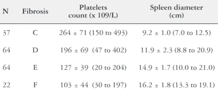
Table 1. Mean platelet count (x 109/L) and spleen diameter (cm) according
to periportal ibrosis pattern (Niamey classiication) in 187 patients with
Schistosomiasis mansoni
N Fibrosis Platelets count (x 109/L)
Spleen diameter (cm)
37 C 264 ± 71 (150 to 493) 9.2 ± 1.0 (7.0 to 12.5)
64 D 196 ± 69 (47 to 402) 11.9 ± 2.3 (8.8 to 20.9)
64 E 127 ± 39 (20 to 204) 14.9 ± 1.7 (10.0 to 21.0)
22 F 103 ± 44 (30 to 197) 16.2 ± 1.8 (13.3 to 19.1)
Figure 1 displays the distribution of the mean platelets counts according to the ibrosis pattern evaluated using ul-trasound in the 187 patients (ANOVAF = 57.26; P<0.001). Tamhane’s test revealed signiicant differences in the platelet count between the different ibrosis patterns (P<0.001), ex-cept between Patterns E and F (P = 0.203).
FIGURE 1. Distribution of mean platelet count (x 109/L) according to
periportal ibrosis pattern (Niamey classiication) in 187 patients with
Figure 2 displays the distribution of the mean spleen diameter (in cm) according to the ibrosis pattern evaluated using ultrasound in the 187 patients (ANOVA F = 108.53;
P<0.001). Tamhane’s test revealed signiicant differences in mean spleen diameter between Patterns C and D and Pat-terns D and E of ibrosis (P<0.001), and between Patterns E and F (P = 0.037).
DISCUSSION
The present indings demonstrate that severe cases of SM continue to occur, as nearly half (45%) of the patients analyzed exhibited advanced patterns of periportal ibrosis (E and F). These patients with more advanced ibrosis pat-terns and with both platelet count and spleen size outside the range of normality have the hepatosplenic form of disease(8).
Additionally, they presented higher levels of portal pressure and a consequently greater risk of digestive bleeding(6, 21).
In clinical practice, propaedeutic methods for the diagno-sis, measurement and follow up of periportal ibrosis continue to be necessary. Therefore, a simple method as platelets count can be taken into the ield for monitoring patient with portal hypertension in endemic areas.
The recent advent of hepatic elastography allows the measurement of the rigidity of the liver parenchyma and offers the same advantages as ultrasound scan in that it is a noninvasive procedure that allows evaluating a large por-tion of the parenchyma and can be employed repeatedly throughout cirrhotic patient follow up. The main advantages of elastography, however, include the determination of a numeric mean for some measurements and the fact that it is less dependent on the operator than an ultrasound exam, thereby allowing its use by healthcare professionals without the need for vast experience(9). Nonetheless, the equipment
is expensive and there are no data on its accuracy regarding the evaluation of periportal ibrosis in SM.
Due to the drawbacks of liver biopsies, blood markers involved in the metabolism of collagen have been investi-gated in periportal ibrosis(10, 19, 27). Using ultrasound with the
Niamey classiication as the gold standard, correlations have been described between patterns of ibrosis and serum levels of IgG and hyaluronic acid(5, 22). Though, the determination
of these substances requires sophisticated methods that are generally not available in simpler laboratories found in endemic regions for schistosomiasis.
An inverse correlation has been described between peri-portal ibrosis and platelet count in patients with SM. Indeed, Koepke-Aguiar et al.(11) found more severe thrombocytopenia
in patients with advanced forms of hepatosplenic schisto-somiasis with portal hypertension.In addition, studying 47 patients with SM and 13 controls, Lambertucci et al.(12) found
a mean number of 194 x 109/L platelets in the controls and
44 x 109/L in patients with severe periportal ibrosis, as
as-sessed by ultrasound. In this same study, the AST to Platelet Ratio Index (APRI) was also calculated to measure ibrosis, which demonstrated similar accuracy to that achieved with the platelet count.
In our study involving 187 patients, the mean platelet count was progressively lower with the increase of the ibrosis severity measured using ultrasound. It should be stressed that this is the irst study to demonstrate a strong correlation between the platelet count and the four patterns of periportal ibrosis of the Niamey classiication, as well as an inverse correlation between the mean platelet number and spleen diameter. Thrombocytopenia in patients with SM is believed FIGURE 2. Distribution of mean spleen diameter (cm) according to
periportal ibrosis pattern (Niamey classiication) in 187 patients with
Schistosomiasis mansoni (ANOVA F = 108.53; P<0.001)
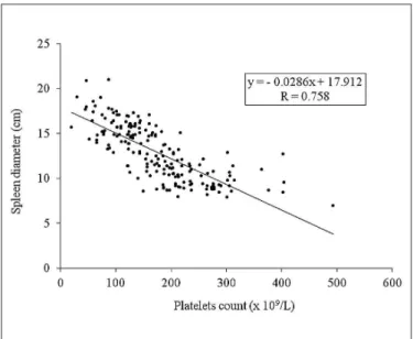
FIGURE 3. Correlation between spleen diameter (cm) and platelet count (x 109/L) in 187 patients with Schistosomiasis mansoni (P<0.001)
to stem from the retention of platelets in the parenchyma of the spleen(5, 15, 23). In fact, in our study, thrombocytopenia was
greater in those patients with a more enlarged spleen, denot-ing more advanced patterns of ibrosis (E + F).
The correlations found in the present study likely stem from the pathophysiology of portal hypertension in SM. There is an increased resistance to blood low, as in cir-rhosis, due to ibrosis secondary to the deposition of eggs in the portal branches, along with an increase in blood low in the portal vein due to the splenomegaly induced by both the parasites and passive congestion(18, 21, 24). Thus, a greater
quantity of eggs (parasite load) may cause both a greater ibrotic reaction around the egg as well as greater dificulty in hepatic blood low and greater splenomegaly due to im-munological stimulation and congestion, which may lead to the greater sequestration of platelets(5).
The strong correlation between the number of platelets and the spleen diameter observed in SM could also be strengthened by the absence of other factors that induces thrombocytopenia, which are found in the cirrhosis(25). For
example, in this parasitic disease the production of throm-bopoietin is higher that described in the cirrhotic patients, as well as in the SM antibodies antiplatelets are not observed as found in the HCV chronic liver disease(11, 25).
Recently, a smaller number of platelets and larger spleen diameter have been described in patients with SM and with esophageal varices in comparison to those without varices(1).
These authors propose an index (platelets count/spleen di-ameter ratio) for the noninvasive diagnosis of esophageal varices in patients with SM(1).
According to the present findings, platelet count in patients with SM was inversely correlated with the sever-ity of periportal ibrosis and spleen diameter. Therefore, the platelet count constitutes a tool for the evaluation of periportal ibrosis severity in patients with SM, offering the advantages of high accuracy, simplicity and low cost. However, further studies are needed to validate these data and compare the platelet count to other markers of peri-portal ibrosis in SM.
Authors’ contributions
Tibério B Medeiros and Ana Lucia C Domingues con-ceive and design the study; acquisition of data; analysis and interpretation of data; Carlos F Luna interpretation of the data and statistical analysis; Edmundo P Lopes analysis and interpretation of data; drafting of the manuscript. All authors read and approved the inal manuscript.
Ethical clearance
Prior to inclusion, the patients received information on the objectives and procedures and agreed to participate by signing a statement of informed consent. This study received approval from the Human Research Ethics Committee of the UFPE Center for Health Sciences.
Medeiros TB, Domingues ALC, Luna CF, Lopes EP. Correlação entre o número de plaquetas e o grau de ibrose hepática e o tamanho do baço em pacientes com Esquistossomose mansoni. Arq Gastroenterol. 2014,51(1):34-8.
RESUMO - Contexto - Estudos vem descrevendo correlação entre o número de plaquetas e o grau de ibrose hepática na hepatite viral crônica, mas poucas publicações estudaram esta correlação em pacientes com Esquistossomose mansoni. Objetivo - Correlacionar a contagem de plaquetas com o padrão de ibrose periportal e com o diâmetro do baço, avaliados pela ultrassonograia em pacientes com Esquistossomose mansoni. Método - Os pacientes com Esquistossomose mansoni foram avaliados pela ultrassonograia abdominal, por um único examinador, para determinação do padrão de ibrose periportal (classiicação de Niamey) e do diâmetro do baço. A contagem de plaquetas foi realizada em contador automatizado. Resultados – Cento e oitenta e sete pacientes com Esquistossomose mansoni com média de idade de 50,2 anos foram incluídos no estudo, 114 (61%) dos quais eram mulheres. De acordo com a classiicação de Niamey, a ultrassonograia revelou que 37, 64, 64 e 22 pacientes exibiam padrões C, D, E e F, respectivamente. Nestes quatro grupos, o número médio de plaquetas foi 264, 196, 127 e 103 x 109/L, respectivamente, e o diâmetro médio do baço foi 9,2, 11,9, 14,9 e 16,2
centímetros, respectivamente. Observou-se, portanto, redução signiicativa na contagem de plaquetas associada à progressão da ibrose periportal e ao aumento do tamanho do baço. Conclusão – Neste estudo veriicou-se que a contagem de plaquetas foi inversamente correlacionada com o padrão de ibrose periportal, como também com o diâmetro do baço nos pacientes com Esquistossomose mansoni.
REFERENCES
1. Agha A, Abdualhadi MM, Marenco S, Bella A, AlSaudi D, El-Haddad A, Inferre-ra S, Savarino V, Giannini EG. Use of the platelet count/spleen diameter ratio for the noninvasive diagnosis of esophageal varices in patients with schistosomiasis.
SaudiJ Gastroenterol. 2011;17:307-11.
2. Bataller R, Brenner DA. Liver ibrosis. J Clin Invest. 2005;115:209-218.
3. Cerri GG, Alves VA, Magalhães A. Hepatosplenic schistosomiasis mansoni.
Radiology. 1984;153:777-80.
4. Correia HST, Domingues ALC, Lopes EPA, Morais CNL, Sarteschi C, Moura IMF. Níveis séricos de globulinas e a intensidade da ibrose hepática em pacientes com esquistossomose mansônica. Arq Gastr. 2009;46:194-8.
5. Correia MCB, Domingues ALC, Lacerda HR, Santos LM, Machado CGF, Hora V, Neves MA, Brito A, Coelho RCD, Silva JLA. Platelet function and the Von Willebrand factor antigen in the hepatosplenic form of schistosomiasis mansoni.
Trans R Soc Med Trop Hyg. 2009;103:1053-8.
6. Dias HS, Domingues ALC, Cordeiro FTM, Jucá NT, Lopes EP. Associating portal congestive gastropathy and hepatic ibrosis in hepatosplenic mansoni schistosomiasis. Acta Trop. 2013;126:240-3.
7. Dimmette RM. Liver biopsy in clinical schistosomiasis; comparison of wedge and needle types. Gastroenterology. 1955;29:219-34.
8. Domingues ALC, Lima ARF, Dias HS, Leão GC, Coutinho A. An ultrasono-graphic study of liver ibrosis in patients infected with Schistosoma mansoni in north-east Brazil. Trans R Soc Trop Med Hyg. 1993;87:555-8.
9. Foucher J, Chanteloup E, Verginol J, Castera L, Le Bail B, Adhoute X, Bertet J, Couzigou P, De Lédinghen V. Diagnosis of cirrhosis by transient elastography (FibroScan): a prospective study. Gut. 2006;55:403-8.
10. Köpke-Aguiar LA, Martins JR, Passerotti CC, Toledo CF, Nader HB, Borges DR. Serum hyaluronic acid as a comprehensive marker to assess severity of liver disease in schistosomiasis. Acta Trop. 2002;84:117-26.
11. Köpke-Aguiar LA, de Leon CP, Shigueoka DC, Lourenço DM, Kouyomdjian M, Borges DR. Reticulated platelets and thrombopoetin in schistosomiasis patients.
Int J LabHematol. 2009;31:69-73.
12. Lambertucci JR, Silva LC, Antunes CM. Aspartate aminotransferase to platelet ratio index and blood platelet count are good markers for ibrosis evaluation in schistosomiasis mansoni. Rev Soc Bras Med Trop. 2007;40:559.
13. Maia MD, Lopes EPA, Ferraz AAB, Barros FMR, Domingues, ALC, Ferraz EM.
Evaluation of splenomegaly in the hepatosplenic form of mansoni schistosomiasis.
Acta Trop. 2007;101:183-6.
14. Morais, CNL Montenegro, SML, Abath, FGC, Souza W, Jucá NT, Domingues, ALC, Lopes EPA, Melo, WG, Carvalho, BM. Preliminar evaluation of cyto-kines in the hepatitis C/schistosomiasis co-infection. Mem Inst Oswaldo Cruz. 2006;101:353-4.
15. Oliveira LFA, Moreno EC, Gazzinelli G, Martins-Filho AO, Silveira MAS, Gazzinelli A, Malaquias LCC, LoVerde P, Leite PM, Correa-Oliveira R. Cytokine production associated with periportal ibrosis during chronic Schistosomiasis mansoni in humans. Infect Immun. 2006;74:1215-21.
16. Parise ER, Rosa H. Serum laminin in hepatic schistosomiasis. Trans R Soc Trop Med Hyg. 1992;86:179–81.
17. Petroianu A, Oliveira AE, Alberti LR. Hypersplenism in schistosomotic portal hypertension. Arch Med Res 2005; 36:496-501.
18. Raia S, Mies S, Alieri JF. Portal hypertension in mansonic schistosomiasis. World J Surg. 1991;15:176-87.
19. Ricard-Blum S, Hartmann DJ, Grenard P, Ravaoalimalala VE, Boisier P, Es-terre P. Relationships between several markers of extracellular matrix turnover and ultrasonography in human schistosomiasis mansoni. Am J Trop Med Hyg. 1999;60:658-63.
20. Richter J, Domingues ALC, Barata CH, Prata AR, Lambertucci JR. Report on the second satellite symposium on ultrasound in schistosomiasis. Mem Inst Oswaldo Cruz. 2001;96:151-6.
21. Richter J, Monteiro Eda S, Braz RM, Abdalla M, Abdel-Rahim IM, Fano U, Huntgeburth U, Feldmeier H. Sonographic organometry in Brazilian and Suda-nese patients with hepatosplenic schistosomiasis mansoni and its relation to the risk of bleeding from oesophageal varices. Acta Trop. 1992;51:281-90.
22. Silva CC, Domingues AL, Lopes EP, Moraes CN, Santos RB, Luna CF, Nader HB, Martins JR. Schistosomiasis mansoni: ultrasound evaluated hepatic ibrosis and serum concentrations of hyaluronic HCID. Ann Trop Med Parasit 2011; 105:233-9.
23. Souza MR, Toledo CF, Borges DR. Thrombocytemia as a predictor of portal hypertension in schistosomiasis. Dig Dis Sci. 2000;45:1964-70.
24. Vezozzo DC, Farias AQ, Cerri GG, Da Silva LC, Carrilho FJ. Assessment of portal hemodynamics by Doppler ultrasound and of liver morphology in the hepatosplenic and hepatointestinal forms of schistosomiasis mansoni. Dig Dis Sci. 2006;51:1413-9.
25. Violi F, Basili S, Raparelli V, Chowdary P, Gatt A, Burroughs AK. Patients with liver cirrhosis suffer from primary haemostatic defects? Fact or iction?J Hepatol. 2011;55:1415-27.
26. World Health Organization (WHO). Ultrasound in schistosomiasis. A pratical guide to the standardized use of ultrasonography for the assessment of schisto-somiasis-related morbidity. Second International Workshop. 22-26 October, 1996, Niamey, Niger.
27. Wyszomirska RM, Nishimura NF, Almeida JR, Yamanaka A, Soares EC. High serum laminin and type IV collagen levels in schistosomiasis mansoni. Arq Gastr. 2005;42:221-5.