Improving breast volume assessment in immediate
reconstruction using permanent expanders
Aperfeiçoando a mensuração do volume mamário na reconstrução imediata
com expansores permanentes
ABSTRACT
Background: The accurate determination of breast volume improves the outcome of reconstructive procedures. The aim of the present study is to correlate the preoperative measurement of breast volume using the plastic shells method with the intraoperative application of the Archimedes principle to assess breast volume in immediate breast reconstruction using permanent expanders in patients having breasts with a small volu me and mild ptosis grade. Methods: Ten patients were enrolled in this retrospective non randomized study. Breast volume was measured preoperatively using fixedvolume plastic shells and intraoperatively using the Archimedes method. An appropriate perma nent expander was selected and inserted in a retromuscular position. The team classified breast symmetry as poor, good, or very good. Results: Differences between pre and in traoperative measurements were statistically significant (P < 0.01, Student’s ttest). Preoperative values were lower than intraoperative values by 15% in 70% of the cases.
Conclusions: Breast volume assessment in immediate reconstruction procedures with permanent expanders can be improved and yield more predictable results by the combi ned use of different pre and intraoperative measuring techniques. The method that uses fixedvolume plastic shells tends to underestimate the resected breast volume compared to the intraoperative method that uses the Archimedes principle.
Keywords: Breast. Mammaplasty. Breast implants. Reconstructive surgical procedures. Sur gery, plastic.
RESUMO
Introdução: A determinação precisa do volume mamário pode melhorar o resultado dos procedimentos de reconstrução. O objetivo deste estudo é correlacionar a mensuração préoperatória do volume mamário utilizando o método de conchas plásticas e a aplicação intraoperatória do princípio de Arquimedes para a avaliação volumétrica das mamas, em pacientes submetidas a reconstrução mamária imediata utilizando expansores permanentes, portadoras de mamas de pequeno volume e grau ptótico leve. Método: Dez pacientes foram incluídas em um estudo retrospectivo não randomizado. O volume da mama foi mensurado, no préoperatório, com conchas plásticas com volumes predeterminados e, no período intra operatório, com o método de Arquimedes. Expansor apropriado permanente foi selecionado e inserido de maneira retromuscular. A equipe classiicou a simetria da mama como pobre,
This study was performed at the Serviço de Microcirurgia Reconstrutiva e Cirurgia Plástica da Santa Casa de Porto Alegre – Paulo Roberto Franco Azambuja, Porto Alegre, RS, Brazil.
Submitted to SGP (Sistema de Gestão de Publicações/Manager Publications System) of RBCP (Revista Brasileira de Cirurgia Plástica/Brazilian Journal of Plastic Surgery).
Article received: October 2, 2012 Article accepted: December 23, 2012
1. Plastic Surgeon, full member of Sociedade Brasileira de Cirurgia Plástica (Brazilian Society of Plastic Surgery – SBCP), Managing Associate of Webster Cirurgia Plástica, Invited Professor at the Serviço de Cirurgia Plástica e Microcirurgia da Santa Casa de Misericórdia de Porto Alegre of the Universidade Federal de Ciências da Saúde de Porto Alegre (UFCSPA), Porto Alegre, RS, Brazil.
2. Resident Physician of the Serviço de Cirurgia Plástica e Microcirurgia da Santa Casa de Misericórdia de Porto Alegre e da UFCSPA, Aspiring Member in Training of the SBCP, Porto Alegre, RS, Brazil.
3. Plastic Surgeon, Full Member of the SBCP, Adjunct Professor at the Serviço de Cirurgia Plástica e Microcirurgia da Santa Casa de Misericórdia de Porto Alegre e da UFCSPA, Porto Alegre, RS, Brazil.
boa ou muito boa. Resultados: As diferenças entre as medidas pré e intraoperatórias foram estatisticamente signiicantes (P < 0,01, teste t de Student). Os valores pré operatórios foram subestimados em 70% dos casos e foram 15% menores que os valores intraoperatórios.
Conclusões: A avaliação do volume da mama em procedimentos de reconstrução imediata utilizando expansores permanentes pode ser aperfeiçoada pela combinação de diferentes técnicas de mensuração pré e intraoperatórias, levando a resultados mais previsíveis. O método de conchas plásticas de volume preestabelecido tende a subestimar o volume ressecado da mama quando comparado aos valores obtidos com método de mensuração intraoperatória utilizando o princípio de Arquimedes.
Descritores: Mama. Mamoplastia. Implantes de mama. Procedimentos cirúrgicos recons trutivos. Cirurgia plástica.
INTRODUCTION
Breast reconstruction using implants following mastec tomy is an option for women who do not have suficient sub cutaneous tissue available for autologous reconstruction. In addition, many women prefer not to undergo autologous reconstruction due to donorsite morbidity, prolonged reco very periods, and associated muscle weakness. Therefore, muscle and skin tissue expansion has become one of the most popular approaches in breast reconstruction1.
Options for implantbased breast reconstruction include singlestage reconstruction using a standard or adjustable im plant, tissue expansion followed by the placement of a per manent implant, or combined autologous tissue/implant reconstruction. The choice of procedure is based on a range of patient variables, including location and type of breast cancer, availability of donor tissue (local, regional, or distant), size and shape of the desired breast, surgical risk, and patient preference2.
The accurate determination of breast volume facilitates reconstructive procedures and helps with the planning of tissue removal in breast reduction surgery. The various methods that are currently used to assess breast size are limited by technical dificulties and unreliable volume de termination3.
The aim of the present study is to correlate preoperati ve measurements of breast volume using the plastic shell me thod with the intraoperative application of the Archimedes prin ciple to assess breast volume in immediate breast reconstruc tion using permanent expanders in patients having breasts with small volume and mild ptosis grade.
METHODS
This study was performed according to the principles of the Declaration of Helsinki on medical research involving human subjects and the guidelines and norms regulating
research involving human subjects of the Brazilian National Health Council (Resolution CNS 196/96) and was based on the Sociedade Brasileira de Cirurgia Plástica guidelines. Patients signed the informed consent document approved by the Ethics Committee of the Santa Casa de Porto Alegre prior to participating.
Ten private clinic patients were enrolled in this retrospec tive nonrandomized study between 2007 and 2011. Patient inclusion criteria were as follows: women with breast cancer who underwent radical modiied mastectomy; an estimated breast volume ≤ 500 mL; and breast ptosis < 2 cm below the inframammary fold. Patient exclusion criteria were as follows: positive lymph node biopsy, adjuvant radiotherapy, and contralateral mastopexy.
The ten patients were diagnosed with breast cancer by the oncology team based on clinical examination and complemented by mammography, mammary ultrasound, and core biopsy findings. Whenever necessary, needle insertion was performed preoperatively to locate the tumor. A preoperative biopsy of the sentinel lymph node was performed in all cases.
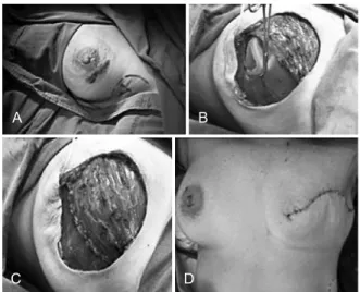
Breast volume was measured preoperatively using fixedvo lume plastic shells (Figure 1). The surgery proce dure was based on a radical modified mastectomy with removal of the skin and fascia covering the muscle. The Archimedes method was applied intraoperatively and involved the total immersion of the mastectomized tissue in a plastic container filled to the top with a 0.9% saline solution (Figure 2). The displaced solution was collected into a second container placed below the first container and was then accurately measured in a 60mL graduated syringe via needle aspiration.
A permanent expander was then selected (Menthor®
RESULTS
The mean time for the surgical procedure was 3.5 hours. In all cases, ductal carcinoma in situ was diagnosed based on anatomopathological studies. Use of the Archimedes procedure increased the mean surgical time by approximately 3 minutes.
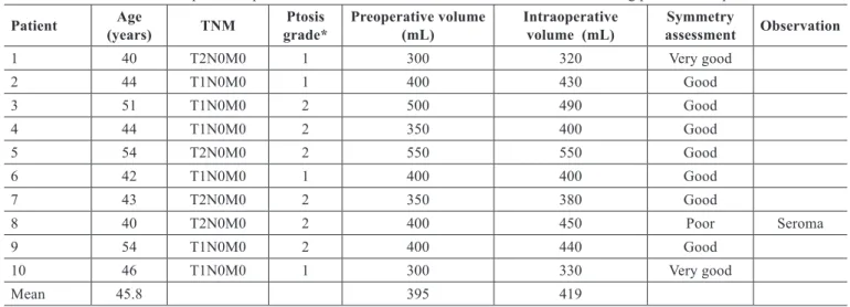
The mean preoperative breast volume measured using the plastic shells method was 395 ml (Table 1). The mean intraoperative breast volume measured using the Archime des method was 419 ml (Table 1). The difference between the pre and intraoperative measurements was statistically significant (P < 0.01, Student’s ttest). Preoperative values were lower than intraoperative values by approximately 15% in 70% of the cases (Tables 1 and 2). In one case, the preoperative value was 2% higher than the intraoperative value. The difference between the pre and intraoperative measurements was not related to ptosis grade (breast ptosis < 1 cm below the inframammary fold, P = 0.102; ptosis of 12 cm below the inframammary fold, P = 0.078).
The team classiied the results as poor, good, or very good in 1, 7, and 2 cases, respectively (Figure 4, Table 1). The results were not related to the degree of ptosis. No serious complications involving loss of the expander were noted. One patient underwent minimal tissue debridement at the scar margin, 1 patient developed recurrent seroma near the axillary lymph node biopsy site, and 1 patient underwent scar revision combined with areolar complex reconstruction. There was no leakage or displacement of the expander du ring
Figure 1 – Mamasizer®: ixed-volume plastic shells used
for the preoperative measurement of breast volume. Available at: http://www.sbcp-sc.org.br/anais/42/frame.htm
Figure 2 – The Archimedes method. In A, the inner container is illed with 0.9% saline solution and the outer container is ready to receive the displaced liquid when the mastectomized tissue is totally immersed. In B, breast tissue to be immersed within the inner container. In C, mastectomized tissue is immersed in the inner
container from which the solution is displaced and collected in the outer container. In D, syringe aspiration for accurate measurement
of the liquid displaced after immersion of the breast specimen within the inner container.
A
C
B
D
All cases were documented with photographs and the team classiied breast symmetry as poor, good, or very good. Independent evaluators were asked to analyze breast volume and shape as well as the aesthetic appearance of the scar. The analysis was performed 90 days after total illing of the expander and removal of the expander valve prior to recons truction of the areolar complex.
Pre and intraoperative breast volumes were compared statistically using the Student’s ttest. The relationship bet ween the degree of ptosis and pre and transoperative volu me differences was assessed using the Wilcoxon test.
A
C
B
D
Figure 3 – Intraoperative appearance of the location of the permanent expander in the retromuscular position behind the pectoralis major and serratus muscles. In A, blue dye is injected for
sentinel lymph node biopsy. In B, pectoralis major and serratus muscles are mobilized for expander insertion. In C,
the postoperative followup period.
DISCUSSION
The use of a permanent expander can achieve high levels of patient satisfaction4. However, immediate breast re construction using a permanent expander places a great responsibility on the surgeon. The main purposes of the procedure should be to achieve the best possible symmetry and minimize surgical trauma to the patient.
In the present study, we assessed the isolated effect of volume measurement on immediate breast reconstruction using permanent expanders. For this purpose, only pa tients with small breast volumes and mild ptosis grades who had not undergone contralateral mammaplasty or areo lar complex reconstruction were examined. These cri teria facilitated the elimination of other variables that could confound the aes thetic results of the transoperati ve
volume assessment using the Archimedes method, and thus, we could optimize singlestage reconstruction using an expander. Patients who underwent radiotherapy or complete axillary node dissection were excluded for the same reason.
Various methods of assessing breast volume in the Table 1 – General proile of patients who underwent immediate breast reconstruction using permanent expanders.
Patient Age
(years) TNM
Ptosis grade*
Preoperative volume (mL)
Intraoperative volume (mL)
Symmetry
assessment Observation
1 40 T2N0M0 1 300 320 Very good
2 44 T1N0M0 1 400 430 Good
3 51 T1N0M0 2 500 490 Good
4 44 T1N0M0 2 350 400 Good
5 54 T2N0M0 2 550 550 Good
6 42 T1N0M0 1 400 400 Good
7 43 T2N0M0 2 350 380 Good
8 40 T2N0M0 2 400 450 Poor Seroma
9 54 T1N0M0 2 400 440 Good
10 46 T1N0M0 1 300 330 Very good
Mean 45.8 395 419
TNM = tumor stage. *Ptosis 1, < 1 cm below the inframammary fold; Ptosis 2, 12 cm below the inframammary fold. Breast asymmetry assessments were made by independent evaluators.
Table 2 – Differences in breast volumes measured pre and intraoperatively.
Volume (mL)
P
Preoperatively Intraoperatively
Mean ± standard deviation 395 ± 79.7 419 ± 69.7
0.006
95% CI 337.9452.1 369.2468.8
Median 400
(337.5425)
415.5 (367.5460)
Variation 300550 320350
CI = conidence interval; p = level of signiicance by Student’s ttest.
A B C
Figure 4 – Aesthetic results of immediate breast reconstruction using permanent expanders and intraoperative volume measurement using the Archimedes method. In A, result classiied as very good. In B, result classiied as good. In C, result classiied
preoperative period have been described, including plaster casts5, GrossmanRoudner devices6, stereophotogram metry7,8, threedimensional imaging912, mammography13, and thermoplastic casts14. A method for measuring differences in breast volume based on the Archimedes principle has been described. In this method, a plastic container is placed on the breast of the patient lying in the supine position during the preoperative period. The remaining portion of the container is illed with water and the volume is then measured. This method allows for the measurement of volume differences of asymmetric breasts and helps the surgeon estimate the size of the implant to be used in augmentation mamoplasty15. The same principle has also been used to accurately and intraope ratively calculate the volume of pedicle laps used in breast reconstruction16.
We believe that the use of the Archimedes principle, as described in the present report, is an accurate method for determining breast volume and leads to more predic table results. Moreover, in procedures in which permanent expanders are used, no additional surgical procedures are required to achieve volume symmetry, thus supporting these findings.
It is generally accepted that a standard method of volume assessment should be used to compare volumes before and after the surgical procedure or between patients and that comparing breast volumes using different methods is not a reliable approach. However, the use of different methods for measuring breast volume pre and intraoperatively is justiied by the suitability of each technique at speciic points during treatment. Moreover, plastic shells cannot be used to intra operatively estimate the volume of the permanent expander. The use of plastic shells is technically more feasible preopera tively and avoids the inconveniences associated with the use of the Archimedes method17. On the other hand, transopera tive volume measurements can be made safely and precisely using the Archimedes method without the need for sterilized plastic shells. In addition, the whole surgical sample can be immersed in the liquid.
The results of this study suggest that surgeons should have ready access to permanent expanders in the operating room that are 515% larger than the preoperatively mea sured volume. This difference in volume is probably due to the additional amount of subcutaneous tissue that is re moved with the surgical specimen. We suggest that, in patients with a high body mass index, a subcutaneous pinch test can subjectively identify the need to further increase the expander volume. However, further studies are needed to confirm this idea.
The present study has certain limitations, including the lack of a patient satisfaction assessment using a qualityof life questionnaire.
CONCLUSIONS
Breast volume assessment in immediate reconstruction procedures with permanent expanders can be improved and yield more predictable results by combining different pre and intraoperative measuring techniques. The method that uses ixedvolume plastic shells tends to underestimate the resected breast volume compared to the intraoperative method using the Archimedes principle.
ACKNOWLEDGEMENTS
The authors thank the plastic surgery resident physicians of the Universidade Federal de Ciências da Saúde, Porto Alegre.
REFERENCES
1. Loustau HD, Mayer HF, Sarrabayrouse M. Pocket work for optimizing
outcomes in prosthetic breast reconstruction. J Plast Reconstr Aesthet
Surg. 2009;62(5):62632.
2. Mesbahi AN, McCarthy CM, Disa JJ. Breast reconstruction with pros
thetic implants. Cancer J. 2008;14(4):2305.
3. Sigurdson LJ, Kirkland SA. Breast volume determination in breast
hypertrophy: an accurate method using two anthropomorphic measu
rements. Plast Reconstr Surg. 2006;118(2):31320.
4. Gui GP, Kadayaprath G, Tan SM, Faliakou EC, Choy C, Ward A, et al. Longterm qualityoflife assessment following onestage immediate
breast reconstruction using biodimensional expander implants: the
patient’s perspective. Plast Reconstr Surg. 2008;121(1):1724. 5. Campaigne BN, Katch VL, Freedson P, Sady S, Katch FI. Measurement
of breast volume in females: description of a reliable method. Ann Hum
Biol. 1979;6(4):3637.
6. Caruso MK, Guillot TS, Nguyen T, Greenway FL. The cost effective ness of three different measures of breast volume. Aesthetic Plast Surg.
2006;30(1):1620.
7. Loughry CW, Sheffer DB, Price TE Jr, Bartfai RG, Morek WM, La ckney MJ, et al. Right and left breast volume and volume distribution comparisons in normal and tumorcontaining breasts. Cancer Detect
Prev. 1987;10(34):21521.
8. Loughry CW, Sheffer DB, Price TE, Einsporn RL, Bartfai RG, Morek
WM, et al. Breast volume measurement of 598 women using bioste
reometric analysis. Ann Plast Surg. 1989;22(5):3805.
9. Kovacs L, Eder M, Hollweck R, Zimmermann A, Settles M, Schneider
A, et al. New aspects of breast volume measurement using 3dimen
sional surface imaging. Ann Plast Surg. 2006;57(6):60210. 10. Kovacs L, Eder M, Hollweck R, Zimmermann A, Settles M, Schnei
der A, et al. Comparison between breast volume measurement using
3D surface imaging and classical techniques. Breast. 2007;16(2):
13745.
11. Eder M, Schneider A, Feussner H, Zimmermann A, Höhnke C, Papa dopulos NA, et al. Breast volume assessment based on 3D surface
geometry: veriication of the method using MR imaging. Biomed Tech. 2008;53(3):11221.
12. Tepper OM, Unger JG, Small KH, Feldman D, Kumar N, Choi M, et
al. Mammometrics: the standardization of aesthetic and reconstructive
breast surgery. Plast Reconstr Surg. 2010;125(1):393400.
14. Bulstrode N, Bellamy E, Shrotria S. Breast volume assessment: compa
ring ive different techniques. Breast. 2001;10(2):11723.
15. Tezel E, Numanoglu A. Practical doityourself device for accurate
volume measurement of breast. Plast Reconstr Surg. 2000;105(3):
101923.
16. Chang KP, Lin SD, Hou MF, Lee SS, Tsai CC. Measurement of the volume of the pedicled TRAM lap in immediate breast reconstruction. Ann Plast Surg. 2001;47(6):594601.
17. Schonauer F, Singh S, La Rusca I, Molea G. Preoperative sizing and breast asymmetry. Plast Reconstr Surg. 2011;127(2):10056.
Correspondence to: Ronaldo Webster