Journal of
Coloproctology
w w w . j c o l . o r g . b r
Original Article
Emergency surgery for complicated colorectal
cancer in central Brazil
Alex Caetano dos Santos
a,b, Lucas Leonardo Tavares Martins
c,
Andressa Machado Santana Brasil
c, Sebastião Alves Pinto
d, Salustiano Gabriel Neto
e,
Enio Chaves de Oliveira
a,e,∗aHospital de Urgências de Goiânia, General Surgery Division, Goiânia, GO, Brazil
bPostgraduation program in Health Sciences, Medical School, Universidade Federal de Goiás, Goiânia, GO, Brazil cFormer General Surgery Resident, Hospital de Urgências de Goiânia, Goiânia, GO, Brazil
dHospital de Urgências de Goiânia, Pathology Division; Pathology Department, Medical School, Universidade Federal de Goiás, Goiânia, GO, Brazil
eSurgery Department, Medical School, Universidade Federal de Goiás, Goiânia, GO, Brazil
a r t i c l e
i n f o
Article history:
Received 3 November 2013 Accepted 12 March 2014 Available online 5 April 2014
Keywords:
Colorectal neoplasms Intestinal obstruction Intestinal perforation Colonic surgery Adenocarcinoma
a b s t r a c t
Objective:to report clinical and pathological features of patients with colorectal cancer diag-nosed during emergency abdominal surgery.
Methods:records of 107 patients operated between 2006 and 2010 were reviewed.
Results:there were 58 women and 49 men with mean age of 59.8 years. The most fre-quent symptoms were: abdominal pain (97.2%), no bowel movements (81.3%), vomiting (76.6%), and anorexia (40.2%). Patients were divided into five groups: obstructive acute abdomen (n= 68), obstructive acute perforation (n= 21), obstructive acute inflammation (n= 13), abdominal sepsis (n= 3), and severe gastrointestinal bleeding (n= 2). Tumors were located in the rectosigmoid (51.4%), transverse colon (19.6%), ascendent colon (12.1%), descendent colon (11.2%), and 5.6% of the cases presented association of two colon tumors (synchronic tumors). The surgical treatment was: tumor resection with colostomy (85%), tumor resection with primary anastomosis (10.3%), and colostomy without tumor resection (4.7%). Immediate mortality occurred in 33.4% of the patients. Bivariate analysis of sex, tumor location and stage showed no relation to death (p> 0.05%).
Conclusions:colorectal cancer may be the cause of colon obstruction or perfuration in patients with nonspecific colonic complaints. Despite the high mortality rate, resection of tumor is feasible in most patients.
© 2014 Sociedade Brasileira de Coloproctologia. Published by Elsevier Editora Ltda. All rights reserved.
∗ Corresponding author.
E-mail: [email protected], [email protected] (E.C. Oliveira). http://dx.doi.org/10.1016/j.jcol.2014.03.001
Cirurgia de emergência para o câncer colorretal complicado no Brasil
central
Palavras chave:
Tumor colorretal Obstruc¸ão intestinal Perfurac¸ão intestinal Cirurgia colonica Adenocarcinoma
r e s u m o
Objetivo: analisar os aspectos clinicos e patológicos de pacientes operados de cancer color-retal diagnosticados durante operac¸ões abdominais de urgencia.
Métodos:foram estudados os prontuários de 107 pacientes operados entre 2006 e 2010. Result-ados: Foram incluidos 58 mulheres e 49 homens com idade media de 59,8 anos. Os sintomas mais frequentes foram: dor abdominal (97,2%), parade de eliminac¸ão de gases e fezes (81,3%), vomitos (76,6%) e anorexia (40,2%). Os pacientes foram divididos em cinco grupos: abdomen agudo obstrutivo (68), abdomen agudo perfurativo (21), abdomen agudo inflamatorio (13), sepsis abdominal (3) e hemorragia digestive baixa (2). Os tumores localizavam-se no rec-tossigmoide (51,4%), colon transverso (19,6%), colon ascendente (12,1%), colon descendente (11,2%) e 5,6% dos pacientes apresentavam tumors sincronicos. O tratamento cirurgico foi: colectomia com colostomy (85%), colectomia com anastomose primaria (10,3%) e colosto-mia sem ressecc¸ao do tumor (4,7%). Mortalidade immediate ocorreu em 33,4% dos pacientes. Analise bivariate de sexo, localizac¸ão do tumor e estadio não foi relacionada a mortalidade (P>0,05%).
Conclusões: o cancer colorretal pode ser a causa de obstruc¸ão colonica ou perfurac¸ão in pacientes com queixas inespecificas. A despeito da alta taxa de mortalidade, a ressecc¸ão do tumor pode ser realizada na maioria dos pacientes.
© 2014 Sociedade Brasileira de Coloproctologia. Publicado por Elsevier Editora Ltda. Todos os direitos reservados.
Introduction
Colorectal cancer (CRC) is one of the most common can-cers in Europe and United States with increasing incidence with aging.1–5 Cancer epidemiology in developing countries
is getting similar to the developed world.6–8 Colorectal
can-cer has been addressed as a preventable cancan-cer7,8 and that
early diagnosis may be achieved with fecal occult blood test and colonoscopy.9–11However, a high proportion of CRC may
present as a surgical emergency. Large bowel obstruction has been reported in approximately 30% while perforation may occur in 3–8% of patients with CRC.12–14
This retrospective study aimed to analyze the clinical pre-sentation and early outcomes of patients presenting with complicated CRC who underwent emergency surgery at the Hospital de Urgências de Goiânia (HUGO), State of Goias, Brazil. To the best of our knowledge there are no similar data available in English medical literature for this region.
Methods
This study was approved by the Research Ethics Committee of the Hospital de Urgências de Goiânia (CEP/HUGO/SES no. 042/09).
This is a cross-sectional, observational, retrospective, and descriptive study carried out between January 2006 and June 2010 at HUGO, using the medical charts as data source.
All patients had the diagnosis of CRC confirmed by histopathology. Exclusion criteria were: benign neoplasias, no histopathology report, intestinal obstruction or perforation due to other causes, and vulnerable groups.
Histopathology report used World Health Organization (WHO) classification.15Histological grading of CRC takes into
consideration the extension of the glandular appearance of the tumor. The tumors were staged using Dukes’ classifi-cation and TNM staging system (tumor, lymph nodes, and metastasis).16
The medical charts of a convenience sample of 1363 patients that underwent emergency large bowel surgeries at HUGO, excluding appendectomies, were reviewed. A total of 143 patients presented suspicious masses for intraoperative malignant colon tumor. Pathology examinations confirmed 107 cases. Excluded cases were: 14 had benign diseases and 22 did not undergo biopsy. Benign diseases mimicking malignant neoplasias were: non-specific chronic inflammatory process (n= 5), pseudotumoral diverticulosis (n= 4), pseudotumoral appendicitis (n= 4), and Crohn’s disease (n= 1).
All the data were imported into MS Excel®worksheets and
posteriorly analyzed using SPSS 13.0. Quantitative variables were analyzed as mean±standard deviation and qualitative variables were described as frequency and percentage.
The comparative analysis was performed using Pearson’s chi-square test (2) at a significance level of p< 0.05. The
variables age group, sex, tumor location and staging were com-pared with the variable death using bivariate analysis.
Results
Table 1 – Signs and symptoms at hospital admission.
Signs and symptoms n %
Abdominal pain 104 97.2
No bowel movements 87 81.3
Vomiting 82 76.6
Anorexia 43 40.2
Weight loss 31 29.0
Tenderness 28 26.2
Bleeding 18 16.8
Tenesmus 7 6.5
The signs and symptoms at hospital admission of patients presenting with CRC are shown in Table 1. Digital rectal examination was performed in 26.2% of the patients and comorbidities were present in 35.5% of the subjects.
In 86% of the patients, the syndromic preoperative diag-nosis was based on the association of clinical history and physical examination, biochemical exams, and plain abdomen X-rays. Patients were divided into five preoperative groups: acute abdomen with obstruction (n= 68), acute abdomen with perforation (n= 21), acute abdomen with inflammation (n= 13), abdominal sepsis (n= 3), and severe gastrointestinal bleeding (n= 2).
Laparotomy showed 70 cases with obstruction and 37 of perforation. Perforations occurred at the tumor site in 24 patients and close to the tumor in 13 patients. The tumors were located: in the rectosigmoid (51.4%), in the transverse colon (19.6%), in the ascendent colon (12.1%), in the descend-ent colon (11.2%), and 5.6% of the cases presdescend-ented association of two bowel tumors (synchronic tumors).
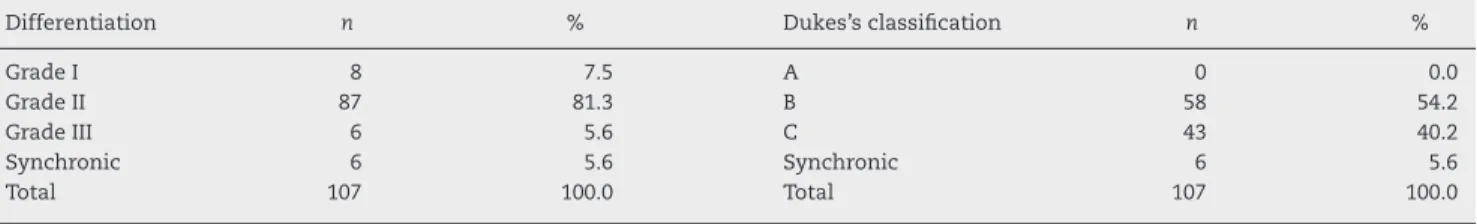
Adenocarcinoma was the most frequent histologic tumor type (n= 105) and two patients presented intestinal lym-phoma. Tumor staging was performed according to tumor differentiation grade and Dukes’ classification (Table 2).
Tumor invasion was locally limited in 28% of the patients while 16.8% had peritoneal tumor deposits. Distant metas-tases were seen in 15.9% and the most commonly affected organ was the liver (n= 16) and one case of cerebral metasta-sis was recorded. TNM staging system for CRC is presented in Table 3.
The mean time between hospital admission and surgery was 17.59±34.84 h. Preoperative water and electrolytes imbal-ance were corrected, antibiotic therapy initiated and clinical support measures stared. Blood transfusion was necessary in 21.5% of patients.
Surgeries were performed using general anesthesia (87.9%), but spinal anesthesia (6.5%), combined anesthesia (3.7%), and continuous epidural anesthesia (1.9%) were also used. The mean time of surgery was 2.39±0.91 h and the surgical
Table 3 – General description, according to TNM staging system.
Diagnosis Individuals
n %
Stage I 2 1.9
Stage IIA 41 38.3
Stage IIIA 8 7.5
Stage IIIB 27 25.2
Stage IIIC 13 12.1
Stage IV 16 15.0
Total 107 100.0
procedures were: tumor resection with colostomy (85%), tumor resection with primary anastomosis (10.3%), and colostomy without tumor resection (4.7%). All tumor resec-tions included partial colectomy with a 3-cm distal and a 5–7-cm proximal safety margin.
The average hospital stay length was 9 days and during this period 66.4% of the patients survived. 36 patients died and the immediate causes of death were: septic shock (61.12%), mul-tisystem organ failure (19.44%), and acute respiratory failure (19.44%). Among patients who died, half presented initially obstruction and the other half perforation (10 cases presented with perforation at the tumor site).
The age group over 60 years was statistically correlated to death (p= 0.002). Based on the results of the bivariate analy-ses, the variables sex, tumor location and staging according to Dukes’ classification did not show statistical significance in relation to death (Table 4).
Discussion
This study reported a high mortality rate for complicated colorectal cancer confirming previous literature data.17–19
Interestingly, complaints of patients in this study were sim-ilar to any other cause of bowel obstruction or perforation.20
Our data did not allow to identify any clinical or pathologi-cal factor to characterize or identify patients more favorable to present an obstructive CRC. Operative mortality due to obstruction or CRC perforation remains controversial and has ranged from 16% to 38%.20,21Although high, the results
pre-sented in this study are within these limits (33.6%). This may be explained by the poor preoperative clinical condi-tion of the patients (malnutricondi-tion, dehydracondi-tion, advanced age). Many patients presented with secondary peritonitis, intestinal obstruction and/or perforation, requiring extensive intestinal resection, which may have led to hydroelectrolictic
Table 2 – General description, according to differentiation grade and Dukes’s classification, of the anatomic pathology characteristics.
Differentiation n % Dukes’s classification n %
Grade I 8 7.5 A 0 0.0
Grade II 87 81.3 B 58 54.2
Grade III 6 5.6 C 43 40.2
Synchronic 6 5.6 Synchronic 6 5.6
Table 4 – Bivariate analyses of age group, sex, tumor location and staging according to Dukes’s classification in relation to death.
Variable n Death % RR IC 95% p
Age group
<60 years 55 11 20.0
0.42 1.09–1.31 0.002
>60 years 52 25 48.1 1.34–1.62
Sex
Female 58 22 37.9
1.33 1.25–1.51 0.208
Male 49 14 28.6 1.15–1.42
Tumor location
Rectosigmoid 55 20 36.4 1.00 1.23–1.49
0.959
Descendant colon 12 3 25.0 0.68 0.96–1.54
Ascendant colon 13 4 30.8 0.85 1.02–1.59
Transverse colon 21 7 33.3 0.91 1.11–1.55
Synchronic 6 2 33.3 0.91 0.79–1.87
Dukes’s classification
B 58 22 37.9 1.00 1.25–1.51
0.574
C 43 12 27.9 0.74 1.14–1.42
Synchronic 6 2 33.3 0.88 0.79–1.87
disorders and acid-base imbalances associated with sepsis and advanced-stage tumors.
Patients, in this series, with a diagnosis of CRC and that could be submitted to elective surgery were referred to another center. Therefore, comparison between emergency and elec-tive surgery was not possible in this series.
In the present study, the mean age of the patients with CRC was 59.8 years, similar to the results obtained in other studies.22,23Our data showed an equal distribution of patients
under 60 years (52%) and with more than 60 years (48%). Some authors report similar results,14but others found a
predomi-nance of patients over 60 years old.14,21Considering age, sex,
tumor site and Dukes’ classification, only age showed statis-tical importance to death with almost half of patients with more than 60 years dead (Table 4). This may be attributable to less coexisting diseases in younger patients. However, the finding in our series of a high proportion of patients under 60 years is not clear when compared to others.22–25
Concerning sex, it may or may not have a predominance in different studies. But, in fact, sex did not influence mortality rate (Table 4). McArdle and Hole26reported a small
predomi-nance of men for elective surgery and of women for emergency surgery.
Obstruction in CRC is not clear. Some factors may be involved as change in colonic flora, inflammatory edema, impaction of solid feces, fatigue of intestinal muscle proxi-mally to stenosis, elasticity of intestinal wall and the amount of fibrosis present.27More studies must be done to clarify such
possible causes of the obstructing mechanism.
When obstruction occurs in right side of the colon the sur-gical treatment is less controversial and a right colectomy with ileo-transverse anastomosis is the choice for the majority of patients. The discussion is what is the best approach to left-sided colonic obstruction.28
In the present study we considered that one-third of patients had a right-sided obstruction (ascendent and trans-verse colon). However only 10% of all patients had primary anastomosis. This surgical option may be due to the fact that the patients were operated by different surgeons with
different experience dealing with CRC. Breitenstein et al.29
reviewing studies of left-sided colonic obstruction concluded that one-stage surgery appears to be superior to two or three-stage procedures considering mortality rates but not morbidity rates. The incidence of obstruction of right colon may vary from 15% to 44% in the world.19,30Our data did not
show statistical difference in mortality rate between right-sided and left-right-sided colonic obstruction. This may be due to a small number of patients with one-stage surgical opera-tion in this series. Two-stage surgical procedures – first, tumor resection with colostomy and, posteriorly, intestinal recon-struction – represented the most frequent choice (85% of the cases) in the present study. This was mainly a consequence of the poor clinical condition of our patients, inappropriate colon preparation, and hemodynamic instability, in some cases.
The choice of surgical procedure for CRC depends mainly on the location of the lesion and the general state of patient to tolerate a specific procedure. Surgical treatment of com-plicated CRC has becoming more radical and the immediate resection of the tumor has been recommended for major-ity of patients. If intraoperative colonic preparation can be performed, in good clinical condition, an one-stage surgical procedure for CRC can be carried out, with tumor resection and primary anastomosis.
Some patients (21.5%) needed blood transfusions, and the mean time of surgical procedure was approximately 140 min. Both variables did not show relation to death rate. But a pos-sible negative effect of blood transfusion on survival rate may occur, despite other variables.
Palliative procedures may be used in selected patients, such as emergency endoscopy, laser coagulation, self-expanding metallic stents, among others. It may be used as a bridge to convert a surgical emergency into an elective surgery or for patients who cannot undergo a surgical procedure due to their present clinical condition.
uncomplicated cases, colon versus rectal location, and early stage.13,26 Although more advanced-stage CRC often results
in higher mortality rates, in this study it was not statistically significant, probably because we had no patients presenting with Dukes’s A tumors and the distribution of patients with Dukes’ B and C tumors was homogeneous.
Conclusions
A higher prevalence of elderly and female individuals pre-senting non-specific complaints was observed among the patients with CRC who underwent emergency surgeries in this series.
The reasons why many patients are asymptomatic until a sudden obstructive onset remain unclear and require further investigation.
Conflicts of interest
The authors declare no conflicts of interest.
r e f e r e n c e s
1. Tan E, Tilney H, Thompson M, Smith J, Tekkis PP. Association of coloproctology of Great Britain and Ireland, The United Kingdom national bowel cancer project – epidemiology and surgical risk in the elderly. Eur J Cancer. 2007;43:2285–94. 2. Albert RH, Clark MM. Cancer screening in the older patient.
Am Fam Physician. 2008;78:1369–79.
3. Zavoral M, Suchanek S, Zavada F, Dusek L, Muzik J, Seifert B, et al. Colorectal cancer screening in Europe. World J Gastroenterol. 2009;15:5907–17.
4. Ferlay J, Bray F, Pisani P, Parkin DM. Globocan 2002: cancer incidence, mortality and prevalence worldwide, version 2.0. IARC Cancer Base No. 5. Lyon: IARC Press; 2004.
5. American Cancer Society. Cancer: facts & figures 2011. Atlanta: American Cancer Society; 2011.
6. Jemal A, Bray F, Center MM, Ferlay J, Ward E, Forman D. Global cancer statistics. CA Cancer J Clin. 2011;61:69–90.
7. Pisani P. The cancer burden and cancer control in developing countries. Environ Health. 2011;10 Suppl. 1:S2.
8. Sharma V, Kerr SH, Kawar Z, Kerr DJ. Challenges of cancer control in developing countries: current status and future perspective. Future Oncol. 2011;7:1213–22.
9. Bresalier RS. Early detection of and screening for colorectal neoplasia. Gut Liver. 2009;3:69–80.
10. Gingras D, Béliveau R. Colorectal cancer prevention through dietary and lifestyle modifications. Cancer Microenviron. 2011;4:133–9.
11. Klabunde CN, Lanier D, Nadel MR, McLeod C, Yuan G, Vernon SW. Colorectal cancer screening by primary care physicians: recommendations and practices, 2006–2007. Am J Prev Med. 2009;37:8–16.
12. Brown SCW, Walsh S, Abraham JS, Sykes PA. Risk factors and operative mortality in surgery for colorectal cancer. Ann R Coll Surg Engl. 1991;73:269–72.
13. Biondo S, Parés D, Frago R, Martí-Ragué J, Kreisler E, De Oca J, et al. Large bowel obstruction: predictive factors for
postoperative mortality. Dis Colon Rectum. 2004;47: 1889–97.
14. Alvarez JA, Baldonedo RF, Bear IG, Truán N, Pire G, Alvarez P. Presentation, treatment, and multivariate analysis of risk factors for obstructive and perforative colorectal carcinoma. Am J Surg. 2005;190:376–82.
15. Bosman FT, Carneiro F, Hruban RH, Theise ND. WHO classification of tumours, vol. 3. IARC WHO Classification of Tumours, No. 3; 2010. Washington.
16. Edge SB, Byrd DR, Compton CC, Fritz AG, Greene FL, Trotti A, editors. AJCC cancer staging manual. London: Springer; 2010.
17. Tentes AA, Mirelis CG, Kakoliris S, Korakianitis OS, Bougioukas IG, Tsalkidou EG, et al. Results of surgery for colorectal carcinoma with obstruction. Langenbecks Arch Surg. 2009;394:49–53.
18. Lopez-Kostner F, Hool GR, Lavery IC. Management and causes of acute large-bowel obstruction. Surg Clin North Am. 1997;77:1265–90.
19. Tan KK, Sim R. Surgery for obstructed colorectal malignancy in an Asian population: predictors of morbidity and comparison between left- and right-sided cancers. J Gastrointest Surg. 2010;14:295–302.
20. Chen HS, Sheen-Chen SM. Obstruction and perforation in colorectal adenocarcinoma: an analysis of prognosis and current trends. Surgery. 2000;127:370–6.
21. Crucitti F, Sofo L, Doglietto GB, Bellantone R, Ratto C, Bossola M, et al. Prognostic factors in colorectal cancer: current status and new trends. J Surg Oncol. 1991;48 Suppl. 2:76–82. 22. Di Gregorio C, Benatti P, Losi L, Roncucci L, Rossi G, Ponti G,
et al. Incidence and survival of patients with Dukes’ A (stages T1 and T2) colorectal carcinoma: a 15 year population-based study. Int J Colorectal Dis. 2005;20:147–54.
23. Fielding LP, Stewart-Brown S, Blesovsky L. Large-bowel obstruction caused by cancer: a prospective study. BMJ. 1979;2:515–7.
24. Basdanis G, Papadopoulos VN, Michalopoulos A, Fahantidis E, Apostolidis S, Berovalis P, et al. Colorectal cancer in patients over 70 year of age: determinants of outcome. Tech Coloproctol. 2004;8 Suppl.:S112–5.
25. O’Connell JB, Maggard MA, Livingston EH, Yo CK. Colorectal cancer in the young. Am J Surg. 2004;187:343–8.
26. McArdle CS, Hole DJ. Emergency presentation of colorectal cancer is associated with poor 5-year survival. Br J Surg. 2004;91:605–9.
27. Phillips RKS, Hittinger R, Fry JS, Fielding LP. Malignant large bowel obstruction. Br J Surg. 1985;72:296–302.
28. Athreya S, Moss J, Urquhart G, Edwards R, Downie A, Poon FW. Colorectal stenting for colonic obstruction: the indications, complications, effectiveness and outcome – 5 year review. Eur J Radiol. 2006;60:91–4.
29. Breitenstein S, Rickenbacher A, Berdajs D, Puhan M, Clavien PA, Demartines N. Systematic evaluation of surgical strategies for acute malignant left-sided colonic obstruction. Br J Surg. 2007;94:1451–60.