Journal of
Coloproctology
w w w . j c o l . o r g . b r
Original Article
Clinicopathological aspects and prevalence of
human papillomavirus in anal cancer
夽
Marina Tayla Mesquita Aguiar
a, Natália Carelli de Castro Bosso
a,
Caio Bruno Quinta de Souza Leal
a, Clerlhan Ferreira de Lira
a,
Lázara Alyne Oliveira Cabral
a, Antonio Márcio Teodoro Cordeiro Silva
a,
Vera Aparecida Saddi
a,b,∗aDepartment of Medicine, Pontifícia Universidade Católica de Goiás (PUC-GO), Goiânia, GO, Brazil
bLaboratory of Bone Marrow Transplant, Hospital Araújo Jorge (HAJ), Associac¸ão de Combate ao Câncer em Goiás (ACCG), Goiânia, GO,
Brazil
a r t i c l e
i n f o
Article history:
Received 13 October 2013 Accepted 15 December 2013 Available online 13 April 2014
Keywords: Anal cancer Characteristics Human papillomavirus HPV
HPV16 HPV18
a b s t r a c t
Anal cancer is relatively rare; however, its incidence has increased in recent years. Several risk factors are associated with the development of anal cancer, including age older than 50 years, low-fiber diet, chronic anal fistulas, smoking, multiple partners, anal intercourse practice, Human Immunodeficiency Virus infection and immunosuppression. However, the presence of human papillomavirus represents the main risk factor for the development of anal cancer. The aim of this study was to evaluate the clinicopathological aspects of a series of patients with anal carcinomas diagnosed in Hospital Araújo Jorge, Goiânia-Goiás, as well as the prevalence of human papillomavirus genome in these tumors. Clinical, pathological and socio-demographic data were collected from the respective medical files and paraffin blocks containing anal carcinomas specimens were used for DNA extraction and detection of human papillomavirus, by means of polymerase chain reaction, using short PCR fragment primers. Forty-three cases were selected and had the data analyzed, while 38 cases were tested for human papillomavirus genome detection. Among the evaluated patients, 62.8% were women; 53.4% of tumors were squamous cell carcinoma and 46.5% of the patients were aged between 60 and 75 years. Risk factors, such as smoking (39.5%) and alcoholism (20.9%) were recorded in the studied group. Lymph node metastases were detected in 30.2% of cases and 7.0% had distant metastasis. The detection of human papillomavirus DNA was positive in 76% of cases assessed and this was significantly associated with squamous cell carcinomas. Aggressive behavior and advanced stage of anal cancer described in this study highlight the need for preventive measures that contemplate these tumors, including vaccination against human papillomavirus.
© 2014 Sociedade Brasileira de Coloproctologia. Published by Elsevier Editora Ltda. All rights reserved.
夽
Study carried out at Hospital Araújo Jorge of Associac¸ão de Combate ao Câncer em Goiás and Genetic Diversity Laboratory of Pontifícia Universidade Católica de Goiás, GO, Brazil. This study was supported by FAPEG - Fundac¸ão de Apoio a Pesquisa do Estado de Goiás (Foundation for Research Suport from the State of Goias).
∗ Corresponding author.
E-mail: [email protected] (V.A. Saddi). http://dx.doi.org/10.1016/j.jcol.2014.03.004
Aspectos clínico-patológicos e prevalência do papilomavírus humano
(HPV) em carcinomas anais
Palavras-chave:
Câncer anal Características Papilomavirus humano HPV
HPV16 HPV18
r e s u m o
O câncer anal é relativamente raro, entretanto, sua incidência aumentou nos últimos anos. Vários fatores de risco são associados ao desenvolvimento do câncer anal, incluindo idade maior que 50 anos, dieta pobre em fibras, fístulas anais crônicas, tabagismo, múltiplos parceiros, prática de intercurso anal, infecc¸ão pelo HIV e imunossupressão. Entretanto, a presenc¸a do Papilomavírus Humano (HPV) representa o principal fator de risco para o desenvolvimento do câncer anal. O objetivo deste estudo consistiu em avaliar os aspectos clínico-patológicos de uma série de pacientes com carcinomas anais diagnosticados no Hos-pital Araújo Jorge, Goiânia/GO, bem como a prevalência do genoma do HPV nesses tumores. Dados clínico-patológicos e sóciodemográficos foram colhidos a partir dos respectivos pron-tuários e blocos de parafina contendo espécimes de carcinomas anais foram usados para extrac¸ão de DNA e detecc¸ão de HPV, por meio da reac¸ão em cadeia da polimerase, usando oligonucleotídeos iniciadores SPF. Quarenta e três casos foram selecionados e tiveram os dados clinico-patológicos analisados, enquanto 38 casos foram testados para a detecc¸ão do genoma do HPV. Dentre os pacientes avaliados, 62,8% eram mulheres; 53,4% dos tumores eram carcinomas de células escamosas e 46,5% dos pacientes estavam na faixa etária entre os 60 e 75 anos. Fatores de risco, como tabagismo (39,5%) e etilismo (20,9%) foram registra-dos no grupo estudado. Metástases linfonodais foram detectadas em 30,2% registra-dos casos e 7,0% apresentaram metástase à distância. A detecc¸ão de HPV foi positiva em 76,0% dos casos analisados e este significativamente associado aos carcinomas de células escamosas. O comportamento agressivo e o estágio avanc¸ado dos carcinomas anais descritos no presente estudo destacam a necessidade de medidas de prevenc¸ão que contemplem esses tumores, incluindo a vacinac¸ão contra o HPV.
© 2014 Sociedade Brasileira de Coloproctologia. Publicado por Elsevier Editora Ltda. Todos os direitos reservados.
Introduction
Anal cancer is relatively rare, accounting for about 30,000 new cases a year worldwide, with a peak incidence between the ages of 58 and 64 years.1 It represents approximately
1.5% of all tumors of the digestive tract and 2–3% of colorec-tal tumors.2 However, its incidence has increased in recent
years.3 In the U.S., an incidence of 1.6 cases/100,000
inhabi-tants was reported in the period 2002–2006.1
Anal tumors can occur in the anal verge or in the anal canal, up to the transition to the rectum. Tumors that arise in the anal verge are dermatological lesions and as such, can be treated with local excision. On the other hand, tumors that arise in the anal canal or in the transition zone of the anal canal with the rectum deserve a more aggressive surgical approach.4
Anal carcinomas have a wide histological variety. Tumors that appear distal to the pectineal line, the border between the anal canal and the rectum, are keratinized squamous cell carcinomas, also called epidermoid carcinomas. The non-keratinized squamous cells carcinomas are located above the pectineal line and are called epithelioid carcinomas. There are other histological types, such as adenocarcinomas (ADC), transitional cell or cloacogenic tumors, basaloid and mucus-epithelioid tumors. The epidermoid carcinoma is the most common cancer of the anus and is responsible for 85% of malignant lesions of this region.4–6
The clinical presentation and severity of anal cancers depend on the size and location of the tumor in the anus. Small, mobile lesions, smaller than 2 cm have a probability of cure in 80% of cases, compared with <50% chance for tumors larger than 5 cm. It is known that 20% of patients are asymp-tomatic; however, anal bleeding and the sensation of a mass occupying the anal canal are the most frequent signs and symptoms, often mistakenly associated with hemorrhoidal disease.4,6
The treatment of anal carcinoma can be surgical and clin-ical. Surgical treatment is based on local excision of the lesion or abdominoperineal resection of the rectum and anus, whereas the medical treatment is based on the anorectal segment preservation through chemoradiotherapy or radio-therapy alone.4
Tumors of the anal canal are more common in females, while anal margin tumors are more frequent in males.7Some
studies have shown that although the prevalence of anal can-cer is higher in women older than 60 years of age, the disease has become very common in men between 30 and 40 years, due to high association with infection by the Human Immu-nodeficiency Virus (HIV).1
human papillomavirus (HPV) shows the main association with the development of anal cancer.2,8
HPVs are small double-stranded DNA viruses, which belong to thePapillomaviridaefamily. Initially, these viruses were iden-tified, cloned and sequenced from samples of cervical tumors, and subsequently established as important etiological agents of carcinogenesis in several human tumors.9,10More than 200
types of HPV have been identified and each of them has par-ticular tropism for specific anatomical sites.11
HPV infection seems to be associated with the majority of anal tumors, especially with squamous cell carcinomas. Among the genotypes most frequently associated with anal tumors, HPV 16 and HPV 18 are considered as high risk for the development of anal cancer. The overall percentage of anal cancers attributed to HPV is 90%, especially for genotypes 16 and 18, which correspond to 92% of cases, with some differ-ences depending on the geographic region (75% in men×91% in women). HPV 16 is the most prevalent type, found in over 70% of cases.1,9
The constant practice of receptive anal sex apparently contributes to a higher frequency of anal lesions and also rep-resents one of the main factors for the acquisition of HIV and HPV coinfection, important risk factors for anal intraepithelial neoplasia (AIN), considered the precursor lesion of anal squa-mous carcinoma. It is believed that the HIV virus is a cofactor that HPV needs to induce neoplasms that can progress to anal carcinoma.2
Objectives
The aim of this study was to evaluate the clinical, pathological and sociodemographic characteristics of patients diagnosed with anal carcinoma in Hospital Araújo Jorge, Goiânia/GO, in the period 2004–2011, as well as the prevalence of HPV genome in these tumors.
Methods
Study type and sample
The study consisted of an epidemiological investigation that used clinicopathological data collected from medical records and analysis of paraffin blocks containing specimens of anal carcinomas. The study was approved by the Ethics Commit-tee on Human Research of Hospital Araújo Jorge (HAJ), under number 272 288 in April 2013. As this was a retrospective study, there was no direct contact with selected patients and thus, it did not require the signing of the free and informed consent form. The prevalence of HPV was investigated in 43 specimens of anal carcinomas diagnosed in HAJ, in Goiânia/GO, in the period 2004–2011.
Patients included in the study were those with confirmed histopathological diagnosis of anal carcinoma and clinico-pathological data available in their medical records. The detection of HPV included the cases in which the paraf-fin blocks were sufficient and were available for molecular analysis. Case selection was made from an active search of the records of the Department of Pathology of HAJ. The slides stained with hematoxylin–eosin related to each
anatomopathological examination were reviewed and sec-tions were prepared for DNA extraction.
DNA extraction
Genomic DNA was purified from tumor samples fixed in formalin and embedded in paraffin, according to standard pro-tocols used in the Laboratory of Genetic Diversity of Pontifícia Universidade Católica Goiás (PUC Goiás). The deparaffiniza-tion and hydradeparaffiniza-tion steps were performed and DNA extracdeparaffiniza-tion and purification were performed using a commercial Wizard Kit (Promega). All purified DNA samples were tested for qual-ity and integrqual-ity via amplification of a fragment of constitutive BRAF gene of 224 bp.
Detection of HPV genome in tumor samples
Detection of HPV DNA was performed by polymerase chain reaction (PCR) using SPF primers,12which amplify a fragment
of 75 base pairs of the viral genome and developed to promote sensitive amplification of the most clinically relevant HPV genotypes. The assay is capable of detecting the genotypes currently known as high risk HPVs (16, 18, 26, 31, 33, 35, 39, 45, 51, 52, 53, 56, 58, 59, 66, 68, 73 e 82), as well as low-risk HPV genotypes (6, 11, 40, 43, 44, 54, 70) and some additional types. HPV detection was performed at the Laboratory of Bone Marrow Transplantation of Associac¸ão de Combate ao Câncer in Goiás and Laboratory of Genetic Diversity of PUC-Goiás. The PCR with SPF primers was performed in a final reaction volume of 25L, containing 2L of purified DNA, 10 mmol/L Tris–HCl, pH 9.0, 50 mmol/L KCl, 2.5 mmol/L MgCl2 200 mmol/L each
of deoxy-nucleotide (dNTP), 10 pmol of each oligonucleotide primer, and 0.25 U of Taq Polymerase (Invitrogen, Brazil). The cycling conditions included: preheating for 1 min at 94◦C fol-lowed by 40 cycles of 1 min at 94◦C, 1 min at 45◦C and 1 min at 72◦C, with a final extension of 5 min at 72◦C.
Each PCR experiment was performed with positive and neg-ative controls, previously tested in the Laboratory of Genetic Diversity of PUC-Goiás. A fragment of human BRAF gene with 224 bp was amplified from DNA extracted from each sample to confirm the presence and the capacity of amplifying the extracted DNA from each sample.
Statistical analyses
The data obtained were coded and stored in a database in Excel software program and analyzed using GraphPad Prism. Clini-copathological and sociodemographic data were processed to descriptive statistical analysis and the data on HPV detection regarding the different variables were analyzed using Fisher’s exact test and Chi-square test, considering statistically signif-icant differences the ones that resulted inpvalue≤0.05.
Results
Clinicopathological and sociodemographic characteristics
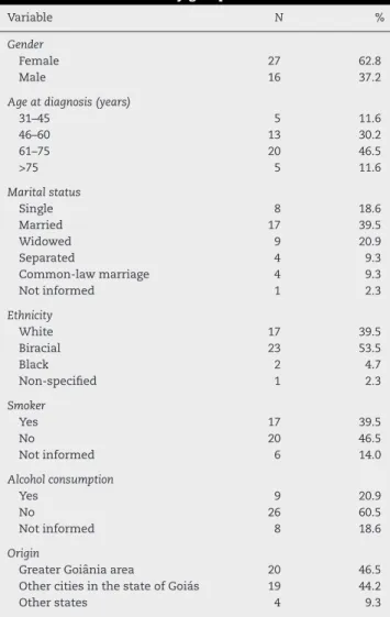
Table 1 – Descriptive analysis of the sociodemographic characteristics of the study group.
Variable N %
Gender
Female 27 62.8
Male 16 37.2
Age at diagnosis (years)
31–45 5 11.6
46–60 13 30.2
61–75 20 46.5
>75 5 11.6
Marital status
Single 8 18.6
Married 17 39.5
Widowed 9 20.9
Separated 4 9.3
Common-law marriage 4 9.3
Not informed 1 2.3
Ethnicity
White 17 39.5
Biracial 23 53.5
Black 2 4.7
Non-specified 1 2.3
Smoker
Yes 17 39.5
No 20 46.5
Not informed 6 14.0
Alcohol consumption
Yes 9 20.9
No 26 60.5
Not informed 8 18.6
Origin
Greater Goiânia area 20 46.5 Other cities in the state of Goiás 19 44.2
Other states 4 9.3
cases received chemotherapy/radiotherapy before surgery and nine were colorectal ADCs extending to the anus. The remaining 43 cases were evaluated regarding clinicopathologi-cal and sociodemographic characteristics, which are described in Tables 1 and 2. All data were collected through medical chart review at HAJ.
Of the assessed patients, 62.8% were females. Approx-imately half of the sample (53.5%) was diagnosed with squamous cell carcinoma (SCC), while 37.2% were ADCs. Most patients (46.5%) were aged between 61 and 75 years and 39.5% were married. Risk factors such as smoking (39.5%) and alco-hol consumption (20.9%) were recorded in the study sample. Data associated with sexual behavior and orientation, such as the number of partners, age at the first sexual intercourse and practice of receptive anal sex were not reported in the medical records.
In 63.2% of cases, the diagnosis was made by biopsy. The treatment chosen for the group was, in general, a combina-tion of surgery, chemotherapy and radiotherapy, with 76.7% of patients being submitted to surgery. Lymph node metas-tases were detected in 13 patients (30.2%) and four (9.3%) had distant metastasis, with the lungs being the most prevalent site (50.0%). Although all patients were not followed for a long period of time, at the end of the data collection period,
Table 2 – Descriptive analysis of clinicopathological characteristics of the study group.
Variable N %
Histological type
SCC 23 53.5
Adenocarcinoma 16 37.2
Basaloid/cloacogenic carcinoma 3 7.0
Neuroendocrine 1 2.3
Diagnostic method
Biopsy 25 58.1
Surgery 18 41.9
Treatment
Surgery 33 76.7
Radiotherapy 28 65.1
Chemotherapy 28 65.1
No treatment 2 4.7
Lymph node metastasis
Yes 13 30.2
No 30 69.8
Distant metastases
Yes 3 7.0
No 40 93.0
Sites of distant metastases
Liver 1 25
Lung 2 50
Brain 1 25
Death record
Yes 11 25.5
No 32 74.4
SCC, squamous cell carcinoma.
11 patients had their deaths recorded in their medical files (25.6%).
HPV detection in anal carcinoma samples
Of the 43 samples initially evaluated, 38 had paraffin-embedded histopathological specimens available and suffi-cient for DNA extraction. All these samples were previously tested for endogenous control amplification, the human BRAF gene and provided DNA suitable for PCR amplification. Of the 38 samples, 29 were positive for detection of HPV DNA (76.3%). Differences between HPV(+) and HPV(−) tumors in relation to
sociodemographic and clinicopathological features were eval-uated and are described in Table 3. Significant differences (p= 0.032) were observed in relation to SCCs and ADCs, con-firming the association between HPV and anal SCCs. The other variables investigated were not statistically significant.
Discussion
Studies carried out in different countries are unanimous in stating that, despite the fact that it is considered a rare tumor, the incidence of anal cancer has increased steadily in recent years.1,13–15 The present study analyzes a
Table 3 – Comparative analysis between the anal carcinomas HPV(+) and HPV(−).
HPV(+) (n= 29) HPV(−) (n= 9)
Variable N % N % p
Histological type
SCC 20 90.9 2 9.1
Adenocarcinoma 7 53.8 6 46.2 0.03218a
Other types 2 66.7 1 33.3
Diagnostic method
Biopsy 16 66.7 8 33.3
Surgery 13 92.9 1 7.1 0.1147
Lymph node metastasis
Yes 6 66.7 3 33.3
No 23 79.3 6 20.7 0.6553
Distant metastasis
Yes 5 62.5 3 37.5
No 24 80.0 6 20.0 0.3631
Death record
Yes 4 57.1 3 42.9
No 25 80.6 6 19.4 0.3225
Gender
Female 16 72.7 6 27.3
Male 13 81.2 3 18.8 0.7060
Marital status
Married 14 87.5 2 12.5
Others 15 68.2 7 31.8 0.2537
Smoker
Yes 14 87.5 2 12.5
No 12 66.7 6 33.3 0.2327
Not informed 3 75.0 1 25.0
Alcohol consumption
Yes 6 66.7 3 33.3
No 18 81.8 4 18.2 0.3841
Not informed 5 71.4 2 28.6
a p≤0.05, statistically significant difference; SCC, squamous cell carcinoma.
characteristics of patients, as well as the detection of HPV genome in the assessed tumor specimens. According to the results of the series, 46.5% of the patients were aged 61–75 years. Different studies have reported that the most prevalent age group for anal cancer is 50–70 years.13,14,16,17 The
accu-mulation of exposure to different risk factors throughout life contributes to the higher prevalence of these tumors at older ages.
A higher proportion of cases of anal carcinomas analyzed in this series was detected in women (62.8%). This information was previously reported by other authors;1,13,14,16–18however,
plausible explanations for this observation are still inconclu-sive. Several hypotheses have been suggested, such as that women are more susceptible to HPV infection, by developing HPV-related lesions in several anatomical sites, such as the vulva, vagina and cervix and developing cervical cancer with a very high frequency.14Men also have frequent anogenital
HPV infection; however, the tumors associated with HPV are significantly less prevalent in males.
Our study showed that 39.5% of patients with anal cancer were married; however, another significant portion of patients were widowed (20.9%) and single (18.6%), a factor that could favor having a higher number of sexual partners. A higher
proportion of married heterosexual men (72.2%) was also found in the DALING study,13which assessed anal cancer in
306 patients from the North-American west coast.
Although smoking is considered a major risk factor for anal cancer, smokers did not represent the majority of cases in our study; however, a significant number reported smoking (20.9%), and moreover, this information was not available in many medical files (18.6%). Among the risk factors for anal cancer, smoking is considered very significant, both in women and men.13
As it is a rare type of cancer, the total number of cases of anal cancer analyzed in different series is, in general, small. The study by Abramowitz et al.1has the largest series, with 362
cases diagnosed in 16 anatomical pathology centers spread throughout France. The second largest series13analyzed 306
patients living in the state of Washington (USA) over a 12 year period, 1986–1998. The histological type of anal cancer most commonly associated to HPV infection is the SCC and, in this study, we found this tumor type in 53.5% of the assessed sam-ples.
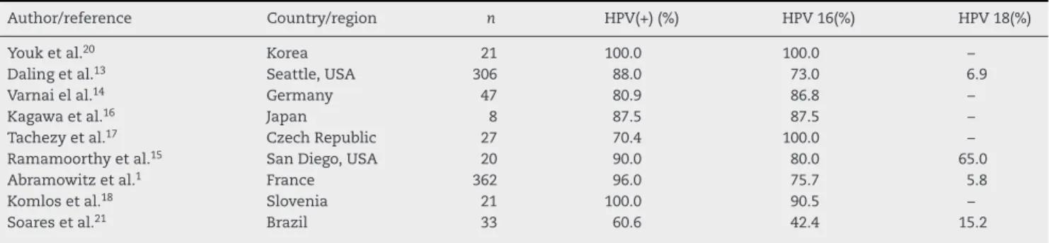
Table 4 – Studies that analyzed the prevalence of HPV in anal carcinomas.
Author/reference Country/region n HPV(+) (%) HPV 16(%) HPV 18(%)
Youk et al.20 Korea 21 100.0 100.0 –
Daling et al.13 Seattle, USA 306 88.0 73.0 6.9
Varnai el al.14 Germany 47 80.9 86.8 –
Kagawa et al.16 Japan 8 87.5 87.5 –
Tachezy et al.17 Czech Republic 27 70.4 100.0 –
Ramamoorthy et al.15 San Diego, USA 20 90.0 80.0 65.0
Abramowitz et al.1 France 362 96.0 75.7 5.8
Komlos et al.18 Slovenia 21 100.0 90.5 –
Soares et al.21 Brazil 33 60.6 42.4 15.2
was made by reviewing articles published in major biblio-graphic databases (PubMed, SciELO, LILACS), using the terms anal cancer, HPV PCR and selecting those published from 2000 in English or Portuguese languages. The search resulted in nine selected studies and the results of HPV prevalence and genotyping in the anal carcinomas are summarized in Table 4. Molecular investigations on the detection of the HPV genome in anal carcinomas indicate that anal cancer resem-bles cervical cancer in relation to HPV, that is, regarding squamous carcinomas, HPV is considered the most important risk factor for anal carcinogenesis.1,15
HPV infection is highly prevalent in patients with anal can-cer, being considered a necessary cause for anal squamous cell carcinoma.19 In this study, an association between the
presence of HPV and anal squamous cells was observed, i.e., HPV DNA was found in 90.9% of the SCCs. Some studies have detected the viral genome in 100% of samples of anal squa-mous cell carcinomas.18,20
However, in the largest series studied, the prevalence of HPV genome in anal carcinomas varied, ranging from 80.9%,14
88%13and 96.7%.1The different molecular methods used in
viral DNA detection in these tumors certainly explain this discrepancy. Our study used generic primers (FPS) capable of amplifying a small fragment (75 bp) of the HPV genome, allow-ing the detection of viral DNA, even in samples of which DNA integrity was not of excellent quality.
Similar to cervical cancer, anal cancer seems to occur in the cells of the squamocolumnar junction, populated by a variety of precursor cells that originate different epithelia.17
Studies available in the literature show that anal carcinoma rarely occurs in the absence of HPV, reinforcing the role of the virus as the main risk factor for this tumor type.1
Regarding the HPV subtypes in anal carcinomas, our study did not include the analysis of present genotypes. According to the available literature, a significant predominance of HPV 16 is shown in relation to other genotypes in anal carcino-mas (Table 4). Youk et al.20and Tachezy et al.17detected the
presence of HPV 16 in 100% of the anal carcinoma samples analyzed. One explanation for this occurrence is that HPV 16 is closely involved in the development of anal SCC by promot-ing changes in the genome still at the phase of micro-invasive carcinoma, according to reports by Kagawa et al.16in Japan.
HPV subtype 18 appears in global studies as the second most prevalent type of HPV in anal carcinomas, although the data are somewhat controversial. In nine studies found in the literature (Table 4), the oncogenic potential of subtype 18
is recognized in the natural history of anal carcinoma; how-ever, only four studies performed HPV 18 genotyping in these tumors.
In the study by Ramamoorthy et al.,15 carried out in San
Diego, USA, the prevalence of HPV subtype 18 was 65%, whereas in the study by Daling et al.13performed in Seattle,
HPV 18 was found in 6.9% of cases. In the study by Abramowitz et al.,1carried out in France, the prevalence of HPV subtype 18
was not greater than 5.8%.
The study by Soares et al.,21carried out in Brazil,
demon-strated the presence of HPV 18 genome in 15.2% of cases. This discrepancy may be related to HPV genotyping method, as the molecular technologies used in the studies were different.
Regarding the prognosis of patients with anal carcinoma, data available in the literature are still very scarce. In the present study, we observed a very aggressive behavior of these tumors, with 13 patients (30.2%) showing lymph node metas-tases and three patients (7.0%) with distant metasmetas-tases: two with lung metastases, one with liver metastasis and one with brain metastasis. The aggressiveness of anal carcinoma can also be observed by the number of deaths recorded during the study period. Of the 43 patients included in the study, 11 died, representing 25.6% of the study sample.
The increased incidence of anal carcinoma and knowl-edge of the main factors involved in the etiology of these tumors stimulate the need to develop preventive measures for anal carcinoma. Such measures include full medical examina-tion, with an investigation of the sexual history of the patient during anamnesis, careful clinical examination of the anogen-ital region, detection of precursor lesions (intraepithelial anal neoplasms), anoscopy and HPV vaccination, especially in the higher-risk groups.16
High-risk patients include promiscuous individuals with a high number of sexual partners, regular practice of anal inter-course, low immunity (HIV, immunosuppressed individuals) and past or current history of infection associated with anal lesions (condylomas, cervical intraepithelial neoplasia (CIN), vulvar intraepithelial neoplasia (VIN), vaginal intraepithelial neoplasia (VAIN) and vulvar, vaginal or cervical SCC). For this group, clinical examination with anal inspection, perianal and intra-anal cytology is recommended, as well as research of HPV genome in samples of anal neoplasia.14
HPV. The studies by Abramowitz et al.1 in France and
Kom-los et al.18in Slovenia emphasize the importance of further
studies on HPV vaccination in the prevention of anal can-cer, as most of these tumors are significantly associated with HPV infection, particularly with HPV subtype 16. Vaccination against high-risk HPV can have a big impact on the preva-lence of anal carcinoma and its precursor lesions; however, more detailed studies on its potential power to prevent such tumors are still necessary.
This study has an important limitation regarding the col-lection of clinical, pathological and behavioral data of the patients. These data were scarce in most medical records, including the practice of receptive anal intercourse, history of HIV infection, presence of sexually transmitted diseases (STDs) or condylomas, among others.
The studies by Daling et al.,13 in the USA and by
Abramowitz et al.,1 in France, also reported difficulties in
obtaining these same data, both in interviews13and during the
review of medical files.1Among the nine studies that
evalu-ated HPV detection in anal carcinomas in the last 10 years, only these two1,13analyzed the risk factors for anal carcinoma, in
addition to HPV infection. The continuation of this study, with the analysis of a greater number of cases of anal carcinomas diagnosed in other oncology centers, as well as the genotyp-ing of HPV in specimens of anal carcinomas is the objective of our team.
Conflicts of interest
The authors declare no conflicts of interest.
r e f e r e n c e s
1. Abramowitz L, Jacquard AC, Jaroud F, et al. Human papillomavirus genotype distribution in anal cancer in France: the EDiTH V study. Int J Cancer. 2011;129:433–9. 2. Nadal LRM, Nadal SR. Citologia anal para rastreamento de
lesões pré-neoplásicas. Rev Assoc Med Bras. 2007;53:147–51. 3. Johnson LG, Madeleine MM, Newcomer LM, Scwartz SM,
Daling JR. Anal cancer incidence and survival: the surveillance, epidemiology, and end results experience. Cancer. 2004;101:281–8.
4. Santos Jr JCM. Câncer ano-reto-cólico: aspectos atuais IV – câncer de cólon – fatores clínicos, epidemiológicos e preventivos. Rev Bras Colo-proctol. 2008;28:378–85. 5. Fenger C. Anal neoplasia and its precursors: facts and
controversiers. Semin Diagn Pathol. 1991;8:190–201. 6. Iarc – International Agency for Research on Cancer; 2010.
www.iarc.fr
7. Torres Neto Jr, Prudente ACL, Santos RL. Estudo demográfico do câncer de canal anal e ânus no estado de Sergipe. Rev Bras Colo-proctol. 2007;27:190–5.
8. Carvalho NS, Ferreira AM, Bueno CC. HPV infection and intraepithelial lesions from the anal region: how to diagnose? Braz J Infect Dis [online]. 2011;15:473–7.
9. Shukla S, Bharti AC, Mahata S, et al. Infection of human papillomaviruses in cancers of different human organ sites. Indian J Med Res. 2009;130:222–33.
10. Insinga RP, Dasbach EJ, Elbasha EH. Epidemiologic natural history and clinical management of human papillomavirus (HPV) disease: a critical and systematic review of the literature in the development of an HPV dynamic transmission model. BMC Infect Dis. 2009;9:119.
11. Cerdeira CR, Cararach M, Alba A. The human papillomavirus (HPV) in human pathology: description, pathogenesis, oncogenic role epidemiology and detection techniques. Open Dermatol J. 2009;3:90–102.
12. Kleter B, Doorn LJV, Schrauwen L, et al. Novel short-fragment PCR assay for highly sensitive broad-spectrum detection of anogenital human papillomaviruses. Am J Pathol.
1998;153:1731–9.
13. Daling JR, Madeleine MM, Johnson LG, et al. Human
papillomavirus, smoking, and sexual practices in the etiology of anal cancer. Cancer. 2004;101:270–80.
14. Varnai AD, Bollmann M, Griefingholt H, et al. HPV in anal squamous cell carcinoma and anal intraepithelial neoplasia (AIN) impact of HPV analysis of anal lesions on diagnosis and prognosis. Int J Colorectal Dis. 2005;21:135–42.
15. Ramamoorthy S, Liu Yt, Luo L, Miyai K, Lu Q, Carethers JM. Detection of multiple human papillomavirus genotypes in anal carcinoma. Infect Agents Cancer. 2010;
5:17.
16. Kagawa R, Yamaguchi T, Furuta R. Histological features of human papilloma virus 16 and its association with the development and progression of anal squamous cell carcinoma. Surg Today. 2006;36:885–91.
17. Tachezy R, Jirasek T, Salakova M, et al. Human papillomavirus infection and tumours of the anal canal: correlation of histology PCR detection in paraffin sections and serology. APMIS. 2007;115:195–203.
18. Komlos KF, Kocjan BJ, Koˇsorok P, et al. Distribution of HPV genotypes in Slovenian patients with anal carcinoma: preliminary results. Acta Dermatovenerol Alp Panonica Adriat. 2011;20:141–3.
19. Palefski JM, Giuliano AR, Goldstone S, et al. N Engl J Med. 2011;365:76–85.
20. Youk EG, Ku JL, Park JG. Detection and typing of human papillomavirus in anal epidermoid carcinomas: sequence variation in the E7 gene of human papillomavirus Type 16. Dis Colon Rectum. 2001;44:236–42.