ORIGINAL RESEARCH
The prevalence of mitochondrial DNA mutations
in Leigh syndrome in a Brazilian series
Suely Kazue Nagahashi Marie,ISueli Mieko Oba-Shinjo,IMaria Joaquina Marques-Dias,ISergio Rosemberg,II Fernando Kok,IUmbertina Conti ReedI
IDepartment of Neurology – Sa˜o Paulo University, Faculty of MedicineIIDepartment of Pathology – Sa˜o Paulo University, Faculty of Medicine, Sao Paulo, Brazil.
OBJECTIVE: To determine the prevalence of mitochondrial DNA (mtDNA) mutations in cases with findings compatible with the diagnosis of Leigh syndrome in a Brazilian Neurological Service, and to compare those findings between the patients presenting or not these mutations.
METHOD:We analyzed six mtDNA point mutations (T8993G, T8993C, T8851C, G1644T, T9176C, and T3308C) by PCR and endonuclease digestion in 32 patients with presumptive diagnosis of Leigh syndrome, according to distribution across different age ranges.
RESULTS: We found two patients, in the subgroup under 4 years of age, presenting T8993G and T8993C mutations. Their clinical symptoms and neuroimaging findings were similar when compared to those patients not harboring these mutations.
CONCLUSION:As the molecular confirmation is pivotal for both the precise genetic counselling and therapeutic guidance, we emphasise the benefit of screening for mtDNA mutation in Leigh syndrome patients under 4 years old. Mitochondrial whole genome and whole exome analysis by next-generation sequencing technology maybe a future alternative for molecular diagnosis of this extensive genetic heterogeneous syndrome.
KEYWORDS: Leigh’s syndrome; T8993G; T8993C; maternal inheritance; earlyinfantile.
Marie SKN, Oba-Shinjo SM, Marques-Dias MJ, Rosemberg S, Kok F, Reed UC. The prevalence of mitochondrial DNA mutations in Leigh syndrome in a Brazilian series. MEDICALEXPRESS. 2014;1(5):239-242.
Received for publication onJune 13 2014;First review completed onJune 25 2014;Accepted for publication onJuly 21 2014
Email: [email protected]
B INTRODUCTION
Subacute Necrotizing Encephalopathy or Leigh Syndrome (LS) is a progressive neurodegenerative disease frequently associated with mitochondrial abnormalities. The preva-lence of LS has been estimated to be 1 in 40,0001 and is characterized by the presence of developmental delay and lactic acidosis. LS was first described in 1951,2 when its distribution and histological aspects were reported, as presenting central nervous lesions resembling those of Wernicke’s disease (athiaminosis), except that the lesions in LS tended to be more extensive, involving the striatum and sparing the mammillary bodies. The pathologic changes are usually bilateral and symmetrical, corresponding to foci of spongy necrosis with myelin degeneration, vascular pro-liferation and gliosis in thalami, midbrain, pons, medulla and spinal cord, as well as changes to basal ganglia, which are characteristically, but not invariably, affected.3The MRI findings correspond generally to the neuropathologic features, and are diagnostic criteria for the disease in children with mental or motor involution. LS is a familial or sporadic disease with a wide variety of clinical
manifestations. Many authors have reported onset of symptoms in the first year of life in more than half of cases, mostly before the sixth month. However, cases with late-onset showing even greater heterogeneity in clinical presentation are also well documented.4
Although LS is relatively rare, the identification of this maternal type of inheritance is very important, calling for specific genetic counseling and therapeutic approach.
The aim of this study is to present the prevalence of mitochondrial DNA (mtDNA) mutations in cases with clinical symptoms and neuroimaging findings compatible with the diagnosis of LS in a Brazilian Neurological Service, and to compare those findings between the patients presenting or not these DNA mutations.
B SUBJECTS AND METHODS
Patients
Thirty-two cases (22 males, 10 females) were included in the present study, and they were referred from Neuropedia-trics, the Neuromuscular Disease Group of Clinical Neurology of the Hospital das Clı´nicas School of Medicine, the Institute of Children of the University of Sa˜o Paulo and from other private and state hospitals. Four cases out of thirty-two were submitted to postmortem examination at the DOI:10.5935/MedicalExpress.2014.05.05
Copyrightq2014MEDICALEXPRESS.Thisisanopenaccessarticledistributedunderthetermsofthecreativecommonsattribution Non-CommercialLicense(creativecommons.org/licenses/by-nc/4.0/)whichpermitsunrestrictednoncommercialuse,distributionand reproductioninanymedium,providedtheoriginalworkisproperlycited.
Department of Neuropathology of the Hospital das Clı´nicas School of Medicine, University of Sa˜o Paulo.
Although the diagnosis of LS is based on neuropatholo-gical findings, it is now common to make a presumptive diagnosis on the basis of characteristic clinical features, radiological abnormalities and lactic acidosis (in blood, in cerebral spine fluid or both). Thus, the inclusion criteria of our study were: 1) progressive neurologic disease with motor and intellectual developmental delay; 2) signs and symptoms of brain stem and/or basal ganglia dysfunction; 3) characteristic features of LS on neuroimaging (i.e., symmetrical lesions in the basal ganglia and/or brain stem) or typical neuropathologic changes at postmortem examination. Patients were classified in five subgroups according to the criteria proposed by van Erven,5based on age of onset of clinical symptoms: neonatal (up to 4 weeks of age), early infantile (1mo to 1y), infantile (.1y to 4y), juvenile (.4y to 16y), and adolescent/young adult (.16y).
Lactate levels
For lactate measurement, blood was collected in sodium fluoride tubes, at rest and after exercise whenever feasible, and cerebral spine fluid, which was kept refrigerated until immediate analysis. Lactate was measured with a Beckman Coulter analyzer by an end-point enzymatic reaction. Blood samples were obtained from 22 patients at rest, from 16 patients after exercise, and cerebral spinal fluid samples were obtained from 17 patients.
Neuroimaging studies
All patients were submitted to CT-scan and/or MRI exams (19 to CT-scan, 27 to MRI, and 14 both to CT-scan and MRI). CT-scan were obtained in transversal images, and MRI on 1.5-Tsystem on transversal and coronal imaging in T2-weighted spin-echo (2400/120/2 [TR/TE/excitations]), T1-weighted spin-echo (400/15/2 [TR/TE/excitations]), and FLAIR sequence (8000/160/2300 [TR/TE/inversion recovery]). Two observers (SKNM, SR), blinded to each other and to the original diagnosis, reviewed all neuroimaging data retrospectively.
Mitochondrial DNA mutation analysis by PCR and endonuclease digestion
Six mtDNA mutation points (T8993G, T8993C, T8851C, G1644T, T9176C, and T3308C) associated with LS were studied in all patients by PCR amplification of genomic DNA extracted from blood samples, and by specific endonuclease digestion of each PCR product. The following primers were used for PCR amplification: T8993GF-ccgactaatcaccacccaac; T8993GR-tgtcgtgcaggtagaggctt;
T8993CF-ccgactaatcaccacc-caac; T8993CR-atgttagcggttaggcgtac; T8851F-tacccgccgcag-tactgatca; T8551R-ctataatcactgtgcccgcta; G1644TF-gtcgaa ggtggatttagcag; G1644TR-cggtcaagttaagttgagat; T9176CF-gg ccacctactcatgcacctaa; T9176CR-tgttgtcgtgcaggtagaggcttcct; T3308CF-ggtttgttaagatggcagagcccggt; T3308CR-tacaatgag-gagtaggaggttggccaccggt.
B RESULTS
Patients and clinical symptoms
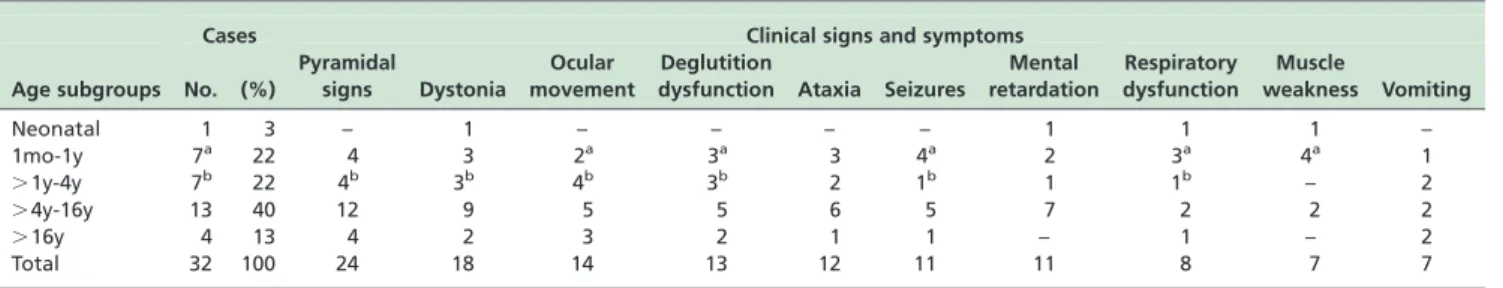
Clinical picture of the 32 patients. According to the classification proposed by van Erven,5based on age of onset of clinical symptoms as neonatal (up to 4 weeks of age), early infantile (1mo to 1y), infantile (.1y to 4y), juvenile (.4y to 16), and adolescent/young adult (.16y), the distribution of our cases was: only one neonatal, seven early infantile, seven infantile, thirteen juvenile, and four adolescent/young adult. Pyramidal signs (75%) were the most frequent clinical sign, followed by dystonia (56%), ocular movement abnormalities (43%), swallowing difficulties (41%), cerebellar signs (37%), seizures and mental developmental delay (34%), respiratory dysfunction (25%), vomiting and muscle weakness (22%) (Table 1). Interestingly, pyramidal signs were predominant after 4 years of age, observed in 16 out of 17 cases (94%); similarly, ocular movement abnormalities, cerebellar signs, swallowing and mental developmental delay were more frequent among those older than 4 years (47% and 41%).
In contrast, respiratory dysfunction and muscle weakness (33%) were more frequent among patients younger than 4 years old, mostly observed in the first year of age.
Lactate levels
Increased serum lactate levels at rest were observed in 19 out of 22 available samples, which were confirmed after exercise in 13 samples out of 16, and also in cerebrospinal fluid in 15 out of 17 samples available. Normal levels were detected in 3 patients, either at rest or after exercise, and also in cerebrospinal fluid of two patients. One of those patients with normal lactate harbored the mtDNA mutation (case b), corroborating that lactate acidosis is not always present in LS, as previously reported.5,6Therefore, the lactate level was
not included among the parameters for presumptive diagnosis of LS in the present study.
Neuroimaging findings
MRI detected lesions mostly in putamen, caudate nucleus, periaqueductal grey matter, white matter, sub-stantia nigra, and globus pallidum, in decreasing order of frequency, as shown in Fig. 1. There was no particular
Table 1 -Clinical signs and symptoms according to the age subgroups
Cases Clinical signs and symptoms
Age subgroups No. (%)
Pyramidal
signs Dystonia
Ocular movement
Deglutition
dysfunction Ataxia Seizures
Mental retardation
Respiratory dysfunction
Muscle
weakness Vomiting
Neonatal 1 3 – 1 – – – – 1 1 1 –
1mo-1y 7a 22 4 3 2a 3a 3 4a 2 3a 4a 1
.1y-4y 7b 22 4b 3b 4b 3b 2 1b 1 1b – 2
.4y-16y 13 40 12 9 5 5 6 5 7 2 2 2
.16y 4 13 4 2 3 2 1 1 – 1 – 2
Total 32 100 24 18 14 13 12 11 11 8 7 7
aindicates patient with T8993G mtDNA mutation;bindicates the patients with T8993C mutation.
DNA mutations in Leigh syndrome
Marie SKN et al. MEDICALEXPRESS 2014;1(5):239-242
pattern of the distribution of lesions for patients with mtDNA mutation.
mtDNA analysis
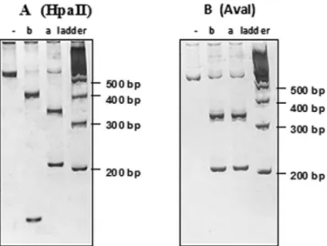
Two patients presented mtDNA mutations: T8993G and T8993C (Fig. 2), and their clinical pictures overlapped with those without mutation (Table 1 and Fig. 1).
B DISCUSSION
This study included 32 patients with clinical symptoms, and neuroimaging findings compatible to LS. The affected brain areas as shown by CT-scanning and MRI were similar, only with a greater number of lesions detected by the MRI, as expected. The MR-FLAIR sequence was more sensitive in demonstrating brainstem involvement than the classical T2-weighted sequence.
There are three different modes of inheritance in LS: X-linked recessive, autosomal recessive, and maternal due to mtDNA mutations.7 Holt et al, in 1990,8 were the first to demonstrate the T8993G mutation in mtDNA, being the most frequently occurring in LS.9 This mutation results in the substitution of leucine, a hydrophilic amino acid by arginine, a hydrophobic amino acid. Arginine is positively charged and blocks proton translocation while impairing the ATP synthesis. A low quantity of this mutation leads to a condition known as NARP, characterized by: peripheral neuropathy, ataxia, retinitis pigmentosa, seizures, and dementia. One of our patients presented this mutation exceeding a 90% level, in proportion to the wild mtDNA. A second patient presented a T8993C point mutation, the second most frequent mtDNA mutation associated with LS, as described by De Vries et al.10 Apparently, this mutation is related to a less severe phenotype. We detected only two out of 32 cases with mtDNA mutation (6%) with clinical and neuroimaging criteria used for the diagnosis of LS. However, when we analyze the same casuistry distributed across different age ranges, we found 13% positivity in the subgroup under 4 years of age (2 out of 15). A thorough phenotype analysis of these 2 patients with mtDNA mutation did not disclose any clinical clue for the presence of mtDNA alteration.
Figure 1 - Upper panel, the main involved areas in MRI. PT: putamen, CN: caudate nucleus, GP: globopallidum, WM: white matter, SN: substantia nigra, CC: corpus callosum, TH: thalamus, PA: periaqueductal grey matter, CBL: cerebellum.a: patient with T8993G mutation,b: patient with T8993C mutation. Lower panel, serial axial MRI of patienta. 1, 2 and 4: T2-weighted images (2400/120) showing hypersignal on midbrain tegmentum, substantia nigra, putamen, head of caudate nucleus and globopallidum, with bilateral and symmetrical distribution. 3: FLAIR sequence (8000/160/2300) better detecting the lesion on midbrain tegmentum.
Figure 2 -The polyacrylamide gel electrophoresis of PCR products digested by HpaII (A) and AvaI (B) endonucleases. HpaII identifies only the T8993G mutation, whereas AvaI identifies both mutations. Note that patientapresents the T8993G mutation, in which HpaII cut the 551bp PCR product into two fragments of 347 and 208 bps. Patientbpresents T8993C mutation, showing the same fragments when cut with AvaI, but not with HpaII. Both patients present a heteroplasmic condition, with high proportion of mtDNA mutant. Patientbpresents, in addition, a non-pathologic polymorphism at position A8784G, confirmed by automatic sequencing, which was first detected by the different pattern of migration when cut with HpaII.
MEDICALEXPRESS 2014;1(5):239-242 DNA mutations in Leigh syndrome Marie SKN et al.
Although four other mtDNA mutations described in LS were also screened in the present casuistry, several other mtDNA mutations have been recently associated with LS.11,12,13,14This stresses the necessity for a more thorough search for mitochondrial genome mutations. To this end, large scale sequencing technologies would be helpful to detect these cases. However, LS may also be a feature of deficiency of any of the mitochondrial respiratory chain complexes: complex I to V, encoded by nuclear genes (OMIM number: 252010, 252011, 124000, 220110, 604273); of components of the pyruvate dehydrogenase complex (OMIN 238331, 300502, 308930); of deficiency of coenzyme Q10;15 and also of COX deficiency associated with the leucine-rich PPR motif-containing protein (LRPPRC) gene mutation.16Therefore, in addition to mitochondrial genome screening, whole exome sequencing analysis would be necessary to identify the molecular defect to provide a reliable genetic counseling and for future therapeutic strategies. Next generation sequencing technology offers flexible and scalable platforms for sequencing several cases at the same time, allowing in-depth coverage to detect mutations and polymorphisms from a set of known genes, up to the whole exome or genome. The procedure can be completed in a few days with a cost per nucleotide much lower than any previous automated sequencing method.17,18 Mostly, the requirement of pico to nanograms of starting DNA/RNA amounts is another crucial advantage when dealing with limited biological material. The cost/effective ratio is decreasing over time and precise molecular diagnosis will be feasible even for an extensive genetic heterogeneous syndrome, such as LS. A continuous collection of systematic clinical, laboratorial, and neuroimaging findings such as those presented here, in spite of any drawbacks of a retrospective study, might be helpful for the establishment of an effective algorithm for such a molecular diagnosis.
B ACKNOWLEDGEMENTS
This study was supported by a grant fromFundaca˜o de Amparo a` Pesquisa do Estado de Sa˜o Paulo(FAPESP, process # 97/3802-6). We sincerely thank the residents of the Department of Neurology for the diagnostic procedures and clinical follow-up of all patients included in this study.
B RESUMO
OBJETIVO:Determinar a prevaleˆncia de mutaco˜es no DNA mitocondrial (DNAmt) em casos com achados compatı´veis com o diagno´stico de sı´ndrome de Leigh em um Servico de Neurologia brasileiro, e comparar essas descobertas entre os pacientes que apresentam ou na˜o essas mutaco˜es. ME´ TODO:Seis pontos de mutaco˜es do DNAmt (T8993G, T8993C, T8851C, G1644T, T9176C e T3308C) foram analisados por PCR e digesta˜o com endonuclease em 32 pacientes com diagno´stico presuntivo de sı´ndrome de Leigh, de acordo com a distribuica˜o em diferentes faixas eta´rias.
RESULTADOS: Dois pacientes no subgrupo abaixo de 4 anos de idade apresentaram as mutaco˜es T8993G e T8993C do DNAmt. Os sintomas clı´nicos
e os achados de neuroimagens destes dois pacientes foram similares aos dos casos sem mutaco˜es detectadas.
CONCLUSA˜ O:Como a confirmaca˜o molecular e´ fundamental tanto para o aconselhamento gene´tico como para a orientaca˜o terapeˆutica, enfatizamos o benefı´cio da pesquisa de mutaco˜es no DNAmt em pacientes com feno´tipo de Sı´ndrome de Liegh abaixo de 4 anos de idade. O sequenciamento em larga escala do genoma mitocondrial e do exoma completo por tecnologia de sequenciamento de nova geraca˜o podera´ ser uma alternativa futura no estabelecimento do diagno´stico molecular nesta sı´ndrome gene´tica extensa-mente heterogeˆnea.
B REFERENCES
1. Rahman S, Blok RB, Dahl HH, Danks DM, Kirby DM, Chow CW, et al. Leigh syndrome: clinical features and biochemical and DNA abnormal-ities. Ann Neurol. 1996;39(3):343-51.
2. Leigh D. Subacute necrotizing encephalomyelopathy in an infant. J NeurolNeurosurg Psychiatry. 1951;14(3):216-21.
3. Montpetit VJ, Andermann F, Carpenter S, Fawcett JS, Zborowska-Sluis D, Giberson HR. Subacute necrotizing encephalomyelopathy. A review and a study of two families. Brain. 1971;94(1):1-30.
4. Vilarinho L, Maia C, Coelho T, Coutinho P, Santorelli FM. Heterogeneous presentation in Leigh syndrome. J InherMetab Dis. 1997;20(5):704-5. 5. Haas RH, Parikh S, Falk MJ, Saneto RP, Wolf NI, Darin N, et al. The
in-depth evaluation of suspected mitochondrial disease. Mol Genet Met. 2008;94(1):16-37.
6. Chow SL, Rooney ZJ, Cleary MA, Clayton PT, Leonard JV. The significance of elevated CSF lactate. Arch Dis Child. 2005;90(11):1188-9. 7. DiMauro S, De Vivo DC. Genetic heterogeneity in Leigh syndrome. Ann
Neurol. 1996;40(1):5-7.
8. Holt IJ, Harding AE, Petty RKH, Morgan-Hughes JA. A new mitochondrial disease associated with mitochondrial DNA heteroplasmy. Am J Hum Genet. 1990;46(3):428-33.
9. Santorelli FM, Shanske S, Macaya A, DeVivo DC, DiMauro S. The mutation at nt 8993 of mitochondrial DNA is a common cause of Leigh’s syndrome. Ann Neurol. 1993;34(6):827.
10. DeVries DD, Engelen BGM, Gabree¨ls FJM, Ruitenbeek W, Oost BA. A second missense mutation in the mitochondrial ATPase 6 gene in Leigh’s Syndrome. Ann Neurol. 1993;34(3):410-2.
11. Blanco-Grau A, Bonaventura-Ibars I, Coll-Cantı´ J, Melia` MJ, Martinez R, Martı´nez-Gallo M, et al. Identification and biochemical characterization of the novel mutation m.8839G.C in the mitochondrial ATP6 gene associated with NARP syndrome. Genes Brain Behav. 2013;12(8):812-20. 12. La Morgia C, Caporali L, Gandini F, Olivieri A, Toni F, Nassetti S, et al.
Association of the mtDNA m.4171C.A/MT-ND1 mutation with both optic neuropathy and bilateral brainstem lesions. BMC Neurol. 2014; 14(1):116.
13. Leng Y, Liu Y, Fang X, Li Y, Yu L, Yuan Y, et al. The mitochondrial DNA 10197 G.A mutation causes MELAS/Leigh overlap syndrome present-ing with acute auditory agnosia. Mitochondrial DNA. 2014; Apr 8 [Epub ahead of print] PubMed PMID: 24708134.
14. Truong HT, Nguyen VA, Nguyen LV, Pham VA, Phan TN. Screening of common point-mutations and discovery of new T14727C change in mitochondrial genome of Vietnamese encephalomyopathy patients. 2014; Mitochondrial DNA. 2014; Apr 8. [Epub ahead of print] PubMed PMID: 24708131.
15. Van Maldergem L, Trijbels F, DiMauro S, Sindelar PJ, Musumeci O, Janssen A, et al. Coenzyme Q-responsive Leigh’s encephalopathy in two sisters. Ann Neurol. 2002;52(6):750-4.
16. Mootha VK, Lepage P, Miller K, Bunkenborg J, Reich M, Hjerrild M, et al. Identification of a gene causing human cytochrome c oxidase deficiency by integrative genomics. Proc Natl Acad Sci USA. 2003;100(2):605-10. 17. Quail M, Smith ME, Coupland P, Otto TD, Harris SR, Connor TR, et al.
A tale of three next generation sequencing platforms: comparison of Ion torrent, pacific biosciences and IlluminaMiSeq sequencers. BMC Genomics. 2012;13(1):341.
18. Liu L, Li Y, Li S, Hu N, He Y, Pong R, et al. Comparison of next-generation sequencing systems. J Biomed Biotechnol. 2012;2012:251364.
DNA mutations in Leigh syndrome
Marie SKN et al. MEDICALEXPRESS 2014;1(5):239-242